- Kundi seeks PM’s intervention against ‘unconstitutional’ restrictions on inter-provincial movement of wheat Dawn
- Punjab urged to lift flour ban The Express Tribune
- Why three states in Pakistan are fighting over wheat and flour Firstpost
Blog
-
Kundi seeks PM’s intervention against ‘unconstitutional’ restrictions on inter-provincial movement of wheat – Dawn
-
Kundi seeks PM’s intervention against ‘unconstitutional’ restrictions on inter-provincial movement of wheat – Dawn
- Kundi seeks PM’s intervention against ‘unconstitutional’ restrictions on inter-provincial movement of wheat Dawn
- Punjab urged to lift flour ban The Express Tribune
- Why three states in Pakistan are fighting over wheat and flour Firstpost
Continue Reading
-
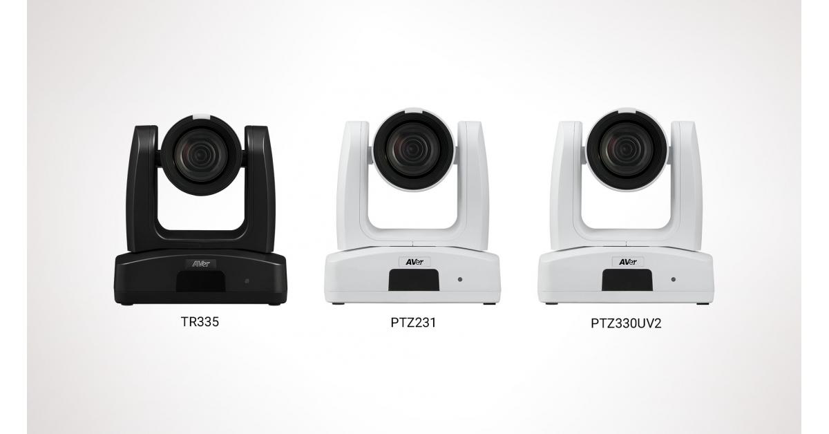
AVer Announces TR335, PTZ231 and PTZ330UV2 Cameras are Certified for Microsoft Teams
TR335, PTZ231 and PTZ330UV2 Cameras are Certified for Microsoft Teams
AVer expands its portfolio with three powerful cameras designed to deliver exceptional video collaboration experiences in Microsoft Teams environments
ROTTERDAM,…
Continue Reading
-
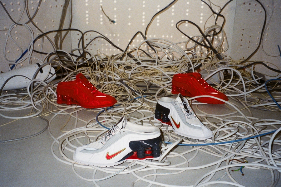
Nike and Martine Rose Expand Their Sporting Universe Into the World of Gaming — NIKE, Inc.
Skill over status. Individuality over conformity. Expression over everything.
It’s with this spirit that Nike and Martine Rose are unveiling the latest product of their longstanding partnership: a vibrant collection of apparel, footwear and…
Continue Reading
-

Mechanisms underlying lichen planus in association with biologic thera
Introduction
Lichen planus (LP) is a chronic, inflammatory, and immune-mediated disorder that may affect the skin, nails, hair, and mucous membranes, with an estimated worldwide prevalence of 0.22–5%.1 The traditional six “P’s” of LP, “Pruritic, Purple, Polygonal, Planar, Papules, and Plaques” describe the typical cutaneous presentation of LP as polygonal, flat-topped, violaceous papules and plaques, though there are many morphological variants.2–4 Although etiology is often idiopathic, LP may be triggered by exogenous factors, including viral infections, such as hepatitis C,5 vaccines,6–9 and pharmacological agents, such as anti-hypertensives, nonsteroidal anti-inflammatory agents, and antimalarials.10
With expanding use of biologic therapies in oncology, rheumatology, dermatology, and gastroenterology, LP and drug-induced lichenoid reactions are increasingly recognized as adverse events. These drug eruptions can complicate the long-term management of patients with chronic inflammatory diseases and necessitate therapeutic reassessment, due to the complexity of these targeted, immune-modulating treatments. Since the mechanisms underlying biologic-associated LP remain incompletely defined, this review aims to summarize current evidence on biologic-induced LP, outline implicated drug classes, and propose mechanistic models for its development.
Materials and Methods
Searches for peer-reviewed journal articles were conducted on August 12, 2025 using the PubMed/MEDLINE database with the search terms “lichen planus”, “lichenoid”, “biologics”, “drug-induced”, and “mechanism.” Additional search terms included “biological therapy”, “TNF inhibitor”, “checkpoint inhibitor”, “IL-17 inhibitor”, “IL-23 inhibitor”, “adalimumab”, “etanercept”, “infliximab”, “ustekinumab”, “secukinumab”, “dupilumab”, “nivolumab”, and “pembrolizumab.” Filters included “Case Reports”, “Clinical Study”, “Clinical Trial”, “Meta-Analysis”, “Review”, and “Systematic Review.” Articles were included if they presented example(s) of patients with lichenoid eruption following biologic therapy or discussed the phenomenon. Exclusion criteria included studies that did not mention lichenoid reactions and biologic therapy, vaccine-induced LP, and studies with discussion of biologic therapy as treatment for LP without presentation of LP after initiation of the therapeutic. Reference lists from these articles were used to find additional articles. Studies without readily obtainable full text in English were excluded (Figure 1).
Figure 1 Flowchart diagram for selection of studies.
Pathogenesis of Lichen Planus
The pathogenesis of LP is incompletely understood, but converging evidence supports that LP is driven by a cytotoxic immune response that induces apoptosis of basal keratinocytes at the dermoepidermal junction.3,4 On immunohistochemistry of both mouse model and human biopsy samples, there is a predominance of CD8+ T lymphocytes in the lichenoid infiltrate, though CD4+ T cells, in particular T-helper-1 (Th1) cells, also play an important role in driving basal keratinocyte apoptosis.4,11–13 Increased expression of intracellular adhesion molecule-1 (ICAM-1) by basal keratinocytes enhances this interaction, which is a unique feature of LP in comparison to other interface dermatoses, such as subacute cutaneous lupus erythematous and erythema multiforme.14,15 Several cytokines are upregulated in this process, including tumor necrosis factor-α (TNF-α), interferon-γ (IFN-γ), nuclear factor kappa B (NFκB), interleukin-1 (IL-1), IL-6, IL-8, and IL-10.16,17 Apoptosis-related molecules, such as Fas/Apo-1 and Bcl-2, are also implicated.18 Plasmacytoid dendritic cells (pDCs) contribute to pathogenesis by producing type I interferons, a pathway which may link viral infections and LP through recruitment of CD8+ lymphocytes.19 Overall, LP likely reflects a breakdown of self-tolerance, with keratinocytes acting as both targets and amplifiers of immune injury. This immune pathogenesis contributes to the histopathological lichenoid tissue reaction pattern characteristic of LP.
Overview of Implicated Biologic Therapies
Biologic-associated LP and lichenoid eruptions have been reported in association with multiple therapeutic classes, most prominently programmed cell death protein-1 (PD-1)/PD-ligand 1 (PD-L1) inhibitors, such as pembrolizumab and nivolumab,20,21 and TNF-α-inhibitors, such as infliximab, adalimumab, and etanercept.22,23 In an observational study of 82 melanoma patients, 17% of patients developed lichenoid drug eruption associated with anti-PD-1 antibody treatment.24 Biologics, such as those targeting IL-17 and IL-23 have also been associated with lichenoid reactions.25 Our literature review identified 157 cases of lichenoid drug eruptions following biologic therapy with 24 distinct medications (Table 1). Clinical presentations were heterogenous, encompassing classic cutaneous LP (Figure 2), 50 cases of oral LP, 22 cases of lichen planus pemphigoides (LPP), 7 cases of nail LP (Figure 3), and 6 cases of lichen planopilaris. The wide spectrum of presentation suggests that the potential disruption of multiple inflammatory pathways may precipitate biologic-induced lichenoid reactions, highlighting the need to examine the immunologic mechanisms.

Figure 2 Multiple flat-topped violaceous papules, some coalescing into plaques, on the forearms and legs.
Notes: Copyright ©2023. Reproduced from Zemlok SK, Buuh S, Brown R et al. Nivolumab-induced lichen planus responsive to dupilumab treatment in a patient with stage III C melanoma. JAAD Case Reports, Volume 38, 23–26.26

Figure 3 (A and B) A 75-year-old male with nail lichen planus secondary to imatinib use. Fingernails atrophic with pterygium on 7/10 fingernails, sparing the right thumbnail and left fourth and fifth fingernails. On dermoscopy, severe nail plate thinning and atrophy is apparent.
Notes: Copyright ©2025. Reproduced from Axler E, Loesch E,Vaidya T et al. Nail lichen planus associated with imatinib mesylate. JAAD Case Reports, Volume 58, 25–28.27

Table 1 Characteristics of Reported Biologic-Associated Lichenoid Eruptions from the Literature, Summarized by Agent and Class
Proposed Mechanisms of Biologic-Induced Lichen Planus
Biologic-associated lichenoid eruptions have been reported across multiple biologic classes, suggesting that these reactions are attributable to nuanced immunological mechanisms. They appear to result from disruptions to immune homeostasis that ultimately favor cytotoxic T cell-mediated injury at the dermoepidermal junction.28 Several mechanistic models emerge from the literature, including cytokine imbalances, immune pathway redirection, unmasking of antigens and epitope spreading, dysregulation of keratinocyte apoptosis pathways, and potential genetic or host susceptibility factors.
Cytokine Imbalances
One of the most widely proposed mechanisms for biologic-induced LP involves cytokine imbalance following targeted inhibition of specific inflammatory pathways. In idiopathic LP, type I interferons, especially IFN-α, play important roles in modulating keratinocyte damage.19 Conversely, TNF-α normally exerts an inhibitory effect on pDCs, limiting IFN-α production. Thus, TNF-α blockade by biologics like adalimumab and etanercept may lead to upregulation of cytokines including IFN-α, activating pDCs and cytotoxic T cells and generating an inflammatory response.29–31 Inhibition of TNF-α has also been hypothesized to increase the expression of IL-17A and IL-23, stimulating the Th17 pathway.32
Similar cytokine shifts have been hypothesized with IL-17 inhibitors, such as secukinumab.33 It has been proposed that IL-17 inhibition may activate pDCs and promote the production of inflammatory cytokines such as IL-23, IL-12, and TNF-α, contributing to LP development.32,33 Additionally, hydroxychloroquine is occasionally used to treat biologic-associated LP, because it interferes with toll-like receptor signaling and decreases proinflammatory cytokine production.34
Although direct mechanistic studies remain limited, cytokine imbalance provides a strong potential mechanism for why certain biologics precipitate lichenoid reactions. Further research using serum cytokine profiling may delineate the precise immune shifts associated with each biologic class.
T Lymphocyte Immune Pathway Shifts
Another proposed mechanism for biologic-associated LP is the activation of a Th1-mediated immune response that stimulates autoreactive CD8+ T cell populations, resulting in basal keratinocyte apoptosis.25 For example, the PD-1 pathway typically inhibits T cell activation, allowing malignant cells to escape the immune response. Therefore, biologic agents that inhibit PD-1, such as pembrolizumab and nivolumab, can remove the block on previously suppressed T cells, inducing T cell activation.35,36 These inflammatory T lymphocytes may then proliferate and attack autoantigens in the skin, resulting in LP.36 Furthermore, it has been proposed that the production of type I IFNs is closely associated with the recruitment of cytotoxic lymphocytes and pDCs via CXCR3 ligands, indicating a Th1-mediated immune response.19,29,30 Furthermore, recent reports indicate that Th2 blockade by dupilumab may lead to a Th1/Th2 imbalance, shifting towards a Th1-dominated immune response that may result in dupilumab-induced LP.37–40
Additionally, narrowband ultraviolet B (NBUVB) phototherapy has successfully been used to treat anti-PD-1-induced LP refractory to steroidal treatment.41,42 Since LP is associated with a Th1/Th17 response,43 NBUVB phototherapy may be used therapeutically to shift from a Th1/Th17 response to a Th2 milieu, promoting immunosuppression.41,42 In addition to hydroxychloroquine’s decreased proinflammatory cytokine production, it also downregulates major histocompatibility class (MHC) II-peptide complex formation, resulting in decreased CD4+ T cell activation and a suppressed immune response.34 Therefore, NBUVB phototherapy and hydroxychloroquine may be used to treat biologic-associated LP by shifting from proinflammatory immune responses to Th2-mediated immune pathways.
Antigen Unmasking & Epitope Spreading
It has also been hypothesized that biologic therapy may precipitate lichenoid eruptions by unmasking a previously suppressed inflammatory response to pre-existing autoantigens localized to specific sites in the body.36,44–46 In anti-PD-1 therapy in particular, biologic-associated LP is proposed to be mediated by an unmasked antigen and/or a neoantigen directly created by the anti-PD-1 antibody.46,47 The resulting lichenoid reactions have been found to contain susceptible mutated keratinocytes that specifically express PD-L1, resulting in infiltration of the dermoepidermal junction by autoreactive T lymphocytes and keratinocyte necrosis.48,49 This suggests that the lichenoid eruption is a target effect of the PD-1/PD-L1 pathway, rather than a nonspecific hypersensitivity reaction.48 Interestingly, biologic-associated LP may therefore serve as a biomarker for efficacy of treatment with immune checkpoint inhibitors, encouraging the resumption or continuation of treatment of patients with immunotherapy once the reaction is managed.47,50
It is also reported that dermoepidermal junction damage in LP may expose previously immune-tolerated membrane antigens, eliciting an immune response through epitope spreading.51 This phenomenon has been associated with LPP in particular, with the blistering response occurring after the formation of lichenoid lesions.51,52 The occurrence of rare variants of biologic-associated LP, such as LPP and lichen planopilaris, suggests that distinct antigenic targets within basement membrane components or hair follicles can be unmasked by biologic agents. Finally, it has been proposed that the oral LP associated with imatinib use may be closely correlated with the altered expression of epidermal markers.53
Keratinocyte Apoptosis Dysregulation
Keratinocyte apoptosis is an essential component in the pathogenesis of idiopathic LP, mediated primarily through Fas/Fas ligand interactions and the perforin-granzyme B pathway.18,54 Biologic therapy may be implicated in heightening keratinocyte vulnerability to immune-mediated injury. For example, the activation of cytotoxic T cells associated with biologic therapy may then enable T lymphocytes to secrete granules containing granulysin, perforin, and granzyme B, leading to keratinocyte apoptosis.33 Additionally, keratinocyte apoptosis results in the release of pro-inflammatory cytokines, perpetuating the disease process.33
Altered apoptotic signaling may particularly be implicated in association with receptor activator of nuclear factor κΒ ligand (RANKL)-inhibitor therapy. Under physiologic conditions, RANK-RANKL signaling supports peripheral immune tolerance by prolonging pDC survival, promoting regulatory T cell development, and eliminating autoreactive T cells through Fas-mediated apoptosis.55,56 Inhibition of this pathway with denosumab therapy disrupts immune tolerance, rendering keratinocytes vulnerable to autoreactive T cell-driven apoptosis, which may promote lichenoid eruptions.56
Genetic & Host Susceptibility Factors
While biologic therapy may provide an inciting stimulus for lichenoid reactions, not all patients exposed to immunotherapeutic agents develop LP, suggesting an important role for underlying host susceptibility. Idiopathic LP has been associated with certain HLA genotypes, such as HLA-DR1,1 but whether similar HLA associations predispose to biologic-associated LP is unclear. However, it has been theorized that in certain genotypes, the effect of TNF-α inhibition can accelerate IFN-α production, pathologically activating T cells and pDCs.30 Overall, biologic-associated LP likely represents an unmasking of preexisting immune vulnerability rather than a uniformly-induced pathogenic response. However, future studies involving HLA typing, immune gene sequencing, and longitudinal patient registries may aid in identifying at-risk individuals.
Implications for Clinical Practice
Recognition of biologic-associated LP has important implications for dermatologists, oncologists, rheumatologists, and other physicians prescribing these agents. Lichenoid eruptions can mimic idiopathic LP both clinically and histopathologically, necessitating a high index of suspicion in patients who develop new cutaneous, oral, or follicular lichenoid lesions while on biologic therapy.57 A careful drug history is essential, as latency from drug initiation to eruption may vary widely, ranging from days to years.21,57
From a management standpoint, most cases of biologic-associated LP respond to withdrawal of the offending biologic and topical or systemic corticosteroids.57,58 Steroid-sparing agents, such as acitretin, cyclosporine, methotrexate, apremilast, hydroxychloroquine, phototherapy, and other biologic agents, such as dupilumab and rituximab are also often used to treat these cutaneous reactions.26,34,36,37,39,41,42,48,57–70 However, in patients with limited or well-controlled disease, continuation of the biologic with adjunctive treatment is often reasonable, especially if the biologic is effectively treating the underlying indication.57,58 Nevertheless, more severe presentations of biologic-associated LP, as well as LPP and lichen planopilaris may often necessitate cessation of biologic therapy.
Importantly, management of lichenoid drug eruptions underscores the need for shared decision-making between physicians and patients. Counseling before initiation of biologic therapy should include discussion of potential cutaneous adverse events and strategies for early recognition. When considering withdrawal of biologic therapy, the balance of risks and benefits must be evaluated. Greater understanding of the mechanisms underlying biologic-associated LP may eventually enable risk stratification.
Research Gaps & Future Directions
Despite the growing number of case reports documenting biologic-associated LP, there remain several key gaps in our understanding of the pathogenesis and clinical implications. Primarily, the true incidence and risk factors remain undefined. Large, multicenter prospective studies are needed to establish incidence rates across different biologic classes and identify patient characteristics that may predispose to lichenoid eruptions, such as HLA genotype and autoimmune history. Additionally, functional studies using in vitro keratinocyte and pDC models, cytokine assays, and lesional transcriptomics may provide direct mechanistic evidence to clarify the immunologic mechanisms underlying biologic-associated LP.
Physicians and patients may also benefit from more standardized treatment algorithms. Optimal approaches to continuing versus withdrawing biologic therapy, the role of adjunctive treatments, and outcomes after rechallenge with the offending biologic remain unclear. Prospective studies evaluating therapeutic decisions and long-term outcomes are needed to streamline management strategies. Finally, studies evaluating HLA typing, immune profiling, and/or genetic testing may guide risk stratification, enabling a more individualized approach to prescribing and surveilling patients initiating biologic therapy.
Conclusion
In sum, biologic therapies are increasingly recognized as potential triggers of LP and related lichenoid eruptions. Evidence to date suggests that biologic-associated LP arise through multiple related mechanisms, including cytokine imbalance, T cell dysregulation, antigen unmasking, keratinocyte apoptosis, and host susceptibility. It is important for physicians to counsel patients regarding potential risks of biologic therapy, and carefully weigh the benefits of continuing vs withdrawing treatment. Future studies aimed at clarifying mechanisms and defining incidence, risk factors, and predictive biomarkers may improve our understanding and help guide patient care.
Abbreviations
LP, lichen planus; Th, T-helper; ICAM-1, intracellular adhesion molecule-1; TNF-α, tumor necrosis factor-α; IFN-γ, interferon-γ; NFκB, nuclear factor kappa B; IL, interleukin; pDC, plasmacytoid dendritic cell; PD-1, programmed cell death protein-1; PD-L1, programmed cell death protein-ligand 1; LPP, lichen planus pemphigoides; NBUVB, narrowband ultraviolet B; MHC, major histocompatibility complex; RANKL, receptor activator of nuclear factor κΒ ligand.
Data Sharing Statement
Data sharing is not applicable to this article as no new data were created or analyzed in this study.
Author Contributions
Ms. Podolsky contributed to formal analysis, investigation, methodology, visualization, and writing – original draft. Dr. Lipner contributed to conceptualization, project administration, supervision, and writing – review and editing. All authors gave final approval of the version to be published; have agreed on the journal to which the article has been submitted; and agree to be accountable for all aspects of the work.
Funding
No funding was obtained for this study.
Disclosure
The authors report no conflicts of interest in this work. Dr. Lipner has served as a consultant for Moberg Pharmaceuticals and BelleTorus Corporation. AI was not used in article composition.
References
1. Gorouhi F, Davari P, Fazel N. Cutaneous and mucosal lichen planus: a comprehensive review of clinical subtypes, risk factors, diagnosis, and prognosis. ScientificWorldJournal. 2014;2014:742826. doi:10.1155/2014/742826
2. Usatine RP, Tinitigan M. Diagnosis and treatment of lichen planus. Am Fam Physician. 2011;84(1):53–60.
3. Gupta MK, Lipner SR. Review of Nail Lichen Planus: epidemiology, Pathogenesis, Diagnosis, and Treatment. Dermatol Clin. 2021;39(2):221–230. doi:10.1016/j.det.2020.12.002
4. Lehman JS, Tollefson MM, Gibson LE. Lichen planus. Int J Dermatol. 2009;48(7):682–694. doi:10.1111/j.1365-4632.2009.04062.x
5. Daoud MS, Gibson LE, Daoud S, el-Azhary RA. Chronic hepatitis C and skin diseases: a review. Mayo Clin Proc. 1995;70(6):559–564. doi:10.4065/70.6.559
6. Agrawal A, Shenoi SD. Lichen planus secondary to hepatitis B vaccination. Indian J Dermatol Venereol Leprol. 2004;70(4):234–235.
7. Hardy C, Glass J. Linear Lichen Planus in the Setting of Annual Vaccination. Mil Med. 2019;184(5–6):e467–e469. doi:10.1093/milmed/usy234
8. Usman A, Kimyai-Asadi A, Stiller MJ, Alam M. Lichenoid eruption following hepatitis B vaccination: first North American case report. Pediatr Dermatol. 2001;18(2):123–126. doi:10.1046/j.1525-1470.2001.018002123.x
9. D’Agostino M, Martora F, Megna M, Napolitano M, Potestio L. Lichen planus following COVID-19 vaccination: a narrative review. Clin Exp Dermatol. 2025;50(2):260–266. doi:10.1093/ced/llae356
10. Thompson DF, Skaehill PA. Drug-induced lichen planus. Pharmacotherapy. 1994;14(5):561–571. doi:10.1002/j.1875-9114.1994.tb02852.x
11. Shiohara T, Mizukawa Y. The immunological basis of lichenoid tissue reaction. Autoimmun Rev. 2005;4(4):236–241. doi:10.1016/j.autrev.2004.11.005
12. Mizukawa Y, Yamazaki Y, Teraki Y, et al. Direct evidence for interferon-gamma production by effector-memory-type intraepidermal T cells residing at an effector site of immunopathology in fixed drug eruption. Am J Pathol. 2002;161(4):1337–1347. doi:10.1016/s0002-9440(10)64410-0
13. Zhou S, Zhang Z, Feng X, Zhao C, Jiang L. Lichenoid mucocutaneous reactions associated with sintilimab therapy in a non-small cell lung adenocarcinoma patient: case report and review. Front Pharmacol. 2023;14:1276788. doi:10.3389/fphar.2023.1276788
14. Norris DA. Cytokine modulation of adhesion molecules in the regulation of immunologic cytotoxicity of epidermal targets. J Invest Dermatol. 1990;95(6 Suppl):111s–120s. doi:10.1111/1523-1747.ep12874977
15. Bennion SD, Middleton MH, David-Bajar KM, Brice S, Norris DA. In three types of interface dermatitis, different patterns of expression of intercellular adhesion molecule-1 (ICAM-1) indicate different triggers of disease. J Invest Dermatol. 1995;105(1 Suppl):71s–79s. doi:10.1111/1523-1747.ep12316107
16. El-Howati A, Thornhill MH, Colley HE, Murdoch C. Immune mechanisms in oral lichen planus. Oral Dis. 2023;29(4):1400–1415. doi:10.1111/odi.14142
17. Lu R, Zhang J, Sun W, Du G, Zhou G. Inflammation-related cytokines in oral lichen planus: an overview. J Oral Pathol Med. 2015;44(1):1–14. doi:10.1111/jop.12142
18. Sklavounou-Andrikopoulou A, Chrysomali E, Iakovou M, Garinis GA, Karameris A. Elevated serum levels of the apoptosis related molecules TNF-alpha, Fas/Apo-1 and Bcl-2 in oral lichen planus. J Oral Pathol Med. 2004;33(7):386–390. doi:10.1111/j.1600-0714.2004.00221.x
19. Wenzel J, Scheler M, Proelss J, Bieber T, Tüting T. Type I interferon-associated cytotoxic inflammation in lichen planus. J Cutan Pathol. 2006;33(10):672–678. doi:10.1111/j.1600-0560.2006.00527.x
20. Alharbi A, Khobrani A, Noor A, et al. Risk of Lichen Sclerosus and Lichen Planus in Patients Receiving Immune Checkpoint Inhibitors. Int J Environ Res Public Health. 2022;20(1):580. doi:10.3390/ijerph20010580
21. Le TK, Brown I, Goldberg R, et al. Cutaneous Toxicities Associated with Immune Checkpoint Inhibitors: an Observational, Pharmacovigilance Study. J Invest Dermatol. 2022;142(11):2896–2908.e4. doi:10.1016/j.jid.2022.04.020
22. Succaria F, Bhawan J. Cutaneous side-effects of biologics in immune-mediated disorders: a histopathological perspective. J Dermatol. 2017;44(3):243–250. doi:10.1111/1346-8138.13762
23. Behrangi E, Moodi F, Jafarzadeh A, Goodarzi A. Paradoxical and bimodal immune-mediated dermatological side effects of TNF-α inhibitors: a comprehensive review. Skin Res Technol. 2024;30(5):e13718. doi:10.1111/srt.13718
24. Hwang SJ, Carlos G, Wakade D, et al. Cutaneous adverse events (AEs) of anti-programmed cell death (PD)-1 therapy in patients with metastatic melanoma: a single-institution cohort. J Am Acad Dermatol. 2016;74(3):455–61.e1. doi:10.1016/j.jaad.2015.10.029
25. Kim PJ, Sachdeva M, Mufti A, Lytvyn Y, Maliyar K, Yeung J. Lichenoid Drug Eruptions Associated With the Use of Biologic Therapy: a Systematic Review. J Cutan Med Surg Sep-Oct. 2022;26(5):521–522. doi:10.1177/12034754221100188
26. Zemlok SK, Buuh S, Brown R, Murphy M, Hegde UP, Mallett JR. Nivolumab-induced lichen planus responsive to dupilumab treatment in a patient with stage III C melanoma. JAAD Case Rep. 2023;38:23–26. doi:10.1016/j.jdcr.2023.05.036
27. Axler E, Loesch E, Vaidya T, Neubauer Z, Lipner SR. Nail lichen planus associated with imatinib mesylate. JAAD Case Rep. 2025;58:25–28. doi:10.1016/j.jdcr.2024.12.039
28. Watanabe T, Yamaguchi Y. Cutaneous manifestations associated with immune checkpoint inhibitors. Front Immunol. 2023;14:1071983. doi:10.3389/fimmu.2023.1071983
29. Asarch A, Gottlieb AB, Lee J, et al. Lichen planus-like eruptions: an emerging side effect of tumor necrosis factor-alpha antagonists. J Am Acad Dermatol. 2009;61(1):104–111. doi:10.1016/j.jaad.2008.09.032
30. Jayasekera PS, Walsh ML, Hurrell D, Parslew RA. Case Report of Lichen Planopilaris Occurring in a Pediatric Patient Receiving a Tumor Necrosis Factor α Inhibitor and a Review of the Literature. Pediatr Dermatol. 2016;33(2):e143–6. doi:10.1111/pde.12768
31. Barrientos N, García-Sánchez S, Domínguez JD. Letter: lichenoid eruption induced by etanercept. Dermatol Online J. 2012;18(7):15. doi:10.5070/D32FW0W71N
32. Liu V, Moiin A, Diaz C. Lichen planus associated with secukinumab treatment for plaque psoriasis. Dermatol Online J. 2023;29(2):60779. doi:10.5070/d329260779
33. Heidari A, Fathabadi F, Ghanavati K, Rasouli A, Soleimani S, Heidari N. A systematic review of the role of interleukin inhibitors in lichen planus: therapeutic and paradoxical effects. Inflammopharmacology. 2025. doi:10.1007/s10787-025-01853-4
34. Alnajjar NF, Tahan SR, Iriarte C. Immunotherapy-associated bullous and oral erosive lichenoid eruption successfully treated with hydroxychloroquine. JAAD Case Rep. 2025;56:63–66. doi:10.1016/j.jdcr.2024.11.010
35. Martos-Cabrera L, Lladó I, Fernández-Rico P, Butrón-Bris B, Rodríguez-Jiménez P. Umbilical lichen planus induced by nivolumab. An Bras Dermatol. 2023;98(5):712–714. doi:10.1016/j.abd.2021.09.022
36. Coscarart A, Martel J, Lee MP, Wang AR. Pembrolizumab-induced pseudoepitheliomatous eruption consistent with hypertrophic lichen planus. J Cutan Pathol. 2020;47(3):275–279. doi:10.1111/cup.13587
37. Kern L, Kleinheinrich L, Feldmann R, Sator P, Stella A, Breier F. Dupilumab-Induced Lichen Planus: a Case with Oral and Cutaneous Eruptions. Case Rep Dermatol. 2022;14(3):356–361. doi:10.1159/000527918
38. Kim TE, Shin MK. De novo case of lichenoid eruption following dupilumab treatment. JAAD Case Rep. 2021;13:71–72. doi:10.1016/j.jdcr.2021.04.035
39. Gajraj FA, Zahir J, Adereti C, Gajraj MH. A Case Report and Literature Review of the Role of Dupilumab in the Management of Lichen Planus: cause or Treatment? Cureus. 2023;15(7):e41274. doi:10.7759/cureus.41274
40. Srivatsa A, Yaldo M, Rehman W, et al. What is the Verdict? A Systematic Review of Dupilumab and Lichen Planus. Int J Dermatol. 2025. doi:10.1111/ijd.17909
41. Donaldson M, Owen JL, Chae YK, Choi JN. Management of Persistent Pruritus and Lichenoid Reaction Secondary to Nivolumab With Narrowband Ultraviolet B Phototherapy. Front Oncol. 2018;8:405. doi:10.3389/fonc.2018.00405
42. Esfahani K, Henderson Berg MH, Zargham H, et al. Narrowband ultraviolet B therapy for refractory immune-related lichenoid dermatitis on PD-1 therapy: a case report. J Immunother Cancer. 2021;9(3):e001831. doi:10.1136/jitc-2020-001831
43. Solimani F, Pollmann R, Schmidt T, et al. Therapeutic Targeting of Th17/Tc17 Cells Leads to Clinical Improvement of Lichen Planus. Front Immunol. 2019;10:1808. doi:10.3389/fimmu.2019.01808
44. Edwards C, Fearfield L. Nivolumab-induced lichenoid dermatitis occurring in a patient with metastatic melanoma successfully treated with alitretinoin. Clin Exp Dermatol. 2018;43(5):609–610. doi:10.1111/ced.13410
45. Uthayakumar AK, Rudd E, Noy M, Weir J, Larkin J, Fearfield L. Severe progressive scarring pembrolizumab-induced lichen planopilaris in a patient with metastatic melanoma. Australas J Dermatol. 2021;62(3):403–406. doi:10.1111/ajd.13660
46. Shi VJ, Rodic N, Gettinger S, et al. Clinical and Histologic Features of Lichenoid Mucocutaneous Eruptions Due to Anti-Programmed Cell Death 1 and Anti-Programmed Cell Death Ligand 1 Immunotherapy. JAMA Dermatol. 2016;152(10):1128–1136. doi:10.1001/jamadermatol.2016.2226
47. Sehbai A, Hamid MA, Ibrahim Z. Pembrolizumab-Induced Lichen Planus in a Patient With Non-Small-Cell Lung Carcinoma (NSCLC) That Correlates to Therapeutic Response. Cureus. 2022;14(11):e31454. doi:10.7759/cureus.31454
48. Bansal A, Singla A, Paul D, Kaur S. Pembrolizumab-Induced Lichen Planus: a Rare Immune-Related Adverse Side Effect. Indian Dermatol Online J. 2023;14(3):391–394. doi:10.4103/idoj.idoj_377_22
49. Tembunde Y, Dika MN. Pembrolizumab-Induced Eruptive Keratoacanthomas and Lichen Planus in a Lung Cancer Patient. Cureus. 2023;15(8):e43402. doi:10.7759/cureus.43402
50. Kwon CW, Murthy RK, Kudchadkar R, Stoff BK. Pembrolizumab-induced lichen planus pemphigoides in a patient with metastatic Merkel cell carcinoma. JAAD Case Rep. 2020;6(10):1045–1047. doi:10.1016/j.jdcr.2020.03.007
51. Buján Bonino C, López-Pardo Rico M, Moreiras Arias N, Suárez Peñaranda JM, Casas Fernández L. Checkpoint inhibitor-induced lichen planus pemphigoides: a case report and literature review of an unusually reported entity. Int J Dermatol. 2023;62(4):e200–e204. doi:10.1111/ijd.16502
52. Hübner F, Langan EA, Recke A. Lichen Planus Pemphigoides: from Lichenoid Inflammation to Autoantibody-Mediated Blistering. Front Immunol. 2019;10:1389. doi:10.3389/fimmu.2019.01389
53. Ena P, Chiarolini F, Siddi GM, Cossu A. Oral lichenoid eruption secondary to imatinib (Glivec). J DermatolTreat. 2004;15(4):253–255. doi:10.1080/09546630410015556
54. Kastelan M, Prpić Massari L, Gruber F, et al. The role of perforin-mediated apoptosis in lichen planus lesions. Arch Dermatol Res. 2004;296(5):226–230. doi:10.1007/s00403-004-0512-1
55. Walsh MC, Choi Y. Regulation of T cell-associated tissues and T cell activation by RANKL-RANK-OPG. J Bone Miner Metab. 2021;39(1):54–63. doi:10.1007/s00774-020-01178-y
56. Dourra M, Mussad S, Qiblawi S, Singer R. Denosumab-induced alopecia areata with lichenoid eruption. JAAD Case Rep. 2021;17:9–11. doi:10.1016/j.jdcr.2021.09.003
57. Pach J, Leventhal JS. Cutaneous Immune-Related Adverse Events Secondary to Immune Checkpoint Inhibitors and Their Management. Crit Rev Immunol. 2022;42(4):1–20. doi:10.1615/CritRevImmunol.2023046895
58. Bhardwaj M, Chiu MN, Pilkhwal Sah S. Adverse cutaneous toxicities by PD-1/PD-L1 immune checkpoint inhibitors: pathogenesis, treatment, and surveillance. Cutan Ocul Toxicol. 2022;41(1):73–90. doi:10.1080/15569527.2022.2034842
59. Tirnanić T, Tiodorović D, Vidović N, et al. Pembrolizumab-induced lichen planus in patients with metastatic melanoma: a report of two cases and prognostic implications of cutaneous immune-related adverse events. Acta Dermatovenerol Alp Pannonica Adriat. 2023;32(3):119–122.
60. Lim O, Maher E, Miller DD. PD-1 Inhibitor Induced Hypertrophic Lichen Planus: a Case Report. Drugs R D. 2024;24(2):353–357. doi:10.1007/s40268-024-00461-x
61. Leme HJ, Ramos J, Magarreiro-Silva A, Gouveia A, Alves J. Nivolumab-Induced Lichenoid Eruption: a Case Report. Cureus. 2025;17(6):e86862. doi:10.7759/cureus.86862
62. Fixsen E, Patel J, Selim MA, Kheterpal M. Resolution of Pembrolizumab-Associated Steroid-Refractory Lichenoid Dermatitis with Cyclosporine. Oncologist. 2019;24(3):e103–e105. doi:10.1634/theoncologist.2018-0531
63. Kou L, Agarwal S, Miceli A, Kolb L, Krishnamurthy K, Schmieder S. Steroid-Refractory Lichenoid Eruption Associated with Pembrolizumab in a Patient with Non-Small Cell Lung Cancer. HCA Healthc J Med. 2021;2(6):397–400. doi:10.36518/2689-0216.1198
64. Mueller KA, Cordisco MR, Scott GA, Plovanich ME. A case of severe nivolumab-induced lichen planus pemphigoides in a child with metastatic spitzoid melanoma. Pediatr Dermatol. 2023;40(1):154–156. doi:10.1111/pde.15097
65. Varma A, Friedlander P, De Moll EH, Desman G, Levitt J. Resolution of pembrolizumab-associated lichenoid dermatitis with a single dose of methotrexate. Dermatol Online J. 2020;26(5). doi:10.5070/D3265048774
66. Kost Y, Mattis D, Muskat A, Amin B, McLellan B. Immune Checkpoint Inhibitor-Induced Psoriasiform, Spongiotic, and Lichenoid Dermatitis: a Novel Clinicopathological Pattern. Cureus. 2022;14(8):e28010. doi:10.7759/cureus.28010
67. Headd VA, Mathien A, Pei S, Kuraitis D. Nivolumab-induced lichenoid penile ulceration and re-emergent mucocutaneous eruption treated with hydroxychloroquine. Australas J Dermatol. 2024;65(4):e97–e99. doi:10.1111/ajd.14223
68. Park JJ, Park E, Damsky WE, Vesely MD. Pembrolizumab-induced lichenoid dermatitis treated with dupilumab. JAAD Case Rep. 2023;37:13–15. doi:10.1016/j.jdcr.2023.05.004
69. Brennan M, Baldissano M, King L, Gaspari AA. Successful Use of Rituximab and Intravenous Gamma Globulin to Treat Checkpoint Inhibitor- Induced Severe Lichen Planus Pemphigoides. Skinmed. 2020;18(4):246–249.
70. Geisler AN, Phillips GS, Barrios DM, et al. Immune checkpoint inhibitor-related dermatologic adverse events. J Am Acad Dermatol. 2020;83(5):1255–1268. doi:10.1016/j.jaad.2020.03.132
Continue Reading
-

Last year’s Galaxy Z Flip 6 plunges to an unbeatable price at a huge $670 off
Remember Woot’s splendid deal on one of the best flip phones from last month? Don’t worry if you don’t — the Galaxy Z Flip 6 is back on sale, and this time, the discount is even…Continue Reading
-
Metabolic-Behavioral Risk Factors for Colorectal Adenomatous Polyps in
Introduction
Colorectal cancer (CRC) remains the third most prevalent malignancy globally, presenting a significant public health burden characterized by distinct age-stratified patterns in both incidence and mortality.1 Historically associated…
Continue Reading
-
A Rare Case of multiple Carpometacarpal Joint Fracture-Dislocations wi
Introduction
Carpometacarpal (CMC) joint dislocations and fracture-dislocations are rare injuries, accounting for less than 1.5% of all hand trauma cases.1 Among these, dorsal fracture-dislocations involving the capitate with the third…
Continue Reading
-
Xbox’s president says that the console’s new competitors are “TikTok and movies,” not Sony and Nintendo
Summary
- Halo remake on PS5 signals the old Xbox vs PlayStation console war is over.
- …
Continue Reading
-
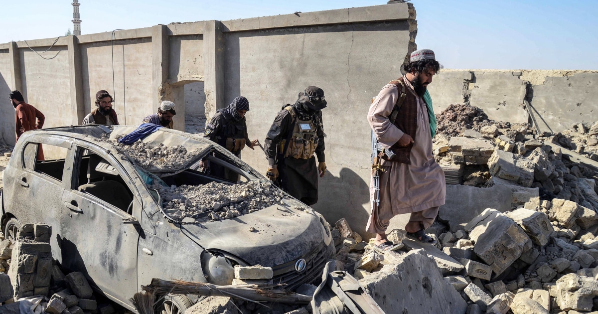
Trump says he will solve Afghanistan-Pakistan crisis ‘very quickly’ as peace talks enter second day
ISLAMABAD — President Donald Trump said Sunday that he will solve the Afghanistan-Pakistan crisis “very quickly,” as peace talks between the warring neighbors entered a second day.
The two countries are embroiled in a bitter security…
Continue Reading