Frieze is pushing into the Gulf, as competition in the wealthy region’s art market heats up.
The London-based art fair and media brand is taking over the existing Abu Dhabi Art fair, which it will relaunch next November as Frieze Abu Dhabi. The…

Frieze is pushing into the Gulf, as competition in the wealthy region’s art market heats up.
The London-based art fair and media brand is taking over the existing Abu Dhabi Art fair, which it will relaunch next November as Frieze Abu Dhabi. The…
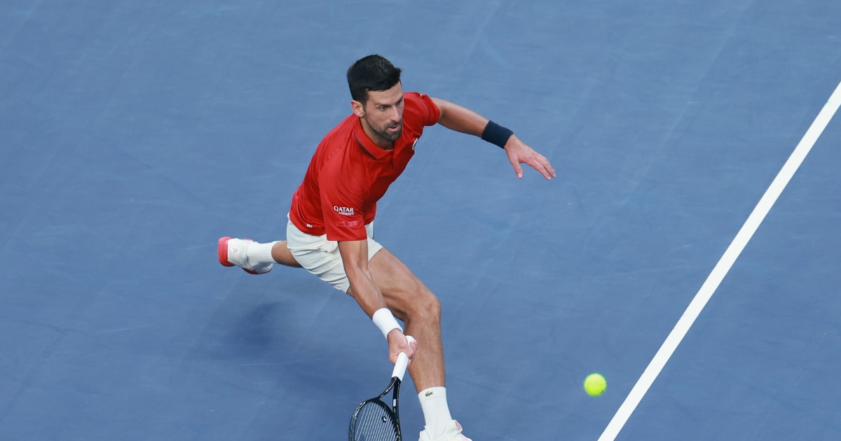
Novak Djokovic gave his all but could not find his way through to the final of the 2025 Shanghai Masters.
The men’s singles tennis Olympics gold medallist was beaten in the semi-finals of the ATP Masters 1000 tournament by world No. 204 Valentin…

Research published this week indicates that North Korean scammers are trying to trick US companies into hiring them for architectural design work, using fake profiles, résumés, and Social Security numbers to pose as legitimate workers. The hustle fits into longstanding campaigns by the hermit kingdom to steal billions of dollars from organizations around the world using careful planning and coordination to pose as professionals in all different fields.
Under pressure from the Department of Justice, Apple removed a series of apps from its iOS App Store this month related to monitoring US Immigration and Customs Enforcement activity and archiving content related to ICE’s actions. As more apps are removed, multiple developers told WIRED this week that they aren’t giving up on fighting Apple over the decisions—and many are still distributing their apps on other platforms in the meantime.
WIRED examined increasing warnings from software supply chain security researchers that the proliferation of AI-generated software in codebases will create an even more extreme version of the code transparency and accountability issues that have come up with widespread integration of open source software components. And Apple announced expansions of its bug bounty program this week, including a maximum $2 million payout for certain exploit chains that could be abused to distribute spyware, and additional bonuses for exploits found in Apple’s Lockdown Mode or in beta versions of new software.
But wait, there’s more! Each week, we round up the security and privacy news we didn’t report in depth ourselves. Click the headlines to read the full stories. And stay safe out there.
The notorious spyware vendor NSO Group, known for developing the Pegasus malware, has faced financial issues since losing a long legal battle against the secure messaging platform WhatsApp as well as a lawsuit filed by Apple. Now, the company, which has long had Israeli ownership, has been purchased by a group of US-based investors led by movie producer Robert Simonds, who helped finance Happy Gilmore, Billy Madison, The Pink Panther, Hustlers, and Ferrari, among many other films. The deal is reportedly worth “several tens of millions of dollars” and is close to completion. Israel’s Defense Export Control Agency (DECA) within the Ministry of Defense will need to approve the sale. Use of mercenary spyware has increased within some US federal government agencies since the beginning of the Trump administration.
Hundreds of national security and cybersecurity specialists who work in the US Department of Homeland Security have faced mandatory reassignment in recent weeks to roles related to President Donald Trump’s mass deportation agenda. Bloomberg reports that affected workers are largely senior staffers who are not union eligible. Workers who refuse to move roles will reportedly be dismissed. Members of DHS’s Cybersecurity and Infrastructure Security Agency (CISA) who have faced reassignment reportedly worked on “issuing alerts about threats against US agencies and critical infrastructure.” For example, CISA’s Capacity Building team has faced a number of reassignments, which could hinder access to emergency recommendations and directives for high-value federal government assets. Workers have been moved to agencies including Immigration and Customs Enforcement, Customs and Border Protection, and the Federal Protective Service.
A recent breach of a third-party customer service provider used by the communication platform Discord included a trove of data from more than 70,000 Discord users that contained identification documents as well as selfies, email addresses, phone numbers, some home location information, and more. The data was collected as part of age verification checks, a mechanism that has long been criticized for centralizing users’ sensitive information. 404 Media reports that the breach was perpetrated by attackers who are attempting to extort Discord. “This is about to get really ugly,” the hackers wrote in a Telegram channel on Wednesday while posting the stolen data.
US Immigration and Customs Enforcement inked a $825,000 contract in May with TechOps Specialty Vehicles (TOSV), a Maryland-based company that manufactures equipment and vehicles for law enforcement. The company provides products including rogue cellphone towers that are used for phone surveillance and sometimes called “stingrays” or “cell-site simulators.” Public records reviewed by TechCrunch show that the agreement describes how the company “provides Cell Site Simulator (CSS) Vehicles to support the Homeland Security Technical Operations program” and is a modification for “additional CSS Vehicles.” TOSV also began a similar $818,000 contract with ICE in September 2024, prior to the start of the Trump administration. In an email to TechCrunch, TOSV president Jon Brianas declined to share details about the contracts but confirmed that the company does provide cell-site simulators. The company does not manufacture them itself, he said.
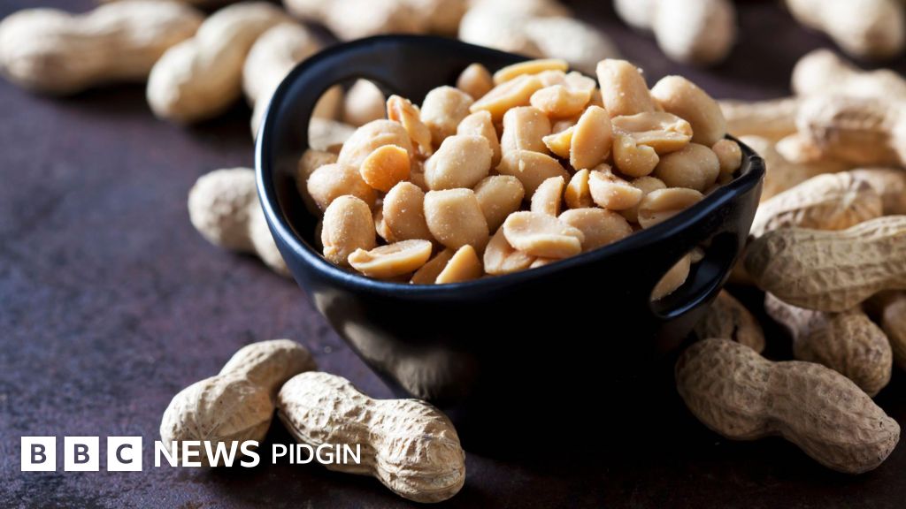
Wia dis foto come from, Westend61/Getty Images
Groundnuts, wey pipo also dey call peanuts, na legume wey full wit nutrients and pipo dey chop am boiled, roasted, or use am…
A venture capital fund is looking for ideas that are out of bounds for traditional investors, seeding technology that may only come to fruition decades down the line, but where researchers can show real results in the lab.
Deep Future…
Migraine is a common neurological disorder impacting about 14% of people worldwide, with a higher reporting frequency in women.1 Triptans, as selective serotonin 5-HT(1B/1D) receptor agonists, are commonly prescribed as the primary…
Breast cancer (BC) has become the most commonly diagnosed cancer and remains a leading cause of cancer-related mortality among women worldwide.1 Ki-67, a nuclear protein associated with cellular proliferation, serves as an essential marker for evaluating tumor growth dynamics.2 Elevated Ki-67 expression is associated with an increased risk of tumor invasion and recurrence.3 Moreover, it correlates with the pathological complete response rate to neoadjuvant therapy (NAT) in BC patients.4 A study by Chen et al found that higher pre-treatment Ki-67 levels were associated with a better clinical response to neoadjuvant chemotherapy in luminal BC subtypes. Specifically, a Ki-67 cutoff value of 25.5% was identified as a predictor of treatment response, indicating that Ki-67 could serve as a valuable biomarker for guiding individualized treatment strategies.5 Furthermore, the POETIC trial demonstrated that changes in Ki-67 levels during preoperative endocrine therapy were predictive of long-term outcomes, underscoring the critical role of Ki-67 in informing treatment strategies and optimizing patient management.6 Currently, Ki-67 expression is assessed via immunohistochemical (IHC) analysis, which typically requires an invasive core needle biopsy (CNB) prior to surgery. However, due to intratumoral heterogeneity and the limited sampling of CNB, discrepancies between CNB and postoperative specimens are frequently observed, with reported inconsistency rates ranging from 10% to 40%.7 Therefore, there is a pressing need for an accurate, comprehensive, and non-invasive method to predict preoperative Ki-67 expression, which is essential for clinical decision-making.
Ultrasound (US) is widely used for the diagnosis of BC due to its simplicity, low cost, and non-invasive nature.8 Compared to conventional US, ABVS enhances reproducibility through automated scanning,9 representing a significant technological advancement in US imaging.10 Its standardized coronal imaging offers detailed lesion information, making it particularly suitable for radiomics analysis.11 Radiomics enables the extraction of high-throughput quantitative features from medical images, allowing for a comprehensive characterization of tumor phenotype.12 It has been increasingly applied in tumor diagnosis, treatment planning, and prognosis prediction.13 The significant potential of radiomics has been fully demonstrated across numerous oncology applications, particularly in improving the predictive performance of ABVS in BC management.11,14–17 This progress marks a major step toward non-invasive tumor biological profiling and further integrates medical imaging with personalized medicine.18 However, tumor heterogeneity, arising from variations in cell composition and spatial distribution, poses a challenge to accurate characterization.19 To better visualize and quantify this heterogeneity, voxel clusters with similar tumor biological characteristics can be grouped into sub-regions.20 Habitat radiomics, an emerging approach, segments tumors into biologically similar sub-regions and extracts features from these areas to enhance the assessment of tumor heterogeneity. A recent study,21 involving multi-modal logistic regression models based on magnetic resonance imaging (MRI), US, and mammography revealed that incorporating peripheral tumor features (within 5 mm) yielded the best performance in distinguishing benign from malignant breast nodules, with an AUC of 0.905 (95% CI: 0.805–1). This highlights the increasing value of multimodal radiomics approaches. Previous US-based radiomics studies have shown promise in predicting Ki-67 expression in BC.16,17 However, these studies were limited by small sample sizes and single-modality imaging, which may not fully capture the complex characteristics of tumors. Additionally, habitat radiomics has not been widely explored in this context. As tumor heterogeneity plays a crucial role in understanding tumor biology, habitat radiomics may enhance predictive accuracy. To address these limitations, our study integrates multimodal radiomics derived from ABVS and 2D US, along with habitat radiomics, to provide a more comprehensive and accurate prediction of Ki-67 expression in BC.
Motivated by these insights, we aim to evaluate the predictive value of radiomics and habitat radiomics features, captured from entire tumors and their sub-regions, using a machine learning (ML) model. Our goal is to establish a robust, non-invasive model for predicting Ki-67 expression in BC, thereby supporting personalized treatment strategies and improving prognostic assessment.
This retrospective study was conducted in accordance with the Declaration of Helsinki and was approved by the Ethics Committee of the First Affiliated Hospital of Anhui Medical University (Approval No. PJ2023-07-11). Given the retrospective nature of the study, which involved the use of previously collected cases and medical records without any new clinical interventions, the requirement for informed consent was waived in accordance with relevant ethical guidelines. To ensure data reliability, strict inclusion and exclusion criteria were applied. The inclusion criteria were as follows: (1) Patients pathologically diagnosed with BC who underwent both ABVS and conventional US examinations within two weeks prior to surgery; (2) No NAT administered before surgery; (3) Availability of complete clinical and pathological information. The exclusion criteria were: (1) Incomplete clinical or pathological data; (2) History of other malignant tumors; (3) Receipt of preoperative NAT; (4) Maximum tumor diameter > 50 mm as measured by US; (5) Presence of bilateral BC. A total of 426 patients with BC met the eligibility criteria. The cases were randomly divided into a training set (n = 297) and a validation set (n = 127), following a 7:3 ratio (Figure 1).
|
Figure 1 Flowchart of the entire research. Abbreviations: ROI, Region of Interest; ABVS, automated breast volume scanner; RadABVS+2D, Radiomics ABVS and 2D model; HadABVS+2D, Habitat radiomics ABVS and 2D model; Rad-HabABVS+2D, Radiomics and Habitat radiomics ABVS and 2D model; CMClinical + Rad-Hab, Clinical–Radiomics–Habitat ABVS+2D Combined Model.
|
Details regarding image acquisition, evaluation procedures, and US equipment specifications are provided in Supplementary Material S1. Breast lesion classification was performed according to the fifth edition of the American College of Radiology (ACR) Breast Imaging Reporting and Data System (BI-RADS).22 The Ki-67 proliferation index was calculated based on the percentage of malignant cells showing positive nuclear staining for Ki-67. A Ki-67 score ≥ 20% was defined as high expression, while a score < 20% was considered low expression.23,24
To segment the lesion regions on ABVS coronal images and 2D US images, the ITK-SNAP software (version 3.8, website: www.itk-snap.org) was used for tumor segmentation. Details of the tumor ROI delineation process are provided in Supplementary Material S2. Before generating habitat sub-regions, the tumors were first localized on both ABVS and 2D images. Each image was paired with a tumor mask of identical dimensions. The bounding box of the tumor region was extracted from the corresponding mask file, and this region was used to define the area for further analysis. Radiomic features were then extracted from these sub-regions. For each pixel within the tumor, a 5 × 5×5 sliding window was applied, expanding outward by two pixels in each direction. Radiomic features within this window were extracted using the pyradiomics library. To enable subsequent clustering analysis, all features were normalized to a range of 0 to 1. This standardization mitigates the influence of features with larger numerical scales and enhances the accuracy and robustness of clustering.25
Although larger windows and a greater number of features may improve robustness to noise, they also significantly increase computational complexity, especially when extracting features at the pixel level. Therefore, in this study, the number of radiomic features was limited to five, specifically those derived from the Gray-Level Co-occurrence Matrix (GLCM). The GLCM effectively captures subtle textural variations in the image, reflecting microscopic irregularity and complexity. It has been widely used to reveal the potential relationship between tumor tissue structure and biological behavior, making it an important tool in the study of tumor heterogeneity25,26 Subsequently, to further quantify image data, each pixel’s local histological features were transformed into a five-dimensional feature vector. This vector integrates multiple aspects of local feature information, such as texture, contrast, and uniformity, facilitating subsequent quantitative analysis and model construction.
A Gaussian mixture model (GMM) clustering algorithm was employed to identify tumor sub-regions composed of biologically similar pixels. Clustering was performed at the cohort level, rather than the individual patient level, to ensure consistent cluster assignment across patients and to allow the propagation of cluster centers from the training set to the test set, ensuring consistent clustering during model application. To determine the optimal number of clusters (ie, habitats), the Silhouette coefficient was used to evaluate clustering performance across a range of k values from 2 to 10. Following clustering, each cluster was assigned a unique color label to generate a cluster label map, which reflected the global distribution of internal regions within the tumor.
Radiomics features were extracted from both tumor regions and sub-regions within the ABVS and 2D US images. Prior to feature extraction, image preprocessing was performed to ensure consistency across datasets. First, the signal intensity of the original images was normalized and standardized to a range of 0–100 to minimize intensity variations between images. Next, spatial resampling was conducted to achieve a uniform in-plane pixel resolution of 2 mm in the XY direction. All feature extraction was confined to the two-dimensional plane. Image gray levels were discretized based on a predefined bin width (eg, grouping every 5 intensity values), enabling standardized texture quantification. All processing was applied exclusively to regions defined by the specified labels in the tumor mask. A wavelet filter was used for feature enhancement prior to extraction. Radiomics features were extracted using the open-source Python package PyRadiomics (https://pyradiomics.readthedocs.io/en/latest/index.html, version 3.0.1), developed by the Computational Imaging Bioinformatics Laboratory at Harvard Medical School. Both unfiltered (from original images) and filtered features were included in the analysis. A total of 464 features were extracted and categorized into the following classes: 90 first-order features, 9 shape features, 110 Gray-Level Co-occurrence Matrix (GLCM) features, 80 gray-level size zone matrix (GLSZM) features, 80 gray-level run length matrix (GLRLM) features, 25 neighboring gray-tone difference matrix (NGTDM) features, 70 gray-level dependence matrix (GLDM) features. PyRadiomics adheres to the standards of the Imaging Biomarker Standardization Initiative (IBSI), ensuring consistent definitions and methodologies for radiomic feature extraction.
The selection of predictive features associated with Ki-67 expression was performed through a multi-step process. This included screening radiomics features, sub-regional radiomics features, and clinical US features. The detailed feature selection workflow is presented in Supplementary Material S3.
After feature selection and fusion, five predictive models were developed, namely the clinical model, the radiomics model (Rad ABVS + 2D), the habitat radiomics model (Hab ABVS + 2D), the combined radiomics model (Rad-Hab ABVS + 2D), and the Clinical–Radiomics–Habitat ABVS+2D Combined Model(CM Clinical + Rad-Hab). The entire model construction workflow is shown in Figure 2. All models were established using radiomics features derived from ABVS and 2D images. Four ML classifiers were employed: logistic regression (LR), ExtraTree (ET), EXtreme Gradient Boosting (XGBoost), and Light Gradient Boosting Machine (LightGBM). To reduce overfitting, 5-fold cross-validation was applied within the training cohort to optimize hyperparameters for each classifier. Model performance was evaluated by plotting receiver operating characteristic (ROC) curves and calculating the area under the curve (AUC). The DeLong test was applied to statistically compare ROC performance across different models, while the Hosmer-Lemeshow test assessed the models’ goodness-of-fit. Clinical utility was further evaluated using the DCA to estimate the net benefit of each model in guiding clinical decision-making.
 |
Figure 2 Workflow of radiomics analysis. This figure illustrates the segmentation, feature extraction, and feature selection process for ABVS and 2D images in breast cancer. Abbreviations: ABVS, automated breast volume scanner; ICC, intraclass correlation coefficient; LASSO, least absolute shrinkage and selection operator; PCC, Pearson correlation coefficient; RadABVS+2D, Radiomics ABVS and 2D model; HadABVS+2D, Habitat radiomics ABVS and 2D model; Rad-HabABVS+2D, Radiomics and Habitat radiomics ABVS and 2D model; CMClinical + Rad-Hab, Clinical–Radiomics–Habitat ABVS+2D Combined Model.
|
All statistical analyses and data visualizations were performed using R software (version 4.4.2) and JD_DCPM (V6.03, Jingding Medical Technology Co., Ltd.) and Python (version 3.8; https://www.python.org). Continuous variables were presented as mean ± standard deviation, while categorical variables were expressed as counts (n) and percentages (%). For quantitative data following a normal distribution, Student’s t-test was used. Levene’s test was employed to assess the homogeneity of variance. The Kruskal–Wallis test was used for non-normally distributed data. The Chi-square test was applied to compare categorical data. The DeLong test was used to compare the ROC performance among different models. All statistical tests were two-sided, and statistical significance was set at P < 0.05.
A total of 426 eligible BC patients were included in this study, with 297 patients assigned to the training set and 129 to the validation set. A summary of baseline clinical and US characteristics is presented in Table 1. Among these variables, multivariate logistic regression analysis identified T-stage and US-ALNs as independent predictors. These two factors were therefore incorporated into the clinical model (Table 2). Using LR, the clinical model achieved an AUC of 0.720 (95% CI: 0.662–0.775) in the training set and 0.648 (95% CI: 0.557–0.734) in the validation set.
 |
Table 1 Baseline Clinical Ultrasound Characteristics in the Training and Validation Sets
|
 |
Table 2 Univariate and Multivariate Logistic Regression Analyses of Clinical Ultrasound Characteristics in the Training Set
|
In the training set, the tumor ROI were delineated on both ABVS and 2D US images, and a total of 464 radiomics features were extracted from each image. After standardization, features with an intraclass correlation coefficient (ICC) > 0.75 were retained for subsequent analysis, resulting in 898 features. Subsequently, univariate t-tests and Pearson correlation coefficient (PCC) analyses were conducted to evaluate the relationships among these features. Finally, the least absolute shrinkage and selection operator (LASSO) algorithm was used to select the most predictive features based on the optimal λ value. The Rad ABVS + 2D model identified 15 radiomics features, 9 from ABVS images and 6 from 2D US images, that were significantly correlated with Ki-67 expression (λ=0.037, Figure S1).
In this study, the Silhouette Coefficient was used to evaluate clustering performance and determine the optimal number of clusters (Figure 3A). The analysis revealed that when the number of clusters was set to 3, the silhouette coefficient reached its highest value, indicating the best clustering performance. Accordingly, the tumor ROI was divided into three habitat sub-regions for subsequent feature extraction and model construction. For each sub-region, 464 radiomics features were extracted, resulting in a total of 2694 features (898 features × 3 sub-regions), following intraclass correlation coefficient (ICC) filtering. Feature selection was then conducted, and the final Hab ABVS + 2D model identified 13 ABVS and 12 2D radiomics features that were significantly correlated with Ki-67 expression (λ=0.018, Figure S2). The habitat feature maps and corresponding sub-region segmentations are shown in Figure 3B and C.
 |
Figure 3 Determination of optimal clusters and visualization of habitat clusters with corresponding feature maps. (A) Silhouette coefficient plot was used to determine the optimal number of clusters (k), indicating that k=3 is optimal. (B) Distribution of habitats under different clustering numbers, with different colors representing different clusters. (C) Feature maps for the following parameters: original_glcm_DifferenceVariance, original_glcm_Idm, original_glcm_InverseVariance, original_glcm_JointAverage, original_glcm_JointEnergy, original_glcm_MaximumProbability.
|
According to the methodology described above, the Rad-Hab ABVS + 2D model initially included a total of 3592 features (898 + 898×3). After feature selection and dimensionality reduction, the final model identified 16 ABVS features and 8 2D radiomics features that were significantly correlated with Ki-67 expression (λ=0.012, Figure S3).
The optimal model for the traditional Rad ABVS + 2D configuration was LR, as detailed in Table S1 and Figure S4A and B. In the training set, the model achieved an AUC of 0.755 (95% CI: 0.688–0.813), with accuracy, sensitivity, specificity, and F1-score of 0.670, 0.824, 0.602, and 0.605, respectively. In the internal validation set, the AUC was 0.603 (95% CI: 0.515–0.690), with corresponding values of 0.682 for accuracy, 0.442 for sensitivity, 0.802 for specificity, and 0.481 for F1-score.
 |
Figure 4 Comparison of the performance of different radiomics models in the training and validation sets. This figure shows the distribution of radiomics model outputs between the high- and low-expression Ki-67 groups. (A and B) RadABVS+2D; (C and D) HabABVS+2D; (E and F) Rad-HabABVS+2D; (G and H) CMClinical + Rad-Hab. Abbrteviations: RadABVS+2D, Radiomics ABVS and 2D model; HadABVS+2D, Habitat radiomics ABVS and 2D model; Rad-HabABVS+2D, Radiomics and Habitat radiomics ABVS and 2D model; CMClinical + Rad-Hab, Clinical–Radiomics–Habitat ABVS+2D Combined Model; ABVS, automated breast volume scanner.
|
For the Hab ABVS + 2D model, the best-performing algorithm was ExtraTree, as detailed in Table S2 and Figure S4C and D). In the training set, the model achieved an AUC of 0.779 (95% CI: 0.712 −0.825), with accuracy, sensitivity, specificity, and F1-score of 0.781, 0.505, 0.903, and 0.586. In the validation set, the AUC was 0.664 (95% CI: 0.579–0.755), and accuracy, sensitivity, specificity, and F1-score were 0.605, 0.721, 0.547, and 0.549, respectively.
The Rad-Hab ABVS + 2D model performed best when using the XGBoost, as detailed in Table S3 and Figure S4E and F. In the training set, the model achieved an AUC of 0.935 (95% CI: 0.910–0.962), with accuracy, sensitivity, specificity, and F1-score of 0.869, 0.868, 0.869, and 0.802, respectively. In the validation set, the AUC was 0.850 (95% CI: 0.789–0.918), with values of 0.806 for accuracy, 0.744 for sensitivity, 0.837 for specificity, and 0.719 for F1-score.
After combining Rad-HabABVS + 2D features with clinical features, the ML model achieving the best performance was based on LightGBM, as detailed in Table S4 and Figure S4G and H. This resulting model was designated CM Clinical + Rad-Hab. In the training set, the model achieved an AUC of 0.951 (95% CI: 0.928–0.973), with accuracy, sensitivity, specificity, and F1-score of 0.886, 0.890, 0.883, and 0.827, respectively. In the validation set, the AUC was 0.884 (95% CI: 0.831–0.949), with corresponding values of 0.783 for accuracy, 0.860 for sensitivity, 0.744 for specificity, and 0.725 for F1-score. Across the radiomics-based models, statistically significant differences in Ki-67 expression were observed between the high- and low-expression groups, with the exception of the Rad ABVS + 2D in the validation set (P <0.01, Figure 4). The differences in radiomics features from specific habitat sub-regions included in the CM Clinical + Rad-Hab model are shown in Figure 5.
 |
Figure 5 Box plot showing differences in ABVS and 2D image habitat radiomics features between the low- and high-expression groups in the CMClinical+Rad_Hab model. The P value indicates statistical significance and is displayed in each feature chart. Black dots represent individual data points; the horizontal line within each box indicates the median (50th percentile), and the upper and lower edges represent the 25th and 75th percentiles, respectively. Abbreviation: ABVS, automated breast volume scanner.
|
In the training set, the DeLong test results indicated that the AUC differences between the CM Clinical + Rad-Hab model and the Clinical, Rad ABVS + 2D, Hab ABVS + 2D, and Rad-Hab ABVS + 2D models were all statistically significant (Z = 7.979, P < 0.001; Z = 7.162, P < 0.001; Z = 6.017, P < 0.001; Z = 2.669, P = 0.007). Similar results were observed in the validation set (Z = 4.829, P < 0.001; Z = 5.665, P < 0.001; Z = 4.885, P < 0.001; Z = 2.662, P = 0.009). These findings indicate that the CM Clinical + Rad-Hab model significantly outperformed the other models in predicting Ki-67 expression, as presented in Figure 6A and B, Table 3. The calibration curves of the CM Clinical + Rad-Hab model exhibited strong agreement between expected and observed outcomes in both the high and low Ki-67 expression groups, surpassing the calibration performance of the other models, as illustrated in Figure 6C and D. The Hosmer-Lemeshow test further confirmed good model calibration for the CM Clinical + Rad-Hab model, with P = 0.645 and 0.587 for the two sets.
 |
Table 3 Comparison of Radiomics and Sub-Region Features Across Different Models Using ABVS and 2D Imaging
|
 |
Figure 6 ROC curves (A and B), calibration curves (C and D), and DCA curves (E and F) for different models in the training and validation sets. Abbreviations: DCA, Decision Curve Analysis; ROC, Receiver Operating Characteristic; ABVS, automated breast volume scanner; RadABVS+2D, Radiomics ABVS and 2D model; HadABVS+2D, Habitat radiomics ABVS and 2D model; Rad-HabABVS+2D, Radiomics and Habitat radiomics ABVS and 2D model; CM Clinical + Rad-Hab, Clinical–Radiomics–Habitat ABVS+2D Combined Model.
|
The DCA demonstrated that CM Clinical + Rad-Hab achieved superior net clinical benefit compared to all- or no-treatment strategies, as shown in Figure 6E and F. Furthermore, incorporating the Rad-Hab ABVS + 2D model with clinical risk factors (T-stage, US-ALNs) significantly improved the predictive performance of the CM Clinical + Rad-Hab. This improvement was confirmed by significant increases in both the net reclassification improvement (NRI) and integrated discrimination improvement (IDI) indicators in the training and validation sets (Table 4). NRI and IDI values for the other models can be found in Table S5.
 |
Table 4 Evaluation of CM Clinical + Rad-Hab and Clinical Models Using NRI and IDI
|
In this study, we proposed a new approach that integrates ABVS- and 2D-based lesion imaging with radiomics and habitat radiomics to predict the expression of Ki-67 in BC. Our results demonstrated that the Rad-Hab ABVS+2D model could accurately predict Ki-67 expression, and that incorporating clinical factors into this model (CM Clinical + Rad-Hab) further enhanced predictive performance. This approach provides a new strategy for constructing Ki-67 prediction models in BC. More importantly, by leveraging ML to integrate radiomics features, our approach addresses tumor heterogeneity and enables complex nonlinear feature interpretation, thereby improving predictive accuracy.
A previous study utilized six ML algorithms to integrate US radiomics with postoperative pathological features to predict Ki-67 expression in breast malignancies, reporting that the LR achieved the best average predictive performance (training AUC: 0.793, validation AUC: 0.798).17 Another similar study also attempted to predict Ki-67 expression, but the predictive performance was only moderate.27 In our study, the Rad-HabABVS + 2D model achieved AUC values of 0.935 in the training cohort and 0.850 in the validation cohort, demonstrating strong predictive capability. Furthermore, by integrating US features (US-ALNs and T-stage), the LightGBM-based CMClinical + Rad-Hab model achieved even higher performance (training AUC: 0.951, validation AUC: 0.884). The calibration curve demonstrated excellent predictive accuracy, while DCA showed good clinical benefits. These findings highlight the potential of ML-based multimodal radiomics as a non-invasive tool for personalized clinical diagnosis and treatment planning.
Radiomics has emerged as a specialized field within medical imaging, enabling the extraction of high-throughput quantitative features from medical images. This approach captures the intrinsic characteristics of breast tumors, and to some extent, provides additional insights into Ki-67 expression in BC.28 By applying various ML or deep learning algorithms to different imaging modalities,2,11,27 key information about Ki-67 expression in BC can be obtained, supplementing conventional pathological assessments. In our study, dimensionality reduction of the Rad-HabABVS + 2D model yielded 24 key radiomics features related to the Ki-67 status. These features, combined with clinical variables, were evaluated using four ML algorithms (LR, ET, XGBoost, and LightGBM). The results showed that LightGBM demonstrated the best predictive performance. As a gradient boosting decision tree algorithm, LightGBM enhances performance by increasing the number of boosting trees and offers efficiency and flexibility in modeling nonlinear relationships. Its ability to handle large datasets with high-dimensional features makes it particularly effective for predictive modeling.29 Numerous studies have confirmed the robustness and reliability of LightGBM in classification and regression tasks,29–31 further proving its robustness and reliability as a powerful classification tool.
The results of this study indicated that, within our developed CM Clinical + Rad-Hab model, the Rad-Hab ABVS + 2D component was the most influential predictive factor. Tumors represent complex ecosystems, and intratumoral heterogeneity is often distributed unevenly throughout the lesion.32 However, regional heterogeneity within the tumor is frequently overlooked. Recent studies have shown that voxels at different spatial locations within an image may share similar imaging features, and these sub-regions may exhibit comparable biological characteristics.33 Therefore, to better study and quantify such regional heterogeneity, habitat radiomics divides tumors into sub-regions consisting of voxel clusters with similar characteristics for unsupervised analysis.34 Recently, this approach has achieved promising results in the assessment of parotid gland tumors, liver cancer, and BC, among others.3,32,35 In our CMClinical+Rad-Hab model, it is noteworthy that most of the 24 selected radiomics features were derived from wavelet features (16 features). These wavelet features effectively capture heterogeneity at multiple spatial scales (Figure S3),36 patterns that are typically imperceptible through visual inspection but can be extracted using radiomics and mathematically correlated with the Ki-67 status. Although the Rad_HabABVS+2D model alone demonstrated strong predictive performance, incorporating clinical parameters (T-stage, OR = 3.078; US-ALNs, OR = 4.759) significantly improved the overall performance of the CMClinical+ABVS+2D model (training set: Z = 2.669, P = 0.007; validation set: Z = 2.662, P = 0.009). This observation result is consistent with the studies,9,11,37 emphasizing that only by fully leveraging multi-dimensional data can the radiomics features reach their full potential in predicting Ki-67 expression (Table 3 and Figure 6).
Although previous studies have used either ABVS or 2D single-modality images to predict Ki-67 expression, to our knowledge, this study is the first to integrate radiomics features from both ABVS and 2D US images, including sub-regional (habitat) features, for Ki-67 prediction. However, several limitations should be noted. First, as a single-center retrospective study, potential selection bias may limit the generalizability of the findings; future multicenter, prospective studies are needed to address this issue. Second, while this study is the first to employ multimodal sub-region radiomics analysis for Ki-67 prediction, model interpretability remains challenging due to the complexity of habitat radiomics, and incorporating explainable AI techniques could improve clinical interpretability. Third, our study was restricted to ABVS and 2D US images, excluding other modalities such as mammography and MRI, which could provide complementary diagnostic information; integrating these modalities in future studies may further enhance model performance. Lastly, radiomics features were extracted only from ABVS coronal images and the maximum 2D tumor section; extending analyses to 3D imaging or multiple planes may provide a more comprehensive characterization of tumor heterogeneity. In the future, we plan to explore correlations between radiomics or deep learning features and biomarkers such as Ki-67 and HER-2 expression, as well as treatment responses to NAT, using multimodal imaging, to provide deeper insights for the precise treatment of BC.
In conclusion, this study demonstrates that integrating ML with ABVS and 2D US tumor- and sub-regional-based radiomics features can effectively predict Ki-67 expression in BC. The developed CMClinical + Rad-Hab model, which combines US indicators with radiomics features, achieves excellent classification performance and shows substantial clinical value. This approach holds significant potential for improving preoperative diagnostic accuracy and facilitating therapeutic efficacy assessment of BC biomarkers.
This retrospective study was approved by the Ethics Committee of the First Affiliated Hospital of Anhui Medical University, with a waiver of informed consent. All research data were de-identified and processed in strict accordance with relevant privacy protection regulations to ensure the confidentiality of participant information.
We sincerely acknowledge all the staff involved in implementing the intervention and assessing the research components. We also acknowledge the PixelMed AI platform and its developers for their valuable assistance with the code used in this revised manuscript.
This study was supported by Anhui Provincial Natural Science Foundation (Grant number: 2308085MH278), Health Research Program of Anhui (Grant number: AHWJ2023A10017), Anhui Provincial Health Commission Scientific Research Project (Grant number: AHWJ2024Aa30096), and Scientific Research Foundation for High-level Talents of First Affiliated Hospital of Wannan Medical College (Grant number: YR202436).
The author declares no potential conflicts of interest with respect to the research, authorship, and publication of this article.
1. Sung H, Ferlay J, Siegel RL, et al. Global cancer statistics 2020: GLOBOCAN estimates of incidence and mortality worldwide for 36 cancers in 185 countries. CA Cancer J Clin. 2021;71(3):209–249. doi:10.3322/caac.21660
2. Li HE, Cheng C. Development and assessment of a predictive model for Ki-67 expression using ultrasound indicators and non-morphological magnetic resonance imaging parameters before breast cancer therapy. Ultrason Imaging. 2024;46(6):332–341. doi:10.1177/01617346241271107
3. Yerushalmi R, Woods R, Ravdin PM, Hayes MM, Gelmon KA. Ki67 in breast cancer: prognostic and predictive potential. Lancet Oncol. 2010;11(2):174–183. doi:10.1016/S1470-2045(09)70262-1
4. Mukai H, Yamaguchi T, Takahashi M, et al. Ki-67 response-guided preoperative chemotherapy for HER2-positive breast cancer: results of a randomised Phase 2 study. Br J Cancer. 2020;122(12):1747–1753. doi:10.1038/s41416-020-0815-9
5. Chen R, Ye Y, Yang C, et al. Assessment of the predictive role of pretreatment Ki-67 and Ki-67 changes in breast cancer patients receiving neoadjuvant chemotherapy according to the molecular classification: a retrospective study of 1010 patients. Breast Cancer Res Treat. 2018;170(1):35–43. doi:10.1007/s10549-018-4730-1
6. Smith I, Robertson J, Kilburn L, et al. Long-term outcome and prognostic value of Ki67 after perioperative endocrine therapy in postmenopausal women with hormone-sensitive early breast cancer (POETIC): an open-label, multicentre, parallel-group, randomised, Phase 3 trial. Lancet Oncol. 2020;21(11):1443–1454. doi:10.1016/S1470-2045(20)30458-7
7. Rossi C, Fraticelli S, Fanizza M, et al. Concordance of immunohistochemistry for predictive and prognostic factors in breast cancer between biopsy and surgical excision: a single-centre experience and review of the literature. Breast Cancer Res Treat. 2023;198(3):573–582. doi:10.1007/s10549-023-06872-9
8. Fang J, Zhao W, Li Q, Zhang B, Pu C, Wang H. Correlation analysis of conventional ultrasound characteristics and strain elastography with Ki-67 status in breast cancer. Ultrasound Med Biol. 2020;46(11):2972–2978. doi:10.1016/j.ultrasmedbio.2020.06.024
9. Li F, Zhu TW, Lin M, et al. Enhancing Ki-67 prediction in breast cancer: integrating intratumoral and peritumoral radiomics from automated breast ultrasound via machine learning. Acad Radiol. 2024;31(7):2663–2673. doi:10.1016/j.acra.2023.12.036
10. Vourtsis A. Three-dimensional automated breast ultrasound: technical aspects and first results. Diagn Interv Imaging. 2019;100(10):579–592. doi:10.1016/j.diii.2019.03.012
11. Wu Y, Ma Q, Fan L, Wu S, Wang J. An automated breast volume scanner-based intra- and peritumoral radiomics nomogram for the preoperative prediction of expression of Ki-67 in breast malignancy. Acad Radiol. 2024;31(1):93–103. doi:10.1016/j.acra.2023.07.004
12. Lambin P, Rios-Velazquez E, Leijenaar R, et al. Radiomics: extracting more information from medical images using advanced feature analysis. Eur J Cancer. 2012;48(4):441–446. doi:10.1016/j.ejca.2011.11.036
13. Mayerhoefer ME, Materka A, Langs G, et al. Introduction to radiomics. J Nucl Med. 2020;61(4):488–495. doi:10.2967/jnumed.118.222893
14. Jiang W, Deng X, Zhu T, Fang J, Li J. ABVS-based radiomics for early predicting the efficacy of neoadjuvant chemotherapy in patients with breast cancers. Breast Cancer. 2023;15:625–636. doi:10.2147/BCTT.S418376
15. Ma Q, Shen C, Gao Y, et al. Radiomics analysis of breast lesions in combination with coronal plane of ABVS and strain elastography. Breast Cancer. 2023;15:381–390. doi:10.2147/BCTT.S410356
16. Wang SJ, Liu HQ, Yang T, et al. Automated Breast Volume Scanner (ABVS)-based radiomic nomogram: a potential tool for reducing unnecessary biopsies of BI-RADS 4 lesions. Diagnostics. 2022;12(1). doi:10.3390/diagnostics12010172
17. Wu J, Fang Q, Yao J, et al. Integration of ultrasound radiomics features and clinical factors: a nomogram model for identifying the Ki-67 status in patients with breast carcinoma. Front Oncol. 2022;12:979358. doi:10.3389/fonc.2022.979358
18. Chen Y, Xie Y, Li B, et al. Automated Breast Ultrasound (ABUS)-based radiomics nomogram: an individualized tool for predicting axillary lymph node tumor burden in patients with early breast cancer. BMC Cancer. 2023;23(1):340. doi:10.1186/s12885-023-10743-3
19. Li J, Qiu Z, Zhang C, et al. ITHscore: comprehensive quantification of intra-tumor heterogeneity in NSCLC by multi-scale radiomic features. Eur Radiol. 2023;33(2):893–903. doi:10.1007/s00330-022-09055-0
20. Jardim-Perassi BV, Huang S, Dominguez-Viqueira W, et al. Multiparametric MRI and coregistered histology identify tumor habitats in breast cancer mouse models. Cancer Res. 2019;79(15):3952–3964. doi:10.1158/0008-5472.CAN-19-0213
21. Wu J, Li Y, Gong W, Li Q, Han X, Zhang T. Multi-modality radiomics diagnosis of breast cancer based on MRI, ultrasound and mammography. BMC Med Imaging. 2025;25(1):265. doi:10.1186/s12880-025-01767-1
22. D’Orsi C, Morris E, Mendelson E. ACR BI-RADS® Atlas, Breast Imaging Reporting and Data System. 2013:121–140.
23. Jiang M, Zhang D, Tang SC, et al. Deep learning with convolutional neural network in the assessment of breast cancer molecular subtypes based on US images: a multicenter retrospective study. Eur Radiol. 2021;31(6):3673–3682. doi:10.1007/s00330-020-07544-8
24. de Gregorio A, Friedl TWP, Hering E, et al. Ki67 as proliferative marker in patients with early breast cancer and its association with clinicopathological factors. Oncology. 2021;99(12):780–789. doi:10.1159/000517490
25. Zhang Y, Ma H, Lei P, Li Z, Yan Z, Wang X. Prediction of early postoperative recurrence of hepatocellular carcinoma by habitat analysis based on different sequence of contrast-enhanced CT. Front Oncol. 2024;14:1522501. doi:10.3389/fonc.2024.1522501
26. Zhang J, Wang X, Zhang L, et al. Radiomics predict postoperative survival of patients with primary liver cancer with different pathological types. Ann Transl Med. 2020;8(13):820. doi:10.21037/atm-19-4668
27. Zhu Y, Dou Y, Qin L, Wang H, Wen Z. Prediction of Ki-67 of invasive ductal breast cancer based on ultrasound radiomics nomogram. J Ultrasound Med. 2023;42(3):649–664. doi:10.1002/jum.16061
28. Fan M, Liu Z, Xu M, et al. Generative adversarial network-based super-resolution of diffusion-weighted imaging: application to tumour radiomics in breast cancer. NMR Biomed. 2020;33(8):e4345. doi:10.1002/nbm.4345
29. Basha SM, Rajput DS, Vandhan V. Impact of gradient ascent and boosting algorithm in classification. Int J Intell Eng Syst. 2018;11(1):41–49. doi:10.22266/ijies2018.0228.05
30. Zhang J, Mucs D, Norinder U, Svensson F. LightGBM: an effective and scalable algorithm for prediction of chemical toxicity-application to the Tox21 and mutagenicity data sets. J Chem Inf Model. 2019;59(10):4150–4158. doi:10.1021/acs.jcim.9b00633
31. Yanagawa R, Iwadoh K, Akabane M, et al. LightGBM outperforms other machine learning techniques in predicting graft failure after liver transplantation: creation of a predictive model through large-scale analysis. Clin Transplant. 2024;38(4):e15316. doi:10.1111/ctr.15316
32. Ma Q, Wang J, Tu Z, et al. Prediction model of axillary lymph node status using an automated breast volume ultrasound radiomics nomogram in early breast cancer with negative axillary ultrasound. Front Immunol. 2025;16:1460673. doi:10.3389/fimmu.2025.1460673
33. O’Connor JP, Rose CJ, Waterton JC, Carano RA, Parker GJ, Jackson A. Imaging intratumor heterogeneity: role in therapy response, resistance, and clinical outcome. Clin Cancer Res. 2015;21(2):249–257. doi:10.1158/1078-0432.CCR-14-0990
34. Kim J, Ryu SY, Lee SH, Lee HY, Park H. Clustering approach to identify intratumour heterogeneity combining FDG PET and diffusion-weighted MRI in lung adenocarcinoma. Eur Radiol. 2019;29(1):468–475. doi:10.1007/s00330-018-5590-0
35. Xiao Y, Huang P, Zhang Y, et al. Component prediction in combined hepatocellular carcinoma-cholangiocarcinoma: habitat imaging and its biologic underpinnings. Abdom Radiol. 2024;49(4):1063–1073. doi:10.1007/s00261-023-04174-8
36. Xie C, Yang P, Zhang X, et al. Sub-region based radiomics analysis for survival prediction in oesophageal tumours treated by definitive concurrent chemoradiotherapy. EBioMedicine. 2019;44:289–297. doi:10.1016/j.ebiom.2019.05.023
37. Liu J, Wang X, Hu M, et al. Development of an ultrasound-based radiomics nomogram to preoperatively predict Ki-67 expression level in patients with breast cancer. Front Oncol. 2022;12:963925. doi:10.3389/fonc.2022.963925