Sung JH, et al. An unusual degenerative disorder of neurons associated with a novel intranuclear hyaline inclusion (neuronal intranuclear hyaline inclusion disease). A clinicopathological study of a case. J Neuropathol Exp Neurol. 1980;39(2):107–30.
Google Scholar
Sone J, et al. Clinicopathological features of adult-onset neuronal intranuclear inclusion disease. Brain. 2016;139(Pt 12):3170–86.
Google Scholar
Sone J, et al. Long-read sequencing identifies GGC repeat expansions in NOTCH2NLC associated with neuronal intranuclear inclusion disease. Nat Genet. 2019;51(8):1215–21.
Google Scholar
Tian Y, et al. Expansion of human-specific GGC repeat in neuronal intranuclear inclusion disease-related disorders. Am J Hum Genet. 2019;105(1):166–76.
Google Scholar
Sun QY, et al. Expansion of GGC repeat in the human-specific NOTCH2NLC gene is associated with essential tremor. Brain. 2020;143(1):222–33.
Google Scholar
Ehrlich ME, Ellerby LM. Neuronal intranuclear inclusion disease: polyglycine protein is the culprit. Neuron. 2021;109(11):1757–60.
Google Scholar
Yu J, et al. CGG repeat expansion in NOTCH2NLC causes mitochondrial dysfunction and progressive neurodegeneration in drosophila model. Proc Natl Acad Sci U S A. 2022;119(41):e2208649119.
Google Scholar
Liu Q, et al. Expression of expanded GGC repeats within NOTCH2NLC causes behavioral deficits and neurodegeneration in a mouse model of neuronal intranuclear inclusion disease. Sci Adv. 2022;8(47):eadd6391.
Google Scholar
Zhong S, et al. Upstream open reading frame with NOTCH2NLC GGC expansion generates polyglycine aggregates and disrupts nucleocytoplasmic transport: implications for polyglycine diseases. Acta Neuropathol. 2021;142(6):1003–23.
Google Scholar
Fan Y, et al. GGC repeat expansion in NOTCH2NLC induces dysfunction in ribosome biogenesis and translation. Brain. 2023;146(8):3373–91.
Google Scholar
Tian Y, et al. Clinical features of NOTCH2NLC-related neuronal intranuclear inclusion disease. J Neurol Neurosurg Psychiatry. 2022;93(12):1289–98.
Google Scholar
Chen H, et al. Re-defining the clinicopathological spectrum of neuronal intranuclear inclusion disease. Ann Clin Transl Neurol. 2020;7(10):1930–41.
Google Scholar
Tai H, et al. Clinical features and classification of neuronal intranuclear inclusion disease. Neurol Genet. 2023;9(2):e200057.
Google Scholar
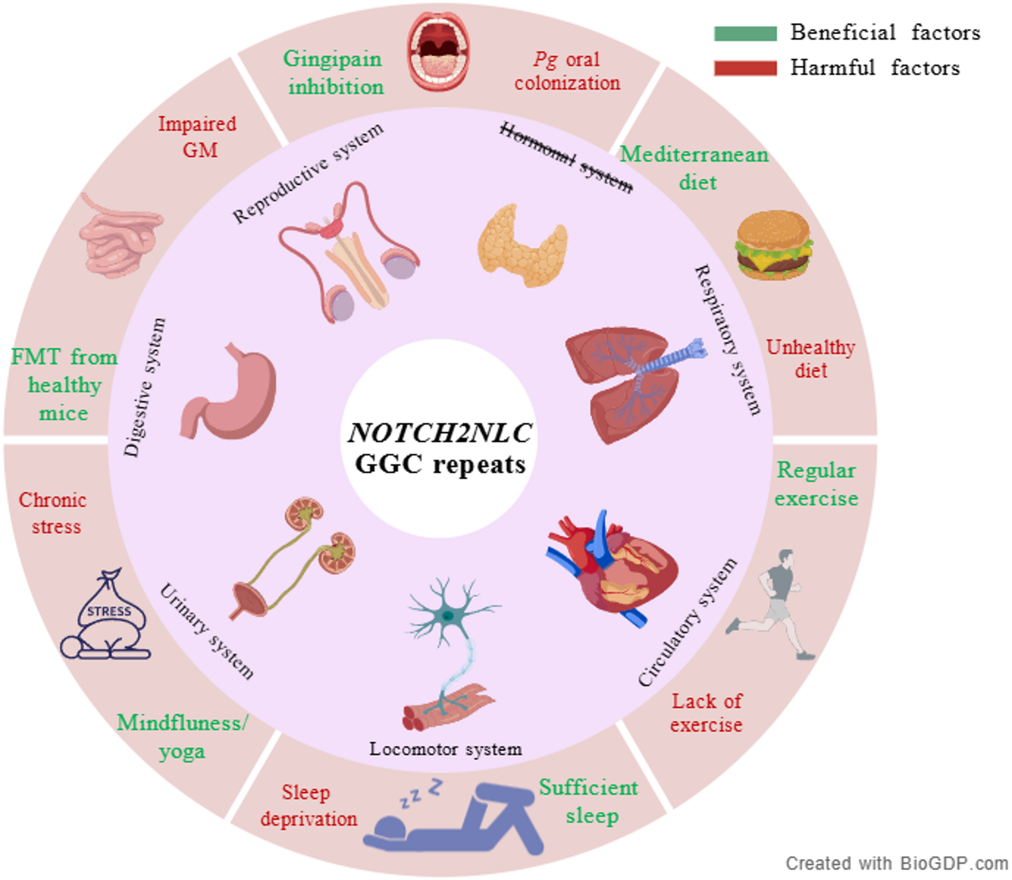
Jiang S, et al. Generic diagramming platform (GDP): a comprehensive database of high-quality biomedical graphics. Nucleic Acids Res. 2025;53(D1):D1670–6.
Google Scholar
Pang J, et al. The value of NOTCH2NLC gene detection and skin biopsy in the diagnosis of neuronal intranuclear inclusion disease. Front Neurol. 2021;12:624321.
Google Scholar
Lee HG, et al. Neuroinflammation: an astrocyte perspective. Sci Transl Med. 2023;15(721):eadi7828.
Google Scholar
Bao L et al. Immune system involvement in neuronal intranuclear inclusion disease. Neuropathol Appl Neurobiol, 2024;50(2):e12976.
Mori K, et al. Imaging findings and pathological correlations of subacute encephalopathy with neuronal intranuclear inclusion disease-case report. Radiol Case Rep. 2022;17(12):4481–6.
Google Scholar
Prinz M, Priller J. The role of peripheral immune cells in the CNS in steady state and disease. Nat Neurosci. 2017;20(2):136–44.
Google Scholar
Yoshii D, et al. An autopsy case of adult-onset neuronal intranuclear inclusion disease with perivascular preservation in cerebral white matter. Neuropathology. 2021;42(1):66–73.
Google Scholar
Tracey KJ, Cerami A. Tumor necrosis factor: a pleiotropic cytokine and therapeutic target. Annu Rev Med. 1994;45:491–503.
Google Scholar
Dinarello CA. Interleukin-1 in the pathogenesis and treatment of inflammatory diseases. Blood. 2011;117(14):3720–32.
Google Scholar
Tanaka T, Narazaki M, Kishimoto T. IL-6 in inflammation, immunity, and disease. Cold Spring Harb Perspect Biol. 2014;6(10):a016295.
Google Scholar
Lu X, Hong D. Neuronal intranuclear inclusion disease: recognition and update. J Neural Transm. 2021;128(3):295–303.
Google Scholar
Bao L, et al. Utility of labial salivary gland biopsy in the histological diagnosis of neuronal intranuclear inclusion disease. Eur J Neurol. 2024;31(1):e16102.
Google Scholar
Zhong S, et al. Spatial and Temporal distribution of white matter lesions in NOTCH2NLC-Related neuronal intranuclear inclusion disease. Neurology. 2025;104(4):e213360.
Google Scholar
Du N et al. Value of neutrophil-to-lymphocyte ratio in neuronal intranuclear inclusion disease. Heliyon, 2024;10(6):e27953.
Yan Y, et al. The clinical characteristics of neuronal intranuclear inclusion disease and its relation with inflammation. Neurol Sci. 2023;44(9):3189–97.
Google Scholar
Shen Y et al. uN2CpolyG-mediated p65 nuclear sequestration suppresses the NF-κB-NLRP3 pathway in neuronal intranuclear inclusion disease. Cell Communication Signal, 2025;23(1):68.
Tateishi J, et al. Intranuclear inclusions in muscle, nervous tissue, and adrenal gland. Acta Neuropathol. 1984;63(1):24–32.
Google Scholar
Platero JL et al. The impact of coconut oil and Epigallocatechin gallate on the levels of IL-6, anxiety and disability in multiple sclerosis patients. Nutrients, 2020;12(2):305.
Casali BT, et al. Omega-3 fatty acids augment the actions of nuclear receptor agonists in a mouse model of alzheimer’s disease. J Neurosci. 2015;35(24):9173–81.
Google Scholar
Chauhan A, et al. Phytochemicals targeting NF-κB signaling: potential anti-cancer interventions. J Pharm Anal. 2022;12(3):394–405.
Google Scholar
Shabab T, et al. Neuroinflammation pathways: a general review. Int J Neurosci. 2017;127(7):624–33.
Google Scholar
Lawrence T. The nuclear factor NF-kappaB pathway in inflammation. Cold Spring Harb Perspect Biol. 2009;1(6):a001651.
Google Scholar
Gleeson M, et al. The anti-inflammatory effects of exercise: mechanisms and implications for the prevention and treatment of disease. Nat Rev Immunol. 2011;11(9):607–15.
Google Scholar
Pedersen BK. Adolph distinguished lecture: muscle as an endocrine organ: IL-6 and other myokines. J Appl Physiol (1985). 2009;107(4):1006–14. Edward F.
Google Scholar
Steensberg A, et al. IL-6 enhances plasma IL-1ra, IL-10, and cortisol in humans. Am J Physiol Endocrinol Metab. 2003;285(2):E433–7.
Google Scholar
Ma Q. Beneficial effects of moderate voluntary physical exercise and its biological mechanisms on brain health. Neurosci Bull. 2008;24(4):265–70.
Google Scholar
Adlard PA, et al. Voluntary exercise decreases amyloid load in a transgenic model of Alzheimer’s disease. J Neurosci. 2005;25(17):4217–21.
Google Scholar
Periasamy S, et al. Sleep deprivation-induced multi-organ injury: role of oxidative stress and inflammation. Excli J. 2015;14:672–83.
Google Scholar
Shearer WT, et al. Soluble TNF-alpha receptor 1 and IL-6 plasma levels in humans subjected to the sleep deprivation model of spaceflight. J Allergy Clin Immunol. 2001;107(1):165–70.
Google Scholar
Ooms S, et al. Effect of 1 night of total sleep deprivation on cerebrospinal fluid β-amyloid 42 in healthy middle-aged men: a randomized clinical trial. JAMA Neurol. 2014;71(8):971–7.
Google Scholar
Reiter RJ, et al. Brain washing and neural health: role of age, sleep, and the cerebrospinal fluid melatonin rhythm. Cell Mol Life Sci. 2023;80(4):88.
Google Scholar
Zhao HY, et al. Chronic sleep restriction induces cognitive deficits and cortical Beta-Amyloid deposition in mice via BACE1-Antisense activation. CNS Neurosci Ther. 2017;23(3):233–40.
Google Scholar
Zhang Y, et al. Quercetin ameliorates memory impairment by inhibiting abnormal microglial activation in a mouse model of Paradoxical sleep deprivation. Biochem Biophys Res Commun. 2022;632:10–6.
Google Scholar
Irwin MR, et al. Tai Chi compared with cognitive behavioral therapy and the reversal of systemic, cellular and genomic markers of inflammation in breast cancer survivors with insomnia: A randomized clinical trial. Brain Behav Immun. 2024;120:159–66.
Google Scholar
Dutcher JM, et al. Smartphone mindfulness meditation training reduces Pro-inflammatory gene expression in stressed adults: A randomized controlled trial. Brain Behav Immun. 2022;103:171–7.
Google Scholar
Walsh E, Eisenlohr-Moul T, Baer R. Brief mindfulness training reduces salivary IL-6 and TNF-α in young women with depressive symptomatology. J Consult Clin Psychol. 2016;84(10):887–97.
Google Scholar
Patel DI, et al. Therapeutic yoga reduces pro-tumorigenic cytokines in cancer survivors. Support Care Cancer. 2022;31(1):33.
Google Scholar
Streeter CC, et al. Effects of yoga on the autonomic nervous system, gamma-aminobutyric-acid, and allostasis in epilepsy, depression, and post-traumatic stress disorder. Med Hypotheses. 2012;78(5):571–9.
Google Scholar
Ozturk FO, Tezel A. Effect of laughter yoga on mental symptoms and salivary cortisol levels in first-year nursing students: A randomized controlled trial. Int J Nurs Pract. 2021;27(2):e12924.
Google Scholar
Nair RG, Vasudev MM, Mavathur R. Role of yoga and its plausible mechanism in the mitigation of DNA damage in Type-2 diabetes: A randomized clinical trial. Ann Behav Med. 2022;56(3):235–44.
Google Scholar
Chen SJ, et al. Association of fecal and plasma levels of Short-Chain fatty acids with gut microbiota and clinical severity in patients with Parkinson disease. Neurology. 2022;98(8):e848–58.
Google Scholar
Chakraborty P, Gamage H, Laird AS. Butyrate as a potential therapeutic agent for neurodegenerative disorders. Neurochem Int. 2024;176:105745.
Google Scholar
Li N, et al. Prebiotic inulin controls Th17 cells mediated central nervous system autoimmunity through modulating the gut microbiota and short chain fatty acids. Gut Microbes. 2024;16(1):2402547.
Google Scholar
Zhang Y, et al. Transmission of alzheimer’s disease-associated microbiota dysbiosis and its impact on cognitive function: evidence from mice and patients. Mol Psychiatry. 2023;28(10):4421–37.
Google Scholar
Kim MS, et al. Transfer of a healthy microbiota reduces amyloid and Tau pathology in an alzheimer’s disease animal model. Gut. 2020;69(2):283–94.
Google Scholar
Beydoun MA, et al. Clinical and bacterial markers of periodontitis and their association with incident All-Cause and alzheimer’s disease dementia in a large National survey. J Alzheimers Dis. 2020;75(1):157–72.
Google Scholar
Ilievski V, et al. Chronic oral application of a periodontal pathogen results in brain inflammation, neurodegeneration and amyloid beta production in wild type mice. PLoS ONE. 2018;13(10):e0204941.
Google Scholar
Wang RP, et al. IL-1β and TNF-α play an important role in modulating the risk of periodontitis and alzheimer’s disease. J Neuroinflammation. 2023;20(1):71.
Google Scholar
Dominy SS, et al. Porphyromonas gingivalis in alzheimer’s disease brains: evidence for disease causation and treatment with small-molecule inhibitors. Sci Adv. 2019;5(1):eaau3333.
Google Scholar
Brettschneider J, et al. Microglial activation correlates with disease progression and upper motor neuron clinical symptoms in amyotrophic lateral sclerosis. PLoS ONE. 2012;7(6):e39216.
Google Scholar
Graves MC, et al. Inflammation in amyotrophic lateral sclerosis spinal cord and brain is mediated by activated macrophages, mast cells and T cells. Amyotroph Lateral Scler Other Motor Neuron Disord. 2004;5(4):213–9.
Google Scholar
Henkel JS, et al. Presence of dendritic cells, MCP-1, and activated microglia/macrophages in amyotrophic lateral sclerosis spinal cord tissue. Ann Neurol. 2004;55(2):221–35.
Google Scholar
Liu YH, et al. Neuronal intranuclear inclusion disease in patients with adult-onset non-vascular leukoencephalopathy. Brain. 2022;145(9):3010–21.
Google Scholar
Chen L, et al. Teaching neuroimages: the zigzag edging sign of adult-onset neuronal intranuclear inclusion disease. Neurology. 2019;92(19):e2295-6.
Google Scholar
Fragoso DC, et al. Imaging of Creutzfeldt-Jakob disease: imaging patterns and their differential diagnosis. Radiographics. 2017;37(1):234–57.
Google Scholar
Muttikkal TJ, Wintermark M. MRI patterns of global hypoxic-ischemic injury in adults. J Neuroradiol. 2013;40(3):164–71.
Google Scholar
Hasebe M, et al. Hypoglycemic encephalopathy. QJM. 2022;115(7):478–9.
Google Scholar
Zhang Z, et al. MRI features of neuronal intranuclear inclusion disease, combining visual and quantitative imaging investigations. J Neuroradiol. 2024;51(3):274–80.
Google Scholar
Okamura S, et al. A case of neuronal intranuclear inclusion disease with recurrent vomiting and without apparent DWI abnormality for the first seven years. Heliyon. 2020;6(8):e04675.
Google Scholar
Liu Y, et al. Clinical and mechanism advances of neuronal intranuclear inclusion disease. Front Aging Neurosci. 2022;14:934725.
Google Scholar
Liang H, et al. Clinical and pathological features in adult-onset NIID patients with cortical enhancement. J Neurol. 2020;267(11):3187–98.
Google Scholar
Shen Y, et al. Encephalitis-like episodes with cortical edema and enhancement in patients with neuronal intranuclear inclusion disease. Neurol Sci. 2024;45(9):4501–11.
Google Scholar
Jacobs AH, Tavitian B. Noninvasive molecular imaging of neuroinflammation. J Cereb Blood Flow Metab. 2012;32(7):1393–415.
Google Scholar
Corica F et al. PET imaging of Neuro-Inflammation with tracers targeting the translocator protein (TSPO), a systematic review: from bench to bedside. Diagnostics (Basel), 2023;13(6):1029.
Banati RB. Visualising microglial activation in vivo. Glia. 2002;40(2):206–17.
Google Scholar
Turner MR, et al. Evidence of widespread cerebral microglial activation in amyotrophic lateral sclerosis: an [11 C](R)-PK11195 positron emission tomography study. Neurobiol Dis. 2004;15(3):601–9.
Google Scholar
Tondo G, et al. (11) C-PK11195 PET-based molecular study of microglia activation in SOD1 amyotrophic lateral sclerosis. Ann Clin Transl Neurol. 2020;7(9):1513–23.
Google Scholar
Malpetti M, et al. Microglial activation in the frontal cortex predicts cognitive decline in frontotemporal dementia. Brain. 2023;146(8):3221–31.
Google Scholar
Zhong S, et al. Microglia contribute to polyG-dependent neurodegeneration in neuronal intranuclear inclusion disease. Acta Neuropathol. 2024;148(1):21.
Google Scholar
Hutton BF. The origins of SPECT and SPECT/CT. Eur J Nucl Med Mol Imaging. 2014;41(Suppl 1):S3-16.
Google Scholar
Benadiba M, et al. New molecular targets for PET and SPECT imaging in neurodegenerative diseases. Braz J Psychiatry. 2012;34(Suppl 2):S125–36.
Google Scholar
Kimachi T, et al. Reversible encephalopathy with focal brain edema in patients with neuronal intranuclear inclusion disease. Neurology and Clinical Neuroscience. 2017;5(6):198–200.
Google Scholar
Fujita K, et al. Neurologic attack and dynamic perfusion abnormality in neuronal intranuclear inclusion disease. Neurol Clin Pract. 2017;7(6):e39-42.
Google Scholar
Pan Y, et al. Expression of expanded GGC repeats within NOTCH2NLC causes cardiac dysfunction in mouse models. Cell Biosci. 2023;13(1):157.
Google Scholar
Heslegrave A, et al. Increased cerebrospinal fluid soluble TREM2 concentration in Alzheimer’s disease. Mol Neurodegener. 2016;11:3.
Google Scholar
Craig-Schapiro R, et al. YKL-40: a novel prognostic fluid biomarker for preclinical alzheimer’s disease. Biol Psychiatry. 2010;68(10):903–12.
Google Scholar
Crols R, et al. Increased GFAP levels in CSF as a marker of organicity in patients with Alzheimer’s disease and other types of irreversible chronic organic brain syndrome. J Neurol. 1986;233(3):157–60.
Google Scholar
Andrés-Benito P, et al. Differential astrocyte and oligodendrocyte vulnerability in murine Creutzfeldt-Jakob disease. Prion. 2021;15(1):112–20.
Google Scholar
Mussbacher M, et al. NF-κB in monocytes and macrophages – an inflammatory master regulator in multitalented immune cells. Front Immunol. 2023;14:1134661.
Google Scholar
Biasizzo M, Kopitar-Jerala N. Interplay between NLRP3 inflammasome and autophagy. Front Immunol. 2020;11:591803.
Google Scholar
Deng Q, et al. Pseudomonas aeruginosa triggers macrophage autophagy to escape intracellular killing by activation of the NLRP3 inflammasome. Infect Immun. 2016;84(1):56–66.
Google Scholar
Geissmann F, et al. Development of monocytes, macrophages, and dendritic cells. Science. 2010;327(5966):656–61.
Google Scholar
Guilliams M, et al. Dendritic cells, monocytes and macrophages: a unified nomenclature based on ontogeny. Nat Rev Immunol. 2014;14(8):571–8.
Google Scholar
Prinz M, Jung S, Priller J. Microglia Biology: One Century Evol Concepts Cell. 2019;179(2):292–311.
Google Scholar
Li Q, Barres BA. Microglia and macrophages in brain homeostasis and disease. Nat Rev Immunol. 2018;18(4):225–42.
Google Scholar
Liddelow SA, et al. Neurotoxic reactive astrocytes are induced by activated microglia. Nature. 2017;541(7638):481–7.
Google Scholar
Keren-Shaul H, et al. A unique microglia type associated with restricting development of alzheimer’s disease. Cell. 2017;169(7):1276–e129017.
Google Scholar
Mathys H, et al. Temporal tracking of microglia activation in neurodegeneration at single-cell resolution. Cell Rep. 2017;21(2):366–80.
Google Scholar
Zhou Y, et al. Human and mouse single-nucleus transcriptomics reveal TREM2-dependent and TREM2-independent cellular responses in Alzheimer’s disease. Nat Med. 2020;26(1):131–42.
Google Scholar
Ginhoux F, Guilliams M. Tissue-resident macrophage ontogeny and homeostasis. Immunity. 2016;44(3):439–49.
Google Scholar
Italiani P, Boraschi D. From monocytes to M1/M2 macrophages: phenotypical vs. functional differentiation. Front Immunol. 2014;5:514.
Google Scholar
Merad M, et al. The dendritic cell lineage: ontogeny and function of dendritic cells and their subsets in the steady state and the inflamed setting. Annu Rev Immunol. 2013;31:563–604.
Google Scholar
Sun R, Jiang H. Border-associated macrophages in the central nervous system. J Neuroinflammation. 2024;21(1):67.
Google Scholar
Goldmann T, et al. Origin, fate and dynamics of macrophages at central nervous system interfaces. Nat Immunol. 2016;17(7):797–805.
Google Scholar
Van Hove H, et al. A single-cell atlas of mouse brain macrophages reveals unique transcriptional identities shaped by ontogeny and tissue environment. Nat Neurosci. 2019;22(6):1021–35.
Google Scholar
Bell RD, Zlokovic BV. Neurovascular mechanisms and blood-brain barrier disorder in Alzheimer’s disease. Acta Neuropathol. 2009;118(1):103–13.
Google Scholar
Faraco G, et al. Perivascular macrophages mediate the neurovascular and cognitive dysfunction associated with hypertension. J Clin Invest. 2016;126(12):4674–89.
Google Scholar
Rua R, et al. Infection drives meningeal engraftment by inflammatory monocytes that impairs CNS immunity. Nat Immunol. 2019;20(4):407–19.
Google Scholar
Dani N, et al. A cellular and Spatial map of the choroid plexus across brain ventricles and ages. Cell. 2021;184(11):3056–e307421.
Google Scholar
Mrdjen D, et al. High-Dimensional Single-Cell mapping of central nervous system immune cells reveals distinct myeloid subsets in Health, Aging, and disease. Immunity. 2018;48(2):380–95. .e6.
Google Scholar
Park L, et al. Brain perivascular macrophages initiate the neurovascular dysfunction of Alzheimer Aβ peptides. Circ Res. 2017;121(3):258–69.
Google Scholar
Schläger C, et al. Effector T-cell trafficking between the leptomeninges and the cerebrospinal fluid. Nature. 2016;530(7590):349–53.
Google Scholar
Herz J, et al. Role of neutrophils in exacerbation of brain injury after focal cerebral ischemia in hyperlipidemic mice. Stroke. 2015;46(10):2916–25.
Google Scholar
Carmeliet P. Angiogenesis in health and disease. Nat Med. 2003;9(6):653–60.
Google Scholar
Greenberg SM, et al. Cerebral microbleeds: a guide to detection and interpretation. Lancet Neurol. 2009;8(2):165–74.
Google Scholar
Charidimou A, Gang Q, Werring DJ. Sporadic cerebral amyloid angiopathy revisited: recent insights into pathophysiology and clinical spectrum. J Neurol Neurosurg Psychiatry. 2012;83(2):124–37.
Google Scholar
Liao YC, et al. NOTCH2NLC GGC repeat expansion in patients with vascular leukoencephalopathy. Stroke. 2023;54(5):1236–45.
Google Scholar
Bao L, et al. GGC repeat expansions in NOTCH2NLC cause uN2CpolyG cerebral amyloid angiopathy. Brain. 2025;148(2):467–79.
Google Scholar
Ryu JK, McLarnon JG. A leaky blood-brain barrier, fibrinogen infiltration and microglial reactivity in inflamed Alzheimer’s disease brain. J Cell Mol Med. 2009;13(9a):2911–25.
Google Scholar
Shan Y, et al. The glucagon-like peptide-1 receptor agonist reduces inflammation and blood-brain barrier breakdown in an astrocyte-dependent manner in experimental stroke. J Neuroinflammation. 2019;16(1):242.
Google Scholar
Orihara A, et al. Acute reversible encephalopathy with neuronal intranuclear inclusion disease diagnosed by a brain biopsy: inferring the mechanism of encephalopathy from radiological and histological findings. Intern Med. 2023;62(12):1821–5.
Google Scholar
Li YY, et al. Interactions between beta-Amyloid and pericytes in Alzheimer’s disease. Front Biosci (Landmark Ed). 2024;29(4):136.
Google Scholar
Huang X, Hussain B, Chang J. Peripheral inflammation and blood-brain barrier disruption: effects and mechanisms. CNS Neurosci Ther. 2021;27(1):36–47.
Google Scholar
Rahimifard M, et al. Targeting the TLR4 signaling pathway by polyphenols: a novel therapeutic strategy for neuroinflammation. Ageing Res Rev. 2017;36:11–9.
Google Scholar
Zhang M, et al. Blockage of VEGF function by bevacizumab alleviates early-stage cerebrovascular dysfunction and improves cognitive function in a mouse model of Alzheimer’s disease. Transl Neurodegener. 2024;13(1):1.
Google Scholar
Wild CP. Complementing the genome with an exposome: the outstanding challenge of environmental exposure measurement in molecular epidemiology. Cancer Epidemiol Biomarkers Prev. 2005;14(8):1847–50.
Google Scholar
Doroszkiewicz J et al. Common and trace metals in alzheimer’s and parkinson’s diseases. Int J Mol Sci, 2023;24(21):15721.
Gu Y, et al. Mediterranean diet, inflammatory and metabolic biomarkers, and risk of Alzheimer’s disease. J Alzheimers Dis. 2010;22(2):483–92.
Google Scholar
Flanagan E, et al. Nutrition and the ageing brain: moving towards clinical applications. Ageing Res Rev. 2020;62:101079.
Google Scholar
Gelpi E, et al. Neuronal intranuclear (hyaline) inclusion disease and fragile X-associated tremor/ataxia syndrome: a morphological and molecular dilemma. Brain. 2017;140(8):e51.
Google Scholar
Tolar M et al. Neurotoxic soluble amyloid oligomers drive alzheimer’s pathogenesis and represent a clinically validated target for slowing disease progression. Int J Mol Sci, 2021;22(12):6355.
Habashi M, et al. Early diagnosis and treatment of alzheimer’s disease by targeting toxic soluble Aβ oligomers. Proc Natl Acad Sci U S A. 2022;119(49):e2210766119.
Google Scholar
Wang ZX, et al. The essential role of soluble Aβ oligomers in alzheimer’s disease. Mol Neurobiol. 2016;53(3):1905–24.
Google Scholar
Besedovsky L, Lange T, Haack M. The sleep-immune crosstalk in health and disease. Physiol Rev. 2019;99(3):1325–80.
Google Scholar
Su WJ, et al. Antidiabetic drug glyburide modulates depressive-like behavior comorbid with insulin resistance. J Neuroinflammation. 2017;14(1):210.
Google Scholar
Hajishengallis G, Darveau RP, Curtis MA. The keystone-pathogen hypothesis. Nat Rev Microbiol. 2012;10(10):717–25.
Google Scholar
Zhang J, et al. Porphyromonas gingivalis lipopolysaccharide induces cognitive dysfunction, mediated by neuronal inflammation via activation of the TLR4 signaling pathway in C57BL/6 mice. J Neuroinflammation. 2018;15(1):37.
Google Scholar
Nie R, et al. Porphyromonas gingivalis infection induces Amyloid-β accumulation in Monocytes/Macrophages. J Alzheimers Dis. 2019;72(2):479–94.
Google Scholar
Kemaladewi DU, et al. Correction of a splicing defect in a mouse model of congenital muscular dystrophy type 1A using a homology-directed-repair-independent mechanism. Nat Med. 2017;23(8):984–9.
Google Scholar
Sarkar S, et al. Rapamycin and mTOR-independent autophagy inducers ameliorate toxicity of polyglutamine-expanded Huntingtin and related proteinopathies. Cell Death Differ. 2009;16(1):46–56.
Google Scholar
Grima JC, et al. Mutant Huntingtin disrupts the nuclear pore complex. Neuron. 2017;94(1):93–e1076.
Google Scholar
