
AirPods are one of those items we all flock to during major sales events. Prime Day, Memorial Day, Black Friday, we want to see AirPods reduced. They’re arguably one of the most popular earbuds on…
Blog
-
Integrative metaprogram analysis reveals transcriptional dysregulation of oxidative stress response in granulosa cells from polycystic ovary syndrome | Journal of Ovarian Research
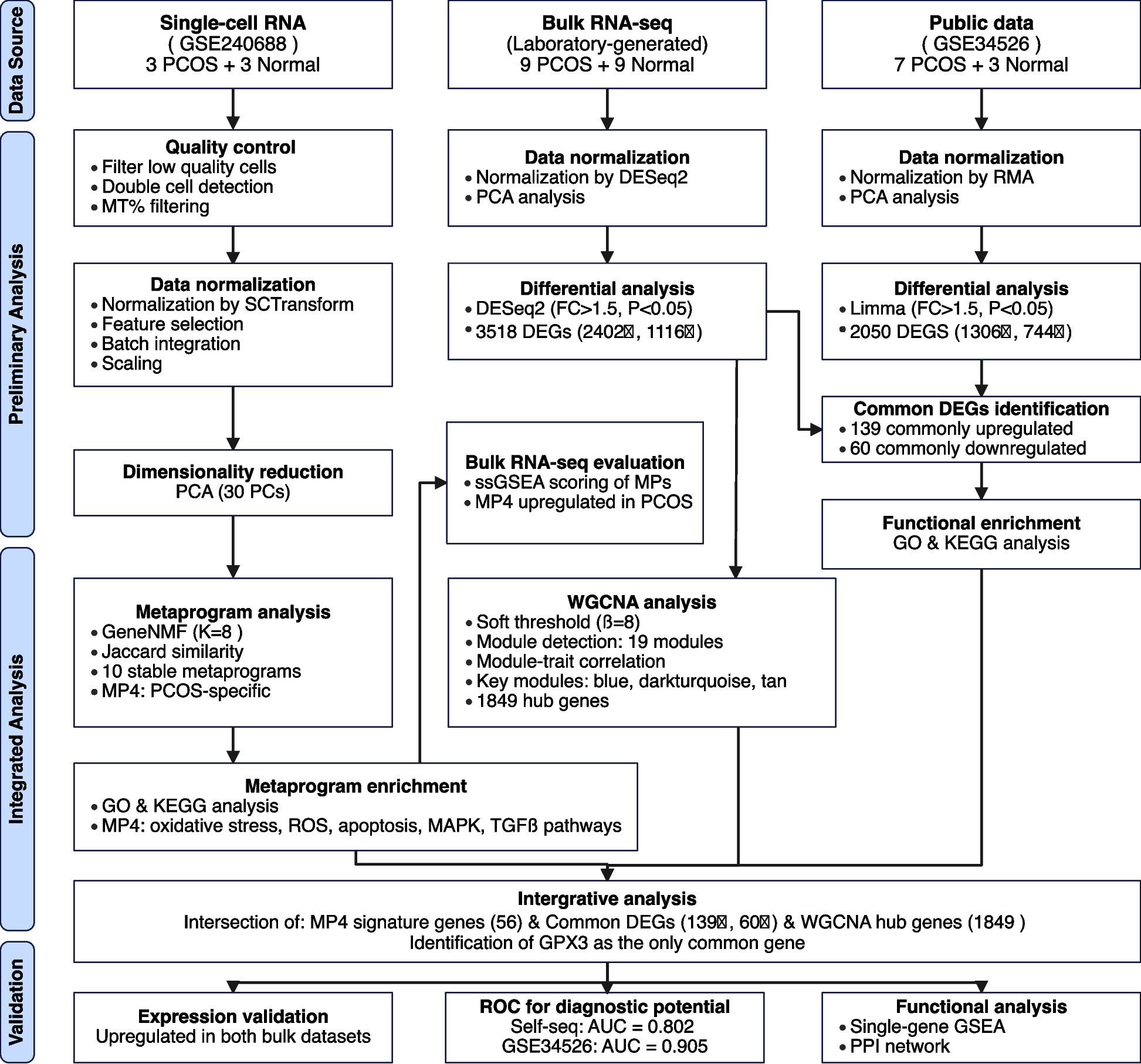
Figure 1 depicts our analytical workflow integrating three datasets: single-cell RNA-seq (GSE240688), bulk RNA-seq from our laboratory cohort, and a validation dataset (GSE34526). This integrative transcriptomic approach combines non-negative matrix factorization, differential expression analysis, and co-expression network analysis to bridge single-cell and bulk transcriptomic findings, ultimately identifying key regulatory genes in PCOS pathophysiology.
Fig. 1 Flowchart of the Study Design and Analytical Workflow
Metaprogram analysis reveals molecular signatures and cellular heterogeneity in PCOS granulosa cells
NMF was applied to deconvolve transcriptional programs in single-cell datasets, yielding 10 stable metaprograms (MPs, Fig. 2). To establish the optimal factorization dimensionality, we systematically scanned k values from 5 to 20, guided by quantitative evaluation of intra-sample cluster separation using silhouette coefficient analysis to ensure robust program delineation. The selected k = 8 demonstrated balanced performance, achieving both high-resolution separation of transcriptional programs within individual samples and preservation of biologically interpretable modules. This parameterization generated 48 sample-specific expression programs (8 per sample across six specimens), which were aggregated into 10 consensus metaprograms via cosine similarity-based hierarchical clustering. The resultant block-diagonal similarity matrix structure revealed conserved transcriptional modules exhibiting cross-sample reproducibility. Metaprogram refinement employed stringent dual criteria: genes recurrently detected in sample-level programs (confidence ≥ 0.5) and accounting for ≥ 80% of cumulative loading variance (weight threshold = 0.8), ensuring both technical robustness and biological coherence.
Fig. 2 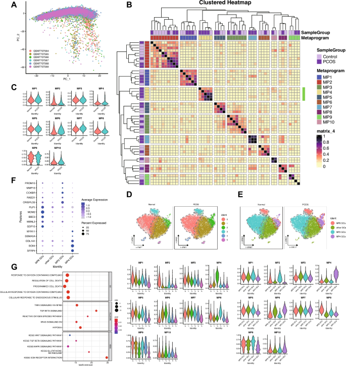
Metaprogram Analysis Reveals Molecular Signatures and Cellular Heterogeneity in PCOS Granulosa Cells. A PCA of single-cell RNA-seq samples. B Heatmap showing the expression of 10 identified MPs across samples. C Differential expression of MPs between PCOS and normal granulosa cells. D t-SNE visualization of six cell clusters and corresponding MP expression patterns in PCOS and normal samples. E Secondary clustering of granulosa cells based on MP expression, identifying four distinct GC subtypes and their corresponding MP expression profiles. F Expression patterns of key markers across the four GC subtypes. G GO, HALLMARK, and KEGG pathway enrichment analysis for MP4, highlighting pathways related to oxidative stress and stress-response signaling
Analysis of metaprogram distribution revealed distinct patterns associated with disease status. Metaprograms MP1-3 and MP8-10 were stably expressed across all samples, indicating their involvement in fundamental cellular processes independent of disease state. In contrast, MP4-7 demonstrated PCOS-specific expression patterns.
Gene set enrichment analysis revealed distinct biological functions for each metaprogram. Notably, MP4 showed significant enrichment in pathways related to cellular response to endogenous stimuli, oxygen-containing compounds, programmed cell death, reactive oxygen species, hypoxia, mitogen-activated protein kinase (MAPK) signaling, transforming growth factor-beta (TGF-β) signaling, and Wingless/Integrated (WNT) signaling pathways. This functional profile strongly implicates MP4 in oxidative stress responses and stress-induced signaling cascades known to be dysregulated in PCOS.
To characterize cellular Heterogeneity, we performed unsupervised clustering using the first 30 principal components with a resolution parameter of 0.2, resulting in distinct cell clusters visualized by t-SNE. UCell score calculation for all 10 metaprograms revealed significant associations between specific metaprograms and cell clusters: MP2 scores were significantly higher in cluster 0 (0.17 ± 0.09 vs 0.05 ± 0.05, p < 0.001); MP4 scores were elevated in cluster 2 (0.25 ± 0.09 vs 0.07 ± 0.05, p < 0.001); and MP8 was predominantly expressed in cluster 3 (0.22 ± 0.06 vs 0.11 ± 0.05, p < 0.001). Based on these patterns, we designated cluster 0 as MP2 granulosa cells (GCs), cluster 2 as MP4 GCs, cluster 3 as MP8 GCs, and remaining cells as other GCs. Notably, the proportion of MP4 GCs was significantly higher in PCOS samples compared to normal controls as determined by Wilcoxon rank-sum test with p = 0.0046, suggesting that expansion of MP4-expressing granulosa cells may be a characteristic feature of PCOS pathophysiology.
Differential expression analysis identifies common dysregulated genes in PCOS
Table 1 presents the clinical and endocrine characteristics of study participants. PCOS and control groups were successfully matched with no significant differences in age (29.00 ± 3.84 vs. 29.78 ± 3.31 years, p = 0.651), BMI (22.03 ± 3.12 vs. 23.12 ± 3.11 kg/m2, p = 0.47), and TSH levels (1.86 ± 1.03 vs. 2.05 ± 1.31 mIU/L, p = 0.732). As expected for PCOS pathophysiology, patients exhibited significantly elevated basal luteinizing hormone (LH, 7.36 ± 2.07 vs. 3.02 ± 0.75 IU/L, p < 0.001), testosterone (1.08 ± 0.43 vs. 0.65 ± 0.19 nmol/L, p = 0.014), anti-Müllerian hormone (AMH) levels (6.96 ± 2.21 vs. 3.80 ± 1.42 ng/mL, p = 0.002), and antral follicle counts (24 vs. 14, p < 0.001) compared to controls. During ovarian stimulation, PCOS patients yielded significantly more oocytes (20 vs. 13, p = 0.024) and mature oocytes (15 vs. 11, p = 0.022), consistent with their enhanced follicular development potential. These findings confirm the distinct hormonal and reproductive characteristics of our PCOS population while validating the effectiveness of our matching strategy for potential confounding variables.
Table 1 Clinical Characteristics and Hormonal Profiles of PCOS Patients and Control Subjects Differential expression analysis was performed on two independent datasets utilizing different transcriptomic platforms: our laboratory-generated bulk RNA-seq dataset (9 PCOS and 9 normal samples) and the publicly available GSE34526 microarray dataset (7 PCOS and 3 normal samples) (Fig. 3). Principal component analysis confirmed clear separation between PCOS and control samples after normalization in both datasets. To account for the fundamental differences between these technologies, we employed platform-specific analytical approaches. For our RNA-seq dataset, DESeq2 analysis with criteria of |FoldChange|> 1.5 and P < 0.05 identified 3,518 differentially expressed genes (DEGs), including 2,402 upregulated and 1,116 downregulated genes in PCOS samples. For the GSE34526 microarray dataset, Limma analysis with the same fold-change and significance thresholds identified 2,050 DEGs (1,306 upregulated and 744 downregulated).
Fig. 3 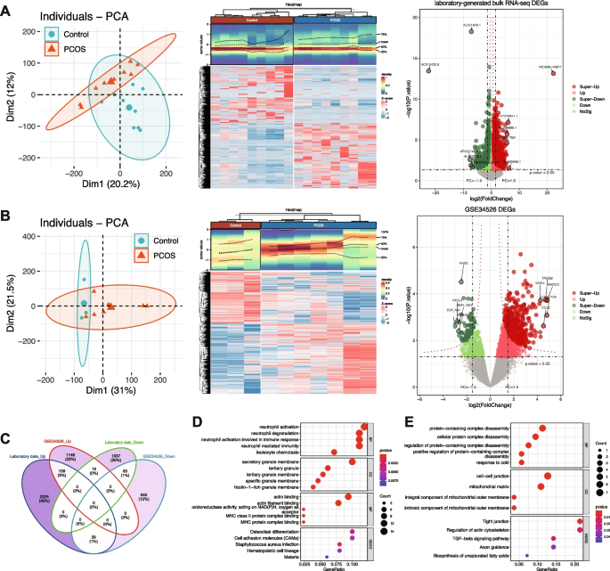
Differential Expression Analysis Identifies Common Dysregulated Genes in PCOS. A Analysis of the laboratory-generated bulk RNA-seq dataset: PCA plot of samples (left), heatmap of DEGs (middle), and volcano plot highlighting upregulated and downregulated genes (right). B Analysis of the GSE34526 dataset: PCA plot of samples (left), heatmap of DEGs (middle), and volcano plot (right). C Venn diagram showing the overlap of DEGs between the two datasets. D GO and KEGG pathway enrichment analysis of commonly upregulated genes. E GO and KEGG pathway enrichment analysis of commonly downregulated genes
To identify consistently dysregulated genes across different patient cohorts, we determined the intersection of DEGs from both datasets, considering the direction of expression changes. This stringent approach yielded 139 commonly upregulated and 60 commonly downregulated genes across both datasets. Functional enrichment analysis of common upregulated genes identified 13 KEGG pathways and 236 GO terms, while common downregulated genes were enriched in 5 KEGG pathways and 21 GO terms. Upregulated genes were significantly associated with pathways related to cellular response to stress, inflammatory processes, and signaling cascades. Downregulated genes were enriched in metabolic pathways and cellular homeostasis processes. These patterns suggest that PCOS is characterized by enhanced stress response mechanisms coupled with impaired metabolic functions.
WGCNA identifies co-expression modules associated with PCOS
To identify co-expressed gene networks associated with PCOS, we performed WGCNA on the laboratory-generated dataset (Fig. 4). Hierarchical clustering of samples confirmed appropriate grouping without outliers. A soft threshold power of 8 was selected based on scale-free topology criteria (R2 = 0.9) and mean connectivity analysis, ensuring optimal network construction while preserving biological relevance. Using dynamic tree cutting with a minimum module size of 100 genes and a cut Height of 0.4, we identified 19 distinct co-expression modules.
Fig. 4 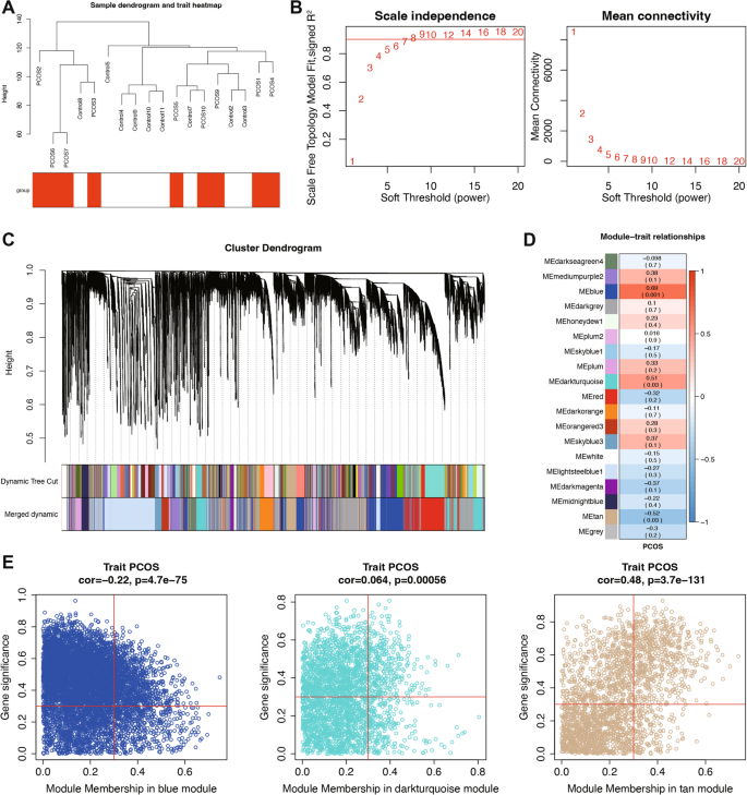
WGCNA Identifies Co-expression Modules Associated with PCOS. A Sample dendrogram and trait heatmap illustrating clustering of samples and their association with PCOS. B Analysis of scale independence and mean connectivity to determine the optimal soft threshold for network construction. C Cluster dendrogram of genes, showing module assignment based on hierarchical clustering. D Module-trait relationships, indicating correlations between module eigengenes and PCOS status. E Scatter plots showing module membership correlation with PCOS status for the blue, darkturquoise, and tan modules
Correlation analysis between module eigengenes and PCOS status revealed significant associations for several modules. Among these, the blue, darkturquoise, and tan modules exhibited the strongest correlations with disease status (correlation coefficient > 0.3, P < 0.05). To identify key regulatory genes within the PCOS-associated modules, we calculated the correlation between individual genes and both module membership (MM) and gene significance for PCOS (GS). By applying thresholds of MM > 0.3 and GS > 0.3, we identified 1,849 hub genes with strong connections to both their respective modules and PCOS status. These hub genes represent potential master regulators of the transcriptional networks dysregulated in PCOS.
Metaprogram validation in bulk RNA-seq data confirms single-cell findings
To validate the relevance of single-cell-derived metaprograms at the tissue level, we first analyzed their distribution across granulosa cell subsets in single-cell RNA-seq data (Fig. 5A). Metaprogram composition varied across different granulosa cell clusters, with distinct enrichment patterns in PCOS and normal samples.
Fig. 5 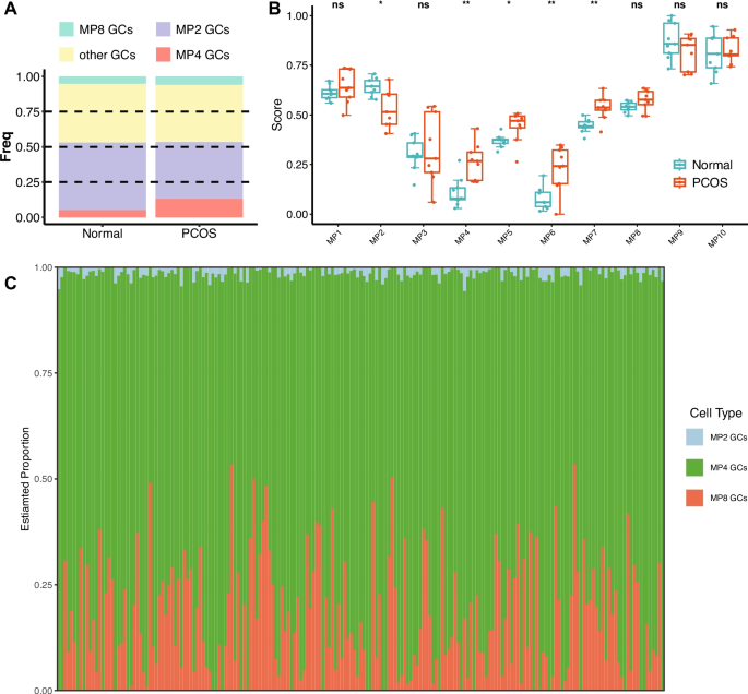
Metaprogram Validation in Bulk RNA-Seq Data Confirms Single-Cell Findings. A Stacked bar plot showing the distribution of MPs across different granulosa cell populations in single-cell RNA-seq data. B Differential expression of MPs in bulk RNA-seq data, comparing PCOS and normal samples. ns = not significant, * p < 0.05, ** p < 0.01. C Deconvolution analysis of 193 GTEx ovary samples showing the relative proportions of MP2 GCs, MP4 GCs, and MP8 GCs
To further bridge the gap between single-cell and bulk transcriptomic analyses, we employed single-sample Gene Set Enrichment Analysis (ssGSEA) to score each metaprogram in bulk RNA-seq samples (Fig. 5B). This approach quantified the activity of each transcriptional program in both PCOS and normal cohorts. Comparative analysis of metaprogram ssGSEA scores revealed significant differences in MP2, MP4, MP5, MP6, and MP7 activity. Consistent with single-cell findings, MP2 exhibited higher activity in normal samples, while MP4, MP5, MP6, and MP7 were upregulated in PCOS samples. The differential activity of these metaprograms in bulk tissue samples corroborates our single-cell findings and further supports the pathological relevance of these transcriptional programs in PCOS. In particular, the consistent upregulation of MP4 across both single-cell and bulk analyses reinforces its potential role as a key driver of PCOS pathophysiology.
To validate that the identified granulosa cell subtypes represent genuine biological entities rather than clustering artifacts, we performed deconvolution analysis on 193 GTEx v10 ovary bulk RNA-seq samples. The analysis successfully detected all three major granulosa cell subtypes (MP2, MP4, and MP8 GCs) across the tissue samples (Fig. 5C).
The deconvolution results revealed consistent patterns of cell type proportions across samples. MP4 GCs constituted the predominant subtype in most samples, typically representing 60–80% of the granulosa cell population. MP8 GCs showed intermediate abundance (approximately 10–30%), while MP2 GCs were consistently detected at lower proportions (5–15%). This abundance hierarchy (MP4 > MP8 > MP2) was remarkably stable across the majority of samples, with only minor variations observed in individual cases.
The successful detection of these cellular subtypes in independent bulk tissue samples, with reproducible relative abundance patterns, provides strong evidence that our single-cell-defined metaprograms correspond to biologically meaningful cell states rather than technical artifacts.
Integrative transcriptomic approach identifies key regulator in PCOS pathophysiology
To identify high-confidence key regulators involved in PCOS pathophysiology, we performed an integrative analysis combining three complementary approaches: MP4 signature genes from single-cell analysis, common differentially expressed genes across bulk datasets, and hub genes from WGCNA modules (Fig. 6). This stringent multi-dimensional filtering strategy identified GPX3 as the only gene that consistently emerged across all three analytical approaches. The convergence of these independent methods strongly suggests its central role in PCOS-associated transcriptional dysregulation, particularly in relation to oxidative stress responses.
Fig. 6 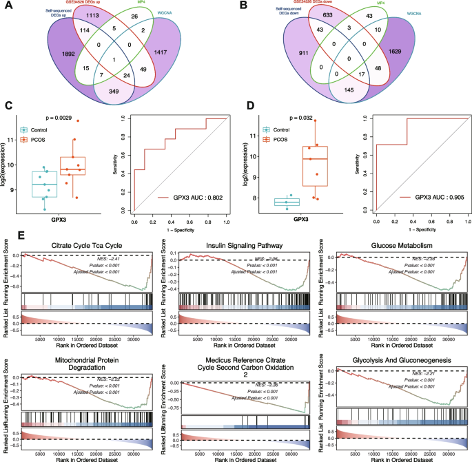
Integrative Transcriptomic Analysis Identifies GPX3 as a Key Regulator in PCOS. A Venn diagram showing the intersection of DEGs, WGCNA hub genes, and MP4 signature genes (upregulated). B Venn diagram showing the intersection of DEGs, WGCNA hub genes, and MP4 signature genes (downregulated). C Left: Box plot displaying GPX3 expression differences between PCOS and normal samples in the laboratory-generated dataset. Right: ROC curve assessing the diagnostic value of GPX3 in the same dataset. D Left: Box plot showing GPX3 expression differences in the GSE34526 dataset. Right: ROC curve from the GSE34526 dataset, validating the diagnostic potential of GPX3. E Single-gene GSEA of GPX3, revealing its association with metabolic and mitochondrial pathways, including the citrate cycle, insulin signaling, glucose metabolism, and mitochondrial protein degradation (NES < 0, adjusted P < 0.001 for all pathways)
Expression analysis confirmed significant upregulation of GPX3 in PCOS samples compared to normal controls across both the laboratory-generated dataset and the GSE34526 validation dataset. ROC curve analysis demonstrated strong discriminatory power of GPX3 between PCOS and normal samples in both the laboratory-generated dataset (AUC = 0.802) and the GSE34526 validation dataset (AUC = 0.905), highlighting its potential as a clinically relevant biomarker for PCOS diagnosis.
Examination of GPX3 expression at the single-cell level revealed specific distribution patterns across granulosa cell subpopulations. Single-gene Gene Set Enrichment Analysis identified 818 significantly enriched pathways (|Normalized Enrichment Score, NES|> 1, p.adjust < 0.05, q.value < 0.2), with those related to glucose metabolism, mitochondrial protein degradation, insulin signaling, citrate cycle, and TCA cycle prominently represented. These enrichment patterns suggest that GPX3 dysregulation may impact fundamental metabolic processes and energy homeostasis, which are known to be perturbed in PCOS.
Multi-level GPX3 regulatory network analysis reveals potential mechanisms in PCOS
To establish a comprehensive understanding of the functional relevance of GPX3 in PCOS pathophysiology, we performed integrative multi-levels analysis constructing a complex regulatory network (Fig. 7). The protein–protein interaction network based on MP4 signature genes revealed GPX3 in a network comprising 49 proteins with multiple functional connections. Within this network, GPX3 demonstrated direct interactions with several proteins involved in redox homeostasis and related cellular processes.
Fig. 7 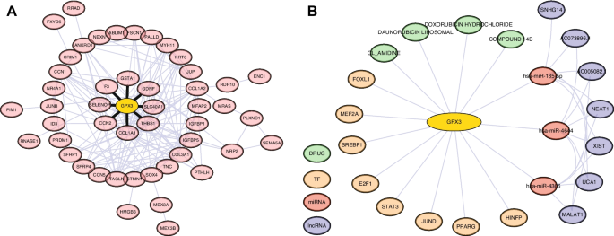
Multi-level Regulatory Network Analysis of GPX3 in PCOS. A PPI network of GPX3 and its associated proteins. B Integrated regulatory elements of GPX3 including: ceRNA network prediction showing GPX3-miRNA-lncRNA interactions; Transcription factor binding site prediction; Drug-gene interaction prediction for potential therapeutic targets
Most notably, GPX3 showed significant connections with selenoprotein P (SELENOP), a major selenium transport protein that works synergistically with GPX3 in the selenium-dependent antioxidant system, providing essential selenium cofactors for glutathione peroxidase activity. Similarly, glutathione S-transferase alpha 1 (GSTA1) exhibited direct interaction with GPX3, suggesting coordinated roles in glutathione metabolism and detoxification of reactive oxygen species. These interactions highlight GPX3’s central position in cellular antioxidant defense mechanisms.
Additionally, GPX3 directly interacts with SLC40A1 (ferroportin), an iron exporter critical for preventing iron-catalyzed oxidative damage, connecting iron homeostasis with antioxidant defense in granulosa cells. Interactions between GPX3 and both THBS1 and F3 suggest linkages between oxidative stress and PCOS-related coagulation and inflammatory pathways. Additionally, the associations with extracellular matrix proteins COL1A1 and CCN2 indicate involvement in oxidative stress-induced matrix remodeling. The connection with GDNF suggests novel neuroendocrine regulatory mechanisms influenced by oxidative status in PCOS pathophysiology.
Our miRNA-mRNA interaction analysis identified several microRNAs potentially regulating GPX3 expression, including has-miR-4644, hsa-miR-4306 and hsa-miR-185-5p, both predicted with high confidence scores. Further exploration through miRNA-lncRNA association analysis uncovered a complex layer of epigenetic regulation, with multiple long non-coding RNAs (lncRNAs) including XIST, UCA1, SNHG14, AC073896.4, MALAT1, NEAT1, and AC005082.1 potentially modulating these miRNA-mediated effects on GPX3 expression.
Transcription factor binding site analysis revealed that GPX3 expression may be regulated by several key transcription factors implicated in ovarian function, including SREBF1, HINFP, E2F1, STAT3, PPARG, MEF2A, FOXL1, and JUND. This suggests multiple potential mechanisms for transcriptional dysregulation of GPX3 in PCOS pathogenesis.
Furthermore, drug-gene interaction queries identified several compounds potentially targeting the GPX3-associated pathway, including DOXORUBICIN HYDROCHLORIDE, DAUNORUBICIN LIPOSOMAL, CL_AMIDINE, and COMPOUND 14B, providing potential therapeutic avenues for further investigation. Collectively, this integrative analysis positions GPX3 within a complex regulatory network essential for redox homeostasis in ovarian function, with multiple layers of regulation that may be disrupted in PCOS pathophysiology.
Continue Reading
-

Kaye Adams says her name was ‘dragged through the mud’ after BBC Scotland suspension | Scotland
The award-winning broadcaster Kaye Adams has spoken of her distress after she was taken off air by BBC Radio Scotland as a result of an unspecified “conduct complaint”, resulting in relentless media speculation.
In her first public comment on…
Continue Reading
-

Liam Lawson: F1 driver says he ‘could have killed’ marshals running on racetrack at Mexico Grand Prix
Formula One driver Liam Lawson said that he narrowly avoided a fatal collision with two marshals who ran across the racetrack at the Mexico Grand Prix.
The incident occurred on the third…
Continue Reading
-

Toxic winds from India choke Lahore, trigger health emergency
Toxic winds from India choke Lahore, trigger health emergency – Daily Times
…Continue Reading
-

Exclusive: Samsung SSDs Power NVIDIA's DGX Spark AI Supercomputer – 조선일보
- Exclusive: Samsung SSDs Power NVIDIA’s DGX Spark AI Supercomputer 조선일보
- How NVIDIA DGX Spark’s Performance Enables Intensive AI Tasks | NVIDIA Technical Blog NVIDIA Developer
- xFusion unveils FusionXpark, a DGX Spark–based mini…
Continue Reading
-
Optimal analgesia after laparoscopic total hysterectomy with pre-pneumoperitoneum phrenic nerve block using different ropivacaine concentrations: randomized controlled trial | BMC Anesthesiology
Study design and ethical approval
This randomized, blinded, controlled trial was conducted at First People’s Hospital of Yichang (The People’s Hospital of China Three Gorges University), approved by its Ethics Committee on December 12, 2022…
Continue Reading
-
Just a moment…
Just a moment… This request seems a bit unusual, so we need to confirm that you’re human. Please press and hold the button until it turns completely green. Thank you for your cooperation!
Continue Reading
-
De Minaur well poised in Live Race To Turin heading to Paris – ATP Tour
- De Minaur well poised in Live Race To Turin heading to Paris ATP Tour
- Chasing Nitto ATP Finals debut, Musetti seeks to fight off chasing pack in Paris ATP Tour
- Turin trio Felix Auger-Aliassime, Casper Ruud, Daniil Medvedev survive Basel, Vienna…
Continue Reading
-
Neuronal gene profiling of tau oligomer-bearing cholinergic nucleus basalis neurons during the onset of Alzheimer’s disease | Acta Neuropathologica Communications
Ahmad F, Das D, Kommaddi RP, Diwakar L, Gowaikar R, Rupanagudi KV, Bennett DA, Ravindranath V (2018) Isoform-specific hyperactivation of calpain-2 occurs presymptomatically at the synapse in Alzheimer’s disease mice and correlates with memory deficits in human subjects. Sci Rep 8:13119. https://doi.org/10.1038/s41598-018-31073-6
Google Scholar
Aldridge JE, Horibe T, Hoogenraad NJ (2007) Discovery of genes activated by the mitochondrial unfolded protein response (mtUPR) and cognate promoter elements. PLoS ONE 2:e874. https://doi.org/10.1371/journal.pone.0000874
Google Scholar
Alldred MJ, Che S, Ginsberg SD (2008) Terminal continuation (TC) RNA amplification enables expression profiling using minute RNA input obtained from mouse brain. Int J Mol Sci 9:2091–2104
Google Scholar
Alldred MJ, Ibrahim KW, Pidikiti H, Chiosis G, Mufson EJ, Stutzmann GE, Ginsberg SD (2024) Down syndrome frontal cortex layer III and layer V pyramidal neurons exhibit lamina specific degeneration in aged individuals. Acta Neuropathol Commun 12:182. https://doi.org/10.1186/s40478-024-01891-z
Google Scholar
Alldred MJ, Penikalapati SC, Lee SH, Heguy A, Roussos P, Ginsberg SD (2021) Profiling basal forebrain cholinergic neurons reveals a molecular basis for vulnerability within the Ts65Dn model of Down syndrome and Alzheimer’s disease. Mol Neurobiol. https://doi.org/10.1007/s12035-021-02453-3
Google Scholar
Allen TG, Abogadie FC, Brown DA (2006) Simultaneous release of glutamate and acetylcholine from single magnocellular “cholinergic” basal forebrain neurons. J Neurosci 26:1588–1595. https://doi.org/10.1523/JNEUROSCI.3979-05.2006
Google Scholar
Anderson MJ (2006) Distance-based tests for homogeneity of multivariate dispersions. Biometrics 62:245–253. https://doi.org/10.1111/j.1541-0420.2005.00440.x
Google Scholar
Anderson MJ (2001) A new method for non-parametric multivariate analysis of variance. Austral Ecol 26:32–46
Arriagada PV, Growdon JH, Hedley-Whyte ET, Hyman BT (1992) Neurofibrillary tangles but not senile plaques parallel duration and severity of Alzheimer’s disease. Neurology 42:631–639
Google Scholar
Baumann K, Mandelkow EM, Biernat J, Piwnica-Worms H, Mandelkow E (1993) Abnormal Alzheimer-like phosphorylation of tau-protein by cyclin-dependent kinases cdk2 and cdk5. FEBS Lett 336:417–424
Google Scholar
Beck JS, Madaj Z, Cheema CT, Kara B, Bennett DA, Schneider JA, Gordon MN, Ginsberg SD, Mufson EJ, Counts SE (2022) Co-expression network analysis of frontal cortex during the progression of Alzheimer’s disease. Cereb Cortex. https://doi.org/10.1093/cercor/bhac001
Google Scholar
Beck JS, Mufson EJ, Counts SE (2015) Evidence for mitochondrial UPR gene activation in familial and sporadic Alzheimer’s disease. Curr Alzheimer Res 13:610
Bennett DA, Buchman AS, Boyle PA, Barnes LL, Wilson RS, Schneider JA (2018) Religious orders study and rush memory and aging project. J Alzheimers Dis 64:S161–S189. https://doi.org/10.3233/JAD-179939
Google Scholar
Bennett DA, Wilson RS, Schneider JA, Evans DA, Beckett LA, Aggarwal NT, Barnes LL, Fox JH, Bach J (2002) Natural history of mild cognitive impairment in older persons. Neurology 59:198–205
Google Scholar
Berchtold NC, Coleman PD, Cribbs DH, Rogers J, Gillen DL, Cotman CW (2013) Synaptic genes are extensively downregulated across multiple brain regions in normal human aging and Alzheimer’s disease. Neurobiol Aging 34:1653–1661. https://doi.org/10.1016/j.neurobiolaging.2012.11.024
Google Scholar
Bonda DJ, Castellani RJ, Zhu X, Nunomura A, Lee HG, Perry G, Smith MA (2011) A novel perspective on tau in Alzheimer’s disease. Curr Alzheimer Res 8:639–642. https://doi.org/10.2174/156720511796717131
Google Scholar
Botella-Lopez A, Burgaya F, Gavin R, Garcia-Ayllon MS, Gomez-Tortosa E, Pena-Casanova J, Urena JM, Del Rio JA, Blesa R, Soriano E et al (2006) Reelin expression and glycosylation patterns are altered in Alzheimer’s disease. Proc Natl Acad Sci U S A 103:5573–5578. https://doi.org/10.1073/pnas.0601279103
Google Scholar
Bowser R, Kordower JH, Mufson EJ (1997) A confocal microscopic analysis of galaninergic hyperinnervation of cholinergic basal forebrain neurons in Alzheimer’s disease. Brain Pathol 7:723–730
Google Scholar
Braak H, Alafuzoff I, Arzberger T, Kretzschmar H, Del Tredici K (2006) Staging of Alzheimer disease-associated neurofibrillary pathology using paraffin sections and immunocytochemistry. Acta Neuropathol 112:389–404. https://doi.org/10.1007/s00401-006-0127-z
Google Scholar
Braak H, Braak E (1991) Neuropathological stageing of Alzheimer-related changes. Acta Neuropathol 82:239–259. https://doi.org/10.1007/bf00308809
Google Scholar
Braak H, Del Tredici K (2004) Alzheimer’s disease: intraneuronal alterations precede insoluble amyloid-beta formation. Neurobiol Aging 25:713–718. https://doi.org/10.1016/j.neurobiolaging.2003.12.015. (discussion 743-716)
Google Scholar
Carvalho C, Santos MS, Oliveira CR, Moreira PI (2015) Alzheimer’s disease and type 2 diabetes-related alterations in brain mitochondria, autophagy and synaptic markers. Biochim Biophys Acta 1852:1665–1675. https://doi.org/10.1016/j.bbadis.2015.05.001
Google Scholar
Castillo-Carranza DL, Gerson JE, Sengupta U, Guerrero-Munoz MJ, Lasagna-Reeves CA, Kayed R (2014) Specific targeting of tau oligomers in Htau mice prevents cognitive impairment and tau toxicity following injection with brain-derived tau oligomeric seeds. J Alzheimers Dis 40(Suppl 1):S97–S111. https://doi.org/10.3233/JAD-132477
Google Scholar
Chu Y, Cochran EJ, Bennett DA, Mufson EJ, Kordower JH (2001) Down-regulation of trkA mRNA within nucleus basalis neurons in individuals with mild cognitive impairment and Alzheimer’s disease. J Comp Neurol 437:296–307
Google Scholar
Clarke MTM, Brinkmalm A, Foiani MS, Woollacott IOC, Heller C, Heslegrave A, Keshavan A, Fox NC, Schott JM, Warren JD et al (2019) CSF synaptic protein concentrations are raised in those with atypical Alzheimer’s disease but not frontotemporal dementia. Alzheimers Res Ther 11:105. https://doi.org/10.1186/s13195-019-0564-2
Google Scholar
Combs B, Mueller RL, Morfini G, Brady ST, Kanaan NM (2019) Tau and axonal transport misregulation in tauopathies. Adv Exp Med Biol 1184:81–95. https://doi.org/10.1007/978-981-32-9358-8_7
Google Scholar
Counts SE, Alldred MJ, Che S, Ginsberg SD, Mufson EJ (2013) Synaptic gene dysregulation within hippocampal CA1 pyramidal neurons in mild cognitive impairment. Neuropharmacology 79C:172–179. https://doi.org/10.1016/j.neuropharm.2013.10.018
Google Scholar
Counts SE, Alldred MJ, Che S, Ginsberg SD, Mufson EJ (2014) Synaptic gene dysregulation within hippocampal CA1 pyramidal neurons in mild cognitive impairment. Neuropharmacology 79:172–179. https://doi.org/10.1016/j.neuropharm.2013.10.018
Google Scholar
Counts SE, Che S, Ginsberg SD, Mufson EJ (2011) Gender differences in neurotrophin and glutamate receptor expression in cholinergic nucleus basalis neurons during the progression of Alzheimer’s disease. J Chem Neuroanat 42:111–117. https://doi.org/10.1016/j.jchemneu.2011.02.004
Google Scholar
Counts SE, He B, Che S, Ginsberg SD, Mufson EJ (2009) Galanin fiber hyperinnervation preserves neuroprotective gene expression in cholinergic basal forebrain neurons in Alzheimer’s disease. J Alzheimers Dis 18:885–896
Google Scholar
Counts SE, He B, Che S, Ginsberg SD, Mufson EJ (2008) Galanin hyperinnervation upregulates choline acetyltransferase expression in cholinergic basal forebrain neurons in Alzheimer’s disease. Neurodegener Dis 5:228
Google Scholar
Counts SE, He B, Che S, Ikonomovic MD, DeKosky ST, Ginsberg SD, Mufson EJ (2007) Alpha7 nicotinic receptor up-regulation in cholinergic basal forebrain neurons in Alzheimer disease. Arch Neurol 64:1771–1776
Google Scholar
Counts SE, He B, Nadeem M, Wuu J, Scheff SW, Mufson EJ (2012) Hippocampal drebrin loss in mild cognitive impairment. Neurodegener Dis 10:216–219. https://doi.org/10.1159/000333122
Google Scholar
Counts SE, Mufson EJ (2005) The role of nerve growth factor receptors in cholinergic basal forebrain degeneration in prodromal Alzheimer disease. J Neuropathol Exp Neurol 64:263–272
Google Scholar
Counts SE, Nadeem M, Lad SP, Wuu J, Mufson EJ (2006) Differential expression of synaptic proteins in the frontal and temporal cortex of elderly subjects with mild cognitive impairment. J Neuropathol Exp Neurol 65:592–601
Google Scholar
Counts SE, Nadeem M, Wuu J, Ginsberg SD, Saragovi HU, Mufson EJ (2004) Reduction of cortical TrkA but not p75(NTR) protein in early-stage Alzheimer’s disease. Ann Neurol 56:520–531
Google Scholar
Cowan CM, Mudher A (2013) Are tau aggregates toxic or protective in tauopathies? Front Neurol 4:114. https://doi.org/10.3389/fneur.2013.00114
Google Scholar
da Rocha TJ, Silva Alves M, Guisso CC, de Andrade FM, Camozzato A, de Oliveira AA, Fiegenbaum M (2018) Association of GPX1 and GPX4 polymorphisms with episodic memory and Alzheimer’s disease. Neurosci Lett 666:32–37. https://doi.org/10.1016/j.neulet.2017.12.026
Google Scholar
Davis KL, Mohs RC, Marin D, Purohit DP, Perl DP, Lantz M, Austin G, Haroutunian V (1999) Cholinergic markers in elderly patients with early signs of Alzheimer disease. JAMA 281:1401–1406
Google Scholar
de Vries LE, Jongejan A, Monteiro Fortes J, Balesar R, Rozemuller AJM, Moerland PD, Huitinga I, Swaab DF, Verhaagen J (2024) Gene-expression profiling of individuals resilient to Alzheimer’s disease reveals higher expression of genes related to metallothionein and mitochondrial processes and no changes in the unfolded protein response. Acta Neuropathol Commun 12:68. https://doi.org/10.1186/s40478-024-01760-9
Google Scholar
DeTure MA, Dickson DW (2019) The neuropathological diagnosis of Alzheimer’s disease. Mol Neurodegener 14:32. https://doi.org/10.1186/s13024-019-0333-5
Google Scholar
Dhar SS, Liang HL, Wong-Riley MT (2009) Nuclear respiratory factor 1 co-regulates AMPA glutamate receptor subunit 2 and cytochrome c oxidase: tight coupling of glutamatergic transmission and energy metabolism in neurons. J Neurochem 108:1595–1606
Google Scholar
Dhar SS, Wong-Riley MT (2009) Coupling of energy metabolism and synaptic transmission at the transcriptional level: role of nuclear respiratory factor 1 in regulating both cytochrome c oxidase and NMDA glutamate receptor subunit genes. J Neurosci 29:483–492
Google Scholar
Du F, Yu Q, Kanaan NM, Yan SS (2022) Mitochondrial oxidative stress contributes to the pathological aggregation and accumulation of tau oligomers in Alzheimer’s disease. Hum Mol Genet 31:2498–2507. https://doi.org/10.1093/hmg/ddab363
Google Scholar
Dujardin S, Commins C, Lathuiliere A, Beerepoot P, Fernandes AR, Kamath TV, De Los Santos MB, Klickstein N, Corjuc DL, Corjuc BT et al (2020) Tau molecular diversity contributes to clinical heterogeneity in Alzheimer’s disease. Nat Med 26:1256–1263. https://doi.org/10.1038/s41591-020-0938-9
Google Scholar
Falke E, Nissanov J, Mitchell TW, Bennett DA, Trojanowski JQ, Arnold SE (2003) Subicular dendritic arborization in Alzheimer’s disease correlates with neurofibrillary tangle density. Am J Pathol 163:1615–1621. https://doi.org/10.1016/S0002-9440(10)63518-3
Google Scholar
Figueiro-Silva J, Gruart A, Clayton KB, Podlesniy P, Abad MA, Gasull X, Delgado-Garcia JM, Trullas R (2015) Neuronal pentraxin 1 negatively regulates excitatory synapse density and synaptic plasticity. J Neurosci 35:5504–5521. https://doi.org/10.1523/JNEUROSCI.2548-14.2015
Google Scholar
Floyd RA (1999) Antioxidants, oxidative stress, and degenerative neurological disorders. Proc Soc Exp Biol Med 222:236–245
Google Scholar
Fukutani Y, Cairns NJ, Shiozawa M, Sasaki K, Sudo S, Isaki K, Lantos PL (2000) Neuronal loss and neurofibrillary degeneration in the hippocampal cortex in late-onset sporadic Alzheimer’s disease. Psychiatry Clin Neurosci 54:523–529. https://doi.org/10.1046/j.1440-1819.2000.00747.x
Google Scholar
Furuya TK, Silva PN, Payao SL, Bertolucci PH, Rasmussen LT, De Labio RW, Braga IL, Chen ES, Turecki G, Mechawar N et al (2012) Analysis of SNAP25 mRNA expression and promoter DNA methylation in brain areas of Alzheimer’s Disease patients. Neuroscience 220:41–46. https://doi.org/10.1016/j.neuroscience.2012.06.035
Google Scholar
Gene Ontology C (2021) The gene ontology resource: enriching a GOld mine. Nucleic Acids Res 49:D325–D334. https://doi.org/10.1093/nar/gkaa1113
Google Scholar
Geula C, Nagykery N, Nicholas A, Wu CK (2008) Cholinergic neuronal and axonal abnormalities are present early in aging and in Alzheimer disease. J Neuropathol Exp Neurol 67:309–318. https://doi.org/10.1097/NEN.0b013e31816a1df3
Google Scholar
Giannakopoulos P, von Gunten A, Kovari E, Gold G, Herrmann FR, Hof PR, Bouras C (2007) Stereological analysis of neuropil threads in the hippocampal formation: relationships with Alzheimer’s disease neuronal pathology and cognition. Neuropathol Appl Neurobiol 33:334–343. https://doi.org/10.1111/j.1365-2990.2007.00827.x
Google Scholar
Ginsberg SD (2008) Transcriptional profiling of small samples in the central nervous system. Methods Mol Biol 439:147–158
Google Scholar
Ginsberg SD, Alldred MJ, Counts SE, Cataldo AM, Neve RL, Jiang Y, Wuu J, Chao MV, Mufson EJ, Nixon RA et al (2010) Microarray analysis of hippocampal CA1 neurons implicates early endosomal dysfunction during Alzheimer’s disease progression. Biol Psychiatry 68:885–893
Google Scholar
Ginsberg SD, Che S (2004) Combined histochemical staining, RNA amplification, regional, and single cell cDNA analysis within the hippocampus. Lab Invest 84:952–962
Google Scholar
Ginsberg SD, Che S, Counts SE, Mufson EJ (2006) Shift in the ratio of three-repeat tau and four-repeat tau mRNAs in individual cholinergic basal forebrain neurons in mild cognitive impairment and Alzheimer’s disease. J Neurochem 96:1401–1408
Google Scholar
Ginsberg SD, Che S, Counts SE, Mufson EJ (2006) Single cell gene expression profiling in Alzheimer’s disease. NeuroRx 3:302–318
Google Scholar
Ginsberg SD, Che S, Wuu J, Counts SE, Mufson EJ (2006) Down regulation of trk but not p75NTR gene expression in single cholinergic basal forebrain neurons mark the progression of Alzheimer’s disease. J Neurochem 97:475–487
Google Scholar
Ginsberg SD, Mufson EJ, Alldred MJ, Counts SE, Wuu J, Nixon RA, Che S (2011) Upregulation of select rab GTPases in cholinergic basal forebrain neurons in mild cognitive impairment and Alzheimer’s disease. J Chem Neuroanat 42:102–110. https://doi.org/10.1016/j.jchemneu.2011.05.012
Google Scholar
Gomez-Isla T, Frosch MP (2022) Lesions without symptoms: understanding resilience to Alzheimer disease neuropathological changes. Nat Rev Neurol 18:323–332. https://doi.org/10.1038/s41582-022-00642-9
Google Scholar
Guerrero-Munoz MJ, Gerson J, Castillo-Carranza DL (2015) Tau oligomers: the toxic player at synapses in Alzheimer’s disease. Front Cell Neurosci 9:464. https://doi.org/10.3389/fncel.2015.00464
Google Scholar
Guo Y, Chen SD, You J, Huang SY, Chen YL, Zhang Y, Wang LB, He XY, Deng YT, Zhang YR et al (2024) Multiplex cerebrospinal fluid proteomics identifies biomarkers for diagnosis and prediction of Alzheimer’s disease. Nat Hum Behav 8:2047–2066. https://doi.org/10.1038/s41562-024-01924-6
Google Scholar
Hondius DC, van Nierop P, Li KW, Hoozemans JJ, van der Schors RC, van Haastert ES, van der Vies SM, Rozemuller AJ, Smit AB (2016) Profiling the human hippocampal proteome at all pathologic stages of Alzheimer’s disease. Alzheimers Dement 12:654–668. https://doi.org/10.1016/j.jalz.2015.11.002
Google Scholar
Hyman BT, Phelps CH, Beach TG, Bigio EH, Cairns NJ, Carrillo MC, Dickson DW, Duyckaerts C, Frosch MP, Masliah E et al (2012) National Institute on Aging-Alzheimer’s Association guidelines for the neuropathologic assessment of Alzheimer’s disease. Alzheimers Dement 8:1–13. https://doi.org/10.1016/j.jalz.2011.10.007
Google Scholar
Hyman BT, Trojanowski JQ (1997) Consensus recommendations for the postmortem diagnosis of Alzheimer disease from the National Institute on aging and the Reagan Institute Working Group on diagnostic criteria for the neuropathological assessment of Alzheimer disease. J Neuropathol Exp Neurol 56:1095–1097
Google Scholar
Johnson ECB, Dammer EB, Duong DM, Ping L, Zhou M, Yin L, Higginbotham LA, Guajardo A, White B, Troncoso JC et al (2020) Large-scale proteomic analysis of Alzheimer’s disease brain and cerebrospinal fluid reveals early changes in energy metabolism associated with microglia and astrocyte activation. Nat Med 26:769–780. https://doi.org/10.1038/s41591-020-0815-6
Google Scholar
Jovaisaite V, Auwerx J (2015) The mitochondrial unfolded protein response-synchronizing genomes. Curr Opin Cell Biol 33:74–81. https://doi.org/10.1016/j.ceb.2014.12.003
Google Scholar
Jovanovic JN, Sihra TS, Nairn AC, Hemmings HC Jr., Greengard P, Czernik AJ (2001) Opposing changes in phosphorylation of specific sites in synapsin I during Ca2+-dependent glutamate release in isolated nerve terminals. J Neurosci 21:7944–7953. https://doi.org/10.1523/JNEUROSCI.21-20-07944.2001
Google Scholar
Kelley CM, Ginsberg SD, Liang WS, Counts SE, Mufson EJ (2022) Posterior cingulate cortex reveals an expression profile of resilience in cognitively intact elders. Brain Commun 4:fcac162. https://doi.org/10.1093/braincomms/fcac162
Google Scholar
Kelly SC, He B, Perez SE, Ginsberg SD, Mufson EJ, Counts SE (2017) Locus coeruleus cellular and molecular pathology during the progression of Alzheimer’s disease. Acta Neuropathol Commun 5:8. https://doi.org/10.1186/s40478-017-0411-2
Google Scholar
Kelly SC, McKay EC, Beck JS, Collier TJ, Dorrance AM, Counts SE (2019) Locus coeruleus degeneration induces forebrain vascular pathology in a transgenic rat model of Alzheimer’s disease. J Alzheimers Dis 70:371–388. https://doi.org/10.3233/JAD-190090
Google Scholar
King D, Holt K, Toombs J, He X, Dando O, Okely JA, Tzioras M, Rose J, Gunn C, Correia A et al (2023) Synaptic resilience is associated with maintained cognition during ageing. Alzheimers Dement 19:2560–2574. https://doi.org/10.1002/alz.12894
Google Scholar
Koffie RM, Hyman BT, Spires-Jones TL (2011) Alzheimer’s disease: synapses gone cold. Mol Neurodegener 6:63. https://doi.org/10.1186/1750-1326-6-63
Google Scholar
Kurbatskaya K, Phillips EC, Croft CL, Dentoni G, Hughes MM, Wade MA, Al-Sarraj S, Troakes C, O’Neill MJ, Perez-Nievas BG et al (2016) Upregulation of calpain activity precedes tau phosphorylation and loss of synaptic proteins in Alzheimer’s disease brain. Acta Neuropathol Commun 4:34. https://doi.org/10.1186/s40478-016-0299-2
Google Scholar
Langfelder P, Horvath S (2008) WGCNA: an R package for weighted correlation network analysis. BMC Bioinf 9:559. https://doi.org/10.1186/1471-2105-9-559
Google Scholar
Lasagna-Reeves CA, Castillo-Carranza DL, Sengupta U, Clos AL, Jackson GR, Kayed R (2011) Tau oligomers impair memory and induce synaptic and mitochondrial dysfunction in wild-type mice. Mol Neurodegener 6:39. https://doi.org/10.1186/1750-1326-6-39
Google Scholar
Lasagna-Reeves CA, Castillo-Carranza DL, Sengupta U, Guerrero-Munoz MJ, Kiritoshi T, Neugebauer V, Jackson GR, Kayed R (2012) Alzheimer brain-derived tau oligomers propagate pathology from endogenous tau. Sci Rep 2:700. https://doi.org/10.1038/srep00700
Google Scholar
Lautrup S, Sinclair DA, Mattson MP, Fang EF (2019) NAD(+) in brain aging and neurodegenerative disorders. Cell Metab 30:630–655. https://doi.org/10.1016/j.cmet.2019.09.001
Google Scholar
Liu Q, Wang X, Hu Y, Zhao JN, Huang CH, Li T, Zhang BG, He Y, Wu YQ, Zhang ZJ et al (2023) Acetylated tau exacerbates learning and memory impairment by disturbing with mitochondrial homeostasis. Redox Biol 62:102697. https://doi.org/10.1016/j.redox.2023.102697
Google Scholar
Marchesan E, Nardin A, Mauri S, Bernardo G, Chander V, Di Paola S, Chinellato M, von Stockum S, Chakraborty J, Herkenne S et al (2024) Activation of Ca(2+) phosphatase calcineurin regulates Parkin translocation to mitochondria and mitophagy in flies. Cell Death Differ 31:217–238. https://doi.org/10.1038/s41418-023-01251-9
Google Scholar
Markesbery WR, Schmitt FA, Kryscio RJ, Davis DG, Smith CD, Wekstein DR (2006) Neuropathologic substrate of mild cognitive impairment. Arch Neurol 63:38–46
Google Scholar
Mary A, Eysert F, Checler F, Chami M (2023) Mitophagy in Alzheimer’s disease: molecular defects and therapeutic approaches. Mol Psychiatry 28:202–216. https://doi.org/10.1038/s41380-022-01631-6
Google Scholar
Mate De Gerando A, Welikovitch LA, Khasnavis A, Commins C, Glynn C, Chun JE, Perbet R, Hyman BT (2023) Tau seeding and spreading in vivo is supported by both AD-derived fibrillar and oligomeric tau. Acta Neuropathol 146:191–210. https://doi.org/10.1007/s00401-023-02600-1
Google Scholar
McKhann GM, Knopman DS, Chertkow H, Hyman BT, Jack CR Jr, Kawas CH, Klunk WE, Koroshetz WJ, Manly JJ, Mayeux R et al (2011) The diagnosis of dementia due to Alzheimer’s disease: recommendations from the National Institute on Aging-Alzheimer’s Association workgroups on diagnostic guidelines for Alzheimer’s disease. Alzheimers Dement 7:263–269. https://doi.org/10.1016/j.jalz.2011.03.005
Google Scholar
Meftah S, Gan J (2023) Alzheimer’s disease as a synaptopathy: evidence for dysfunction of synapses during disease progression. Front Synaptic Neurosci 15:1129036. https://doi.org/10.3389/fnsyn.2023.1129036
Google Scholar
Mesulam M, Shaw P, Mash D, Weintraub S (2004) Cholinergic nucleus basalis tauopathy emerges early in the aging-MCI-AD continuum. Ann Neurol 55:815–828
Google Scholar
Mesulam MM (2013) Cholinergic circuitry of the human nucleus basalis and its fate in Alzheimer’s disease. J Comp Neurol 521:4124–4144. https://doi.org/10.1002/cne.23415
Google Scholar
Mesulam MM, Mufson EJ, Levey AI, Wainer BH (1983) Cholinergic innervation of cortex by the basal forebrain: cytochemistry and cortical connections of the septal area, diagonal band nuclei, nucleus basalis (substantia innominata), and hypothalamus in the rhesus monkey. J Comp Neurol 214:170–197
Google Scholar
Mirra SS, Heyman A, McKeel D, Sumi SM, Crain BJ, Brownlee LM, Vogel FS, Hughes JP, van Belle G, Berg L (1991) The consortium to establish a registry for Alzheimer’s disease (CERAD). Part II. Standardization of the neuropathologic assessment of Alzheimer’s disease. Neurology 41:479–486
Google Scholar
Montine TJ, Cholerton BA, Corrada MM, Edland SD, Flanagan ME, Hemmy LS, Kawas CH, White LR (2019) Concepts for brain aging: resistance, resilience, reserve, and compensation. Alzheimers Res Ther 11:22. https://doi.org/10.1186/s13195-019-0479-y
Google Scholar
Montine TJ, Phelps CH, Beach TG, Bigio EH, Cairns NJ, Dickson DW, Duyckaerts C, Frosch MP, Masliah E, Mirra SS et al (2012) National institute on aging-Alzheimer’s Association guidelines for the neuropathologic assessment of Alzheimer’s disease: a practical approach. Acta Neuropathol 123:1–11. https://doi.org/10.1007/s00401-011-0910-3
Google Scholar
Mufson EJ (1997) NGF, p75NTR and trkA in Alzheimer’s disease. Promega Neural Notes, City, pp 16–19
Mufson EJ, Bothwell M, Hersh LB, Kordower JH (1989) Nerve growth factor receptor immunoreactive profiles in the normal, aged human basal forebrain: colocalization with cholinergic neurons. J Comp Neurol 285:196–217
Google Scholar
Mufson EJ, Bothwell M, Kordower JH (1989) Loss of nerve growth factor receptor-containing neurons in Alzheimer’s disease: a quantitative analysis across subregions of the basal forebrain. Exp Neurol 105:221–232
Google Scholar
Mufson EJ, Counts SE, Fahnestock M, Ginsberg SD (2007) Cholinotrophic molecular substrates of mild cognitive impairment in the elderly. Curr Alzheimer Res 4:340–350
Google Scholar
Mufson EJ, Counts SE, Ginsberg SD (2002) Gene expression profiles of cholinergic nucleus basalis neurons in Alzheimer’s disease. Neurochem Res 27:1035–1048
Google Scholar
Mufson EJ, Counts SE, Perez SE, Binder L (2005) Galanin plasticity in the cholinergic basal forebrain in Alzheimer’s disease and transgenic mice. Neuropeptides 39:232–236
Mufson EJ, Ginsberg SD, Ikonomovic MD, DeKosky ST (2003) Human cholinergic basal forebrain: chemoanatomy and neurologic dysfunction. J Chem Neuroanat 26:233–242
Google Scholar
Mufson EJ, Ikonomovic MD, Counts SE, Perez SE, Malek-Ahmadi M, Scheff SW, Ginsberg SD (2016) Molecular and cellular pathophysiology of preclinical Alzheimer’s disease. Behav Brain Res 311:54–69. https://doi.org/10.1016/j.bbr.2016.05.030
Google Scholar
Mufson EJ, Ma SY, Dills J, Cochran EJ, Leurgans S, Wuu J, Bennett DA, Jaffar S, Gilmor ML, Levey AI et al (2002) Loss of basal forebrain P75(NTR) immunoreactivity in subjects with mild cognitive impairment and Alzheimer’s disease. J Comp Neurol 443:136–153
Google Scholar
Mufson EJ, Ward S, Binder L (2014) Prefibrillar tau oligomers in mild cognitive impairment and Alzheimer’s disease. Neurodegener Dis 13:151–153. https://doi.org/10.1159/000353687
Google Scholar
Neff RA, Wang M, Vatansever S, Guo L, Ming C, Wang Q, Wang E, Horgusluoglu-Moloch E, Song WM, Li A et al (2021) Molecular subtyping of Alzheimer’s disease using RNA sequencing data reveals novel mechanisms and targets. Sci Adv. https://doi.org/10.1126/sciadv.abb5398
Google Scholar
Niewiadomska G, Niewiadomski W, Steczkowska M, Gasiorowska A (2021) Tau oligomers neurotoxicity. Life. https://doi.org/10.3390/life11010028
Google Scholar
Nixon RA, Saito KI, Grynspan F, Griffin WR, Katayama S, Honda T, Mohan PS, Shea TB, Beermann M (1994) Calcium-activated neutral proteinase (calpain) system in aging and Alzheimer’s disease. Ann N Y Acad Sci 747:77–91
Google Scholar
Ohrfelt A, Brinkmalm A, Dumurgier J, Brinkmalm G, Hansson O, Zetterberg H, Bouaziz-Amar E, Hugon J, Paquet C, Blennow K (2016) The pre-synaptic vesicle protein synaptotagmin is a novel biomarker for Alzheimer’s disease. Alzheimers Res Ther 8:41. https://doi.org/10.1186/s13195-016-0208-8
Google Scholar
Patel AO, Caldwell AB, Ramachandran S, Subramaniam S (2023) Endotype characterization reveals mechanistic differences across brain regions in sporadic Alzheimer’s disease. J Alzheimers Dis Rep 7:957–972. https://doi.org/10.3233/ADR-220098
Google Scholar
Pellegrino MW, Nargund AM, Haynes CM (2013) Signaling the mitochondrial unfolded protein response. Biochim Biophys Acta 1833:410–416. https://doi.org/10.1016/j.bbamcr.2012.02.019
Google Scholar
Pelucchi S, Gardoni F, Di Luca M, Marcello E (2022) Synaptic dysfunction in early phases of Alzheimer’s disease. Handb Clin Neurol 184:417–438. https://doi.org/10.1016/B978-0-12-819410-2.00022-9
Google Scholar
Perez CM, Gong Z, Yoo C, Roy D, Deoraj A, Felty Q (2024) Inhibitor of DNA binding protein 3 (ID3) and nuclear respiratory factor 1 (NRF1) mediated transcriptional gene signatures are associated with the severity of cerebral amyloid angiopathy. Mol Neurobiol 61:835–882. https://doi.org/10.1007/s12035-023-03541-2
Google Scholar
Perez-Nievas BG, Stein TD, Tai HC, Dols-Icardo O, Scotton TC, Barroeta-Espar I, Fernandez-Carballo L, de Munain EL, Perez J, Marquie M et al (2013) Dissecting phenotypic traits linked to human resilience to Alzheimer’s pathology. Brain 136:2510–2526. https://doi.org/10.1093/brain/awt171
Google Scholar
Price JL, McKeel DW Jr., Buckles VD, Roe CM, Xiong C, Grundman M, Hansen LA, Petersen RC, Parisi JE, Dickson DW et al (2009) Neuropathology of nondemented aging: presumptive evidence for preclinical Alzheimer disease. Neurobiol Aging 30:1026–1036
Google Scholar
Quntanilla RA, Tapia-Monsalves C (2020) The role of mitochondrial impairment in Alzheimer s disease neurodegeneration: the Tau connection. Curr Neuropharmacol 18:1076–1091. https://doi.org/10.2174/1570159X18666200525020259
Google Scholar
Reiner A, Yekutieli D, Benjamini Y (2003) Identifying differentially expressed genes using false discovery rate controlling procedures. Bioinformatics 19:368–375
Google Scholar
Ritchie ME, Phipson B, Wu D, Hu Y, Law CW, Shi W, Smyth GK (2015) Limma powers differential expression analyses for RNA-sequencing and microarray studies. Nucleic Acids Res 43:e47. https://doi.org/10.1093/nar/gkv007
Google Scholar
Salehi A, Verhaagen J, Dijkhuizen PA, Swaab DF (1996) Co-localization of high-affinity neurotrophin receptors in nucleus basalis of Meynert neurons and their differential reduction in Alzheimer’s disease. Neuroscience 75:373–387
Google Scholar
Samluk L, Ostapczuk P, Dziembowska M (2022) Long-term mitochondrial stress induces early steps of tau aggregation by increasing reactive oxygen species levels and affecting cellular proteostasis. Mol Biol Cell 33:ar67. https://doi.org/10.1091/mbc.E21-11-0553
Google Scholar
Sassin I, Schultz C, Thal DR, Rub U, Arai K, Braak E, Braak H (2000) Evolution of Alzheimer’s disease-related cytoskeletal changes in the basal nucleus of Meynert. Acta Neuropathol (Berl) 100:259–269
Google Scholar
Saura CA, Parra-Damas A, Enriquez-Barreto L (2015) Gene expression parallels synaptic excitability and plasticity changes in Alzheimer’s disease. Front Cell Neurosci 9:318. https://doi.org/10.3389/fncel.2015.00318
Google Scholar
Scheff SW, DeKosky ST, Price DA (1990) Quantitative assessment of cortical synaptic density in Alzheimer’s disease. Neurobiol Aging 11:29–37
Google Scholar
Scheff SW, Price DA (2003) Synaptic pathology in Alzheimer’s disease: a review of ultrastructural studies. Neurobiol Aging 24:1029–1046
Google Scholar
Scheff SW, Price DA, Schmitt FA, DeKosky ST, Mufson EJ (2007) Synaptic alterations in CA1 in mild Alzheimer disease and mild cognitive impairment. Neurology 68:1501–1508
Google Scholar
Schneider JA, Arvanitakis Z, Leurgans SE, Bennett DA (2009) The neuropathology of probable Alzheimer disease and mild cognitive impairment. Ann Neurol 66:200–208. https://doi.org/10.1002/ana.21706
Google Scholar
Seyfried NT, Dammer EB, Swarup V, Nandakumar D, Duong DM, Yin L, Deng Q, Nguyen T, Hales CM, Wingo T et al (2017) A multi-network approach identifies protein-specific co-expression in asymptomatic and symptomatic Alzheimer’s disease. Cell Syst 4(60–72):e64. https://doi.org/10.1016/j.cels.2016.11.006
Google Scholar
Shekari A, Fahnestock M (2019) Retrograde axonal transport of BDNF and proNGF diminishes with age in basal forebrain cholinergic neurons. Neurobiol Aging 84:131–140. https://doi.org/10.1016/j.neurobiolaging.2019.07.018
Google Scholar
Sheng B, Wang X, Su B, Lee HG, Casadesus G, Perry G, Zhu X (2012) Impaired mitochondrial biogenesis contributes to mitochondrial dysfunction in Alzheimer’s disease. J Neurochem 120:419–429. https://doi.org/10.1111/j.1471-4159.2011.07581.x
Google Scholar
Smiley JF, Mesulam MM (1999) Cholinergic neurons of the nucleus basalis of Meynert receive cholinergic, catecholaminergic and GABAergic synapses: an electron microscopic investigation in the monkey. Neuroscience 88:241–255
Google Scholar
Sorrentino V, Romani M, Mouchiroud L, Beck JS, Zhang H, D’Amico D, Moullan N, Potenza F, Schmid AW, Rietsch S et al (2017) Enhancing mitochondrial proteostasis reduces amyloid-beta proteotoxicity. Nature 552:187–193. https://doi.org/10.1038/nature25143
Google Scholar
Sultana R, Banks WA, Butterfield DA (2010) Decreased levels of PSD95 and two associated proteins and increased levels of BCl2 and caspase 3 in hippocampus from subjects with amnestic mild cognitive impairment: insights into their potential roles for loss of synapses and memory, accumulation of Abeta, and neurodegeneration in a prodromal stage of Alzheimer’s disease. J Neurosci Res 88:469–477. https://doi.org/10.1002/jnr.22227
Google Scholar
Swanson E, Breckenridge L, McMahon L, Som S, McConnell I, Bloom GS (2017) Extracellular tau oligomers induce invasion of endogenous tau into the somatodendritic compartment and axonal transport dysfunction. J Alzheimers Dis 58:803–820. https://doi.org/10.3233/JAD-170168
Google Scholar
Swerdlow RH, Burns JM, Khan SM (2010) The Alzheimer’s disease mitochondrial cascade hypothesis. J Alzheimers Dis 20(Suppl 2):S265-279
Google Scholar
Sze CI, Troncoso JC, Kawas C, Mouton P, Price DL, Martin LJ (1997) Loss of the presynaptic vesicle protein synaptophysin in hippocampus correlates with cognitive decline in Alzheimer disease. J Neuropathol Exp Neurol 56:933–944
Google Scholar
Szklarczyk D, Kirsch R, Koutrouli M, Nastou K, Mehryary F, Hachilif R, Gable AL, Fang T, Doncheva NT, Pyysalo S et al (2023) The STRING database in 2023: protein-protein association networks and functional enrichment analyses for any sequenced genome of interest. Nucleic Acids Res 51:D638–D646. https://doi.org/10.1093/nar/gkac1000
Google Scholar
Terry RD, Masliah E, Salmon DP, Butters N, DeTeresa R, Hill R, Hansen LA, Katzman R (1991) Physical basis of cognitive alterations in Alzheimer’s disease: synapse loss is the major correlate of cognitive impairment. Ann Neurol 30:572–580
Google Scholar
Tiernan CT, Combs B, Cox K, Morfini G, Brady ST, Counts SE, Kanaan NM (2016) Pseudophosphorylation of tau at S422 enhances SDS-stable dimer formation and impairs both anterograde and retrograde fast axonal transport. Exp Neurol 283:318–329. https://doi.org/10.1016/j.expneurol.2016.06.030
Google Scholar
Tiernan CT, Ginsberg SD, Guillozet-Bongaarts AL, Ward SM, He B, Kanaan NM, Mufson EJ, Binder LI, Counts SE (2016) Protein homeostasis gene dysregulation in pretangle bearing nucleus basalis neurons during the progression of Alzheimer’s disease. Neurobiol Aging 42:80–90
Google Scholar
Tiernan CT, Ginsberg SD, He B, Ward SM, Guillozet-Bongaarts AL, Kanaan NM, Mufson EJ, Counts SE (2018) Pretangle pathology within cholinergic nucleus basalis neurons coincides with neurotrophic and neurotransmitter receptor gene dysregulation during the progression of Alzheimer’s disease. Neurobiol Dis 117:125–136. https://doi.org/10.1016/j.nbd.2018.05.021
Google Scholar
Tiernan CT, Mufson EJ, Kanaan NM, Counts SE (2017) Tau oligomer pathology in nucleus basalis neurons during the progression of Alzheimer’s disease. J Neuropathol Exp Neurol 77:246
Utz J, Berner J, Munoz LE, Oberstein TJ, Kornhuber J, Herrmann M, Maler JM, Spitzer P (2021) Cerebrospinal fluid of patients with Alzheimer’s disease contains increased percentages of synaptophysin-bearing microvesicles. Front Aging Neurosci 13:682115. https://doi.org/10.3389/fnagi.2021.682115
Google Scholar
Vana L, Kanaan NM, Ugwu IC, Wuu J, Mufson EJ, Binder LI (2011) Progression of tau pathology in cholinergic basal forebrain neurons in mild cognitive impairment and Alzheimer’s disease. Am J Pathol 179:2533–2550. https://doi.org/10.1016/j.ajpath.2011.07.044
Google Scholar
Wang X, Su B, Lee HG, Li X, Perry G, Smith MA, Zhu X (2009) Impaired balance of mitochondrial fission and fusion in Alzheimer’s disease. J Neurosci 29:9090–9103. https://doi.org/10.1523/JNEUROSCI.1357-09.2009
Google Scholar
Ward SM, Himmelstein DS, Lancia JK, Fu Y, Patterson KR, Binder LI (2013) TOC1: characterization of a selective oligomeric tau antibody. J Alzheimers Dis 37:593–602. https://doi.org/10.3233/JAD-131235
Google Scholar
Ward SM, Himmelstein DS, Ren Y, Fu Y, Yu XW, Roberts K, Binder LI, Sahara N (2014) TOC1: a valuable tool in assessing disease progression in the rTg4510 mouse model of tauopathy. Neurobiol Dis 67:37–48. https://doi.org/10.1016/j.nbd.2014.03.002
Google Scholar
Whitehouse PJ, Price DL, Clark AW, Coyle JT, DeLong MR (1981) Alzheimer disease: evidence for selective loss of cholinergic neurons in the nucleus basalis. Ann Neurol 10:122–126
Google Scholar
Wiener HW, Perry RT, Chen Z, Harrell LE, Go RC (2007) A polymorphism in SOD2 is associated with development of Alzheimer’s disease. Genes Brain Behav 6:770–775. https://doi.org/10.1111/j.1601-183X.2007.00308.x
Google Scholar
Wojtas AM, Dammer EB, Guo Q, Ping L, Shantaraman A, Duong DM, Yin L, Fox EJ, Seifar F, Lee EB et al (2024) Proteomic changes in the human cerebrovasculature in Alzheimer’s disease and related tauopathies linked to peripheral biomarkers in plasma and cerebrospinal fluid. Alzheim Dement 20:4043–4065. https://doi.org/10.1002/alz.13821
Google Scholar
Wu H, Williams J, Nathans J (2014) Complete morphologies of basal forebrain cholinergic neurons in the mouse. Elife 3:e02444. https://doi.org/10.7554/eLife.02444
Google Scholar
Wu M, Zhang M, Yin X, Chen K, Hu Z, Zhou Q, Cao X, Chen Z, Liu D (2021) The role of pathological tau in synaptic dysfunction in Alzheimer’s diseases. Transl Neurodegener 10:45. https://doi.org/10.1186/s40035-021-00270-1
Google Scholar
Zheng J, Akbari M, Schirmer C, Reynaert ML, Loyens A, Lefebvre B, Buee L, Croteau DL, Galas MC, Bohr VA (2020) Hippocampal tau oligomerization early in tau pathology coincides with a transient alteration of mitochondrial homeostasis and DNA repair in a mouse model of tauopathy. Acta Neuropathol Commun 8:25. https://doi.org/10.1186/s40478-020-00896-8
Google Scholar
Zhou L, McInnes J, Wierda K, Holt M, Herrmann AG, Jackson RJ, Wang YC, Swerts J, Beyens J, Miskiewicz K et al (2017) Tau association with synaptic vesicles causes presynaptic dysfunction. Nat Commun 8:15295. https://doi.org/10.1038/ncomms15295
Google Scholar
Zhu X, Perry G, Smith MA, Wang X (2013) Abnormal mitochondrial dynamics in the pathogenesis of Alzheimer’s disease. J Alzheimers Dis 33(Suppl 1):S253-262. https://doi.org/10.3233/JAD-2012-129005
Google Scholar
Continue Reading