A new eye implant paired with augmented reality glasses has allowed patients with previously untreatable blindness to read again
The results of a European clinical trial, published in The New…

The results of a European clinical trial, published in The New…

Astronomers have found complex organic ice outside the Milky Way for the first time. The discovery shows that the building blocks of life could arise early in the universe – and under a variety of conditions, said Leiden astronomer Will…

According to the latest news from the Zhoushan High-tech Industrial Park on the morning of October 18, Liyang Tianmu Pioneer Battery Material Technology Co., Ltd. officially signed an agreement with Zhejiang Zhoushan High-tech Industrial Park to…
Age-related macular degeneration (AMD) is a leading cause of irreversible vision loss among individuals aged 50 years and older worldwide.1 The global prevalence of AMD was estimated at approximately 196 million in 2020, with projections rising to 288 million by 2040 due to increasing life expectancy and aging populations in both developed and developing regions.1,2 In India, the burden of AMD is similarly rising: recent population-based studies have reported an overall prevalence of any AMD of 1.4–2.7%, with neovascular AMD (nAMD) accounting for a substantial proportion of vision-threatening disease.3 As life expectancies increase and lifestyles change, the number of individuals at risk for AMD in India is expected to grow significantly over the next decade, underscoring the need for effective management strategies.
Since the introduction of intravitreal anti-VEGF therapy in the mid-2000s, the management of nAMD has been revolutionized. Pegaptanib was the first approved agent, but ranibizumab (Accentrix®, Novartis India), bevacizumab, and aflibercept (Eylea®, Regeneron) showed superior efficacy in pivotal trials.4–7 Ranibizumab improved or maintained vision in over 90% of eyes at one year (MARINA, ANCHOR),4,5 while aflibercept demonstrated non-inferiority with fewer injections (VIEW 1, 2).6 More recently, brolucizumab (Beovu®, Novartis)7 offered longer dosing intervals, though safety concerns, particularly intraocular inflammation (IOI), have limited its uptake.7,8 Despite these advances, a significant subset of eyes exhibits persistent fluid or recurrent exudation, indicating “refractory” nAMD.9,10 Such cases often require monthly injections with suboptimal anatomical and functional outcomes, highlighting the need for therapies that target additional pathways involved in disease pathogenesis.
One such emerging option is faricimab (Vabysmo®, Roche/Genentech, Basel, Switzerland), a bispecific monoclonal antibody that simultaneously inhibits VEGF-A and angiopoietin-2 (Ang-2).11 By dual targeting, faricimab aims not only to suppress angiogenesis but also to stabilize the retinal vasculature and reduce inflammation and vascular leakage mediated by Ang-2/Tie-2 dysregulation.11 In the Phase III TENAYA and LUCERNE trials, faricimab administered every 8- or 16-weeks achieved visual and anatomical outcomes non-inferior to aflibercept dosed every 8 weeks, with a median durability of 12 weeks in treatment-naïve nAMD eyes.12 These results suggested that dual pathway inhibition could potentially improve durability and efficacy over monotherapy. Importantly, faricimab’s distinct mechanism offers a theoretical advantage in eyes that have demonstrated suboptimal response to conventional anti-VEGF-A monotherapy.
Real-world studies of faricimab are emerging, but data specifically in refractory nAMD eyes remain limited.13–15 A retrospective series in the United States reported that a subset of refractory nAMD eyes switched to faricimab after inadequate response to aflibercept achieved decreased central retinal thickness and stability in vision over twelve months, suggesting potential benefits in a real-world context.13 A study from Japan reported that while 40% of aflibercept-resistant eyes could be extended to a bimonthly regimen after switching to faricimab, 59.2% ultimately discontinued the therapy for various reasons.14 To date, no published data have described the use of faricimab in refractory nAMD patients from India, where treatment access, patient demographics, and disease characteristics may differ from Western populations. This lack of local evidence creates a knowledge gap, as socioeconomic factors, genetic predispositions, and treatment adherence patterns can influence outcomes in the Indian setting.15,16
nAMD represents a growing public health challenge, particularly in aging populations such as India’s. While anti-VEGF agents have revolutionized the management of nAMD, a subset of eyes remains refractory to standard therapies, leading to ongoing vision loss and treatment burden. Faricimab’s dual inhibition of VEGF-A and Ang-2 offers a promising therapeutic alternative in these eyes. Given the limited real-world evidence in refractory eyes and the absence of data from India, there is a clear need to evaluate faricimab’s effectiveness in this subgroup. Our study was therefore designed to assess the anatomical and functional outcomes of faricimab in eyes with refractory nAMD in a real-world Indian cohort.
This retrospective, multicenter investigation included patients managed between January 2024 and December 2025 at two tertiary care centers in India: B B Eye Foundation, Kolkata, India and Shantilal Shanghvi Eye Institute, Mumbai, India. The protocol received ethical clearance from both institutions’ review boards (BB Eye Foundation Ethics Committee and Shantilal Shanghvi Foundation Ethics Committee). All procedures adhered to the tenets of the Declaration of Helsinki, and written informed consent was obtained from each participant for treatment and data usage.
Eligible eyes were those diagnosed with nAMD that had demonstrated a refractory response to prior anti-VEGF therapy; specifically, eyes that had received at least three consecutive monthly injections of aflibercept or brolucizumab yet continued to exhibit persistent intraretinal fluid (IRF) and/or subretinal fluid (SRF) on spectral-domain OCT. Patients were required to be 50 years or older, have a confirmed diagnosis of nAMD in the study eye, and have completed a minimum of six months of follow-up at one of the two participating centers after switching to faricimab.
Eyes were excluded if any concurrent retinal or choroidal pathology could confound the diagnosis or treatment response; for example, macular neovascularization (MNV) secondary to high myopia, inflammatory causes, and other. Additional exclusions included significant media opacities (such as dense cataract or vitreous hemorrhage) that precluded reliable OCT imaging, a history of intraocular surgery (other than uncomplicated cataract extraction) within the preceding three months, concurrent diabetic retinopathy requiring treatment, advanced glaucoma, or any other ocular condition that, in the investigator’s judgment, would interfere with outcome assessment or patient safety.
At enrollment, each patient underwent a comprehensive ophthalmic evaluation performed by fellowship-trained retina specialists. Best-corrected visual acuity (BCVA) was recorded using a Snellen chart and converted to logarithm of the minimum angle of resolution (logMAR) for analysis. Intraocular pressure was measured by Goldmann applanation tonometry. Anterior segment examination was carried out with slit-lamp biomicroscopy, and dilated fundus evaluation employed 90D and 20D lenses. SD-OCT (Cirrus HD-6000; Carl Zeiss Meditec, Dublin, CA, USA) captured macular volume scans (6×6 mm, 512×128 scans) to quantify IRF, SRF, and pigment epithelial detachment (PED).
Faricimab (6.0 mg/0.05 mL) was administered on a pro re nata (PRN) basis. Injections were performed under sterile conditions in a designated minor procedure room. After topical anesthesia (proparacaine), 5% povidone-iodine was applied to the ocular surface and periocular area. The pars plana was entered with a 30-gauge needle 3.5 mm posterior to the limbus in phakic eyes (4.0 mm in pseudophakic eyes). No routine prophylactic topical antibiotics were prescribed.
Patients were evaluated monthly for the first three months post-injection and then at physician discretion, based on disease activity. At each visit, BCVA, intraocular pressure, slit-lamp biomicroscopy, and dilated fundus examination were repeated. SD-OCT scans were acquired at every follow-up to document changes in IRF, SRF, and PED. Any unscheduled visits prompted additional assessments if patients reported new symptoms (eg, diminished vision, pain, photopsia).
The primary efficacy endpoint was the change in BCVA from baseline to the final follow-up visit. Secondary endpoints included changes in the central foveal thickness, the proportion of eyes showing complete resolution of IRF, SRF, and PED on SD-OCT. Imaging at each center was evaluated by a single experienced grader each (RB and JS). In the event of any uncertainty or discrepancy in interpretation, findings were jointly reviewed between them, and a final consensus was reached to ensure consistency in assessment.
All statistical analyses were performed using IBM SPSS Statistics for Windows, version 23.0 (IBM Corp., Armonk, NY, USA). Continuous variables, such as BCVA and CFT, were expressed as mean ± standard deviation (SD). Changes from baseline at each follow-up visit were evaluated using paired t-tests, with significance set at P<0.05.
For categorical variables including IRF, SRF, PED, and any fluid, McNemar’s test was employed to compare paired proportions at baseline and at 6 months. A two-sided P<0.05 was considered statistically significant.
A total of 24 eyes from 24 patients with refractory nAMD were included in this study. The mean age of patients was 68.1 (± 10.6) years. Prior to switching to faricimab, the eyes had received an average of 11.4 (± 9.1) anti-VEGF injections, primarily aflibercept or brolucizumab. Over the six-month study period, eyes received a mean of 2.63 ± 1.34 faricimab injections (range, 1–5) on a pro-re-nata (PRN) basis: 25.0% (n=6) of eyes received one injection, 29.2% (n=7) two injections, 16.7% (n=4) three injections, 16.7% (n=4) four injections, and 12.5% (n=3) five injections. Table 1 demonstrates the demographic characteristics and treatment profile of the study eyes.
|
Table 1 Demographic Characteristics and Treatment-Profile of the Study Population
|
The mean BCVA at baseline was 0.66 (± 0.4) logMAR. Statistically significant improvements in BCVA were noted at all follow-up time points. At 1 month, mean BCVA improved to 0.47 (± 0.34) logMAR (mean change: −0.19 [± 0.26]; P=0.0003). At 2 months, further improvement was observed (0.35 [± 0.32]; mean change: −0.3 [± 0.3]; P<0.0001), with continued gains at 3 and 6 months (0.27 [± 0.26] and 0.27 [± 0.27], respectively; mean change from baseline: −0.38 [± 0.31]; P<0.0001 for both) (Table 2).
 |
Table 2 Changes in the Best-Corrected Visual Acuity (BCVA) and Central Foveal Thickness (CFT) in the Study Population
|
In terms of anatomical response, the mean CFT at baseline was 471.1 (± 246.4) µm. CFT showed a statistically significant reduction at each follow-up visit: 337.3 ± 198.3 µm at 1 month (mean change −133.8 [± 133.9] µm; P<0.0001), 265.1 ± 90.7 µm at 2 months (−206.0 [± 184.2] µm; P<0.0001), 217.7 ± [41.3] µm at 3 months (−253.4 [± 205.6] µm; P<0.0001), and 209.4 [± 36] µm at 6 months (−261.7 [± 208.3] µm; P<0.0001) (Table 2).
At baseline, SRF was present in 22 of 24 eyes (91.66%), IRF in 16 eyes (66.67%), and PED in 11 eyes (45.83%). Over the six-month follow-up period, significant anatomical improvements were noted across most parameters. By month 6, SRF had completely resolved in 20 of the 22 affected eyes (90.9%), with only 2 eyes showing persistent SRF. Importantly, no new cases of SRF developed during follow-up. Similarly, IRF resolved in 14 of the 16 eyes (87.5%) in which it was initially present. The remaining two eyes exhibited persistent IRF, and no new cases were noted in the previously unaffected cohort. PED demonstrated a comparatively modest response. Of the 11 eyes with PED at baseline, 6 eyes (54.5%) showed complete resolution, while 5 eyes continued to exhibit persistent PED at the end of 6 months. There were no instances of new PED development in eyes that were initially PED-free. Using McNemar’s test for paired binary outcomes, the reduction in both SRF and IRF was found to be statistically significant (P <0.001 and P=0.0006, respectively), while the change in PED did not reach statistical significance (P=0.32). Complete resolution of fluid was noted in 20/24 eyes (83.33%) at the end of six-months, which was statistically significant (P=0.00002). Table 3 demonstrates the changes in the fluid and PED status of the study eyes.
 |
Table 3 Proportion of Eyes with Resolution of Fluid and Pigment Epithelial Detachment (PED)
|
No ocular or systemic adverse events were reported during the study period.
In this retrospective real-world analysis of 24 eyes with treatment-refractory nAMD, switching to faricimab on a PRN regimen was associated with meaningful functional and anatomical improvements over six months. Visual acuity gains were both early and sustained, with mean BCVA improving from 0.66 logMAR at baseline to 0.27 logMAR at six months. Central retinal thickness decreased steadily, accompanied by high rates of fluid resolution: over 90% of eyes with baseline SRF and nearly 88% of eyes with IRF achieved complete resolution, while PED showed more modest improvement. Overall, 83% of eyes were fluid-free at six months, and no unexpected safety issues were observed. These findings suggest that faricimab may provide anatomical stability and functional benefit in patients with chronic, previously treated nAMD under real-world conditions.
The current study adds to mounting evidence that faricimab can meaningfully improve outcomes in eyes with nAMD that have proven refractory to prior anti-VEGF therapy. In our real-world cohort of refractory nAMD eyes, conversion to faricimab was associated with significant anatomical improvements; notably reductions in retinal thickness, fluid, and PED, while visual acuity was generally maintained. These findings are consistent with previous reports, including those by Tamiya R et al,17 who observed significant anatomical improvements along with preservation of visual acuity in patients with anti-VEGF resistant nAMD. Similarly, Bantounou et al18 reported favorable anatomical outcomes and stable visual acuity, achieved with a reduced number of injections. Together, these data suggest that faricimab can resolve persistent edema that has failed to clear with other agents, even when short‐term functional gains are modest. The current study’s outcomes thus align with the emerging consensus that faricimab may rescue patients in whom prior anti-VEGF therapy has plateaued, reducing fluid burden without compromising safety.
Mechanistically, faricimab’s efficacy in this setting is readily explained by its unique dual-target action. Faricimab is a bispecific monoclonal antibody that simultaneously binds vascular endothelial growth factor A (VEGF-A) and angiopoietin-2 (Ang-2).11,12 The VEGF pathway is the well-known driver of neovascular growth and leakage in AMD, and all prior first-line treatments (bevacizumab, ranibizumab, aflibercept, brolucizumab) target VEGF‐A or its family. Angiopoietins (primarily Ang-1 and Ang-2) regulate vascular stability via the Tie2 receptor: Ang-1/Tie2 signaling promotes quiescence and tight endothelial junctions, whereas elevated Ang-2 (usually released from hypoxic or stressed endothelium) competes with Ang-1 and effectively destabilizes vessels, making them more permeable and prone to inflammation.19 In nAMD, chronic hypoxia and inflammation drive Ang-2 upregulation, so that even if VEGF is neutralized, ongoing Ang-2–mediated permeability and inflammatory signaling can sustain fluid. By simultaneously inhibiting Ang-2 and VEGF-A, faricimab promotes vascular stabilization and mitigates inflammatory processes.17–19 In practical application, this results in a more comprehensive inhibition of both angiogenic signaling and vascular permeability pathways. Thus, faricimab’s mechanism directly addresses a hypothesized contributor to refractory fluid: elevated Ang-2 and persistent vascular leak despite prior VEGF blockade. If a patient’s persistent edema is partly driven by Ang-2–mediated inflammation and microvascular instability, faricimab is the first available therapy that can counteract both pathogenic arms simultaneously.
Prior strategies for refractory AMD, including lateral switches among VEGF agents or the use of higher-dose or longer-acting molecules, have had variable and often incomplete success.20,21 While newer agents like brolucizumab showed potent drying effects and extended durability in pivotal trials, concerns over intraocular inflammation (IOI) and rare but severe instances of retinal vasculitis have limited their adoption in clinical practice.8,9 Faricimab, by targeting an additional angiogenic pathway without a marked increase in inflammatory risk, offers an appealing alternative.17–19 We hypothesize that by targeting a complementary angiogenic pathway, one not addressed by earlier agents, faricimab underlies the superior visual and anatomical outcomes we observed, all while avoiding the immune-mediated toxicity profile characteristic of brolucizumab.
Beyond its dual-target action, faricimab offers practical advantages that are especially relevant in a high-burden setting. The pivotal TENAYA and LUCERNE trials showed that faricimab dosed up to every 16 weeks achieved non-inferior visual outcomes compared to aflibercept every 8 weeks.12 By two years, ~60–80% of patients on faricimab could be extended to 12- or 16-week intervals.12 This durability was mirrored in DME trials (YOSEMITE/RHINE) and small real-world studies;22 for example, Penha et al23 report that faricimab treated patients often achieved 12-week or longer dosing schedules in practice. In our study, over six months, more than half of eyes (54.2%) required two or fewer injections after switching; 25.0% received a single injection and 29.2% received two, underscoring the potential to reduce treatment burden. In India, where adherence is often compromised by travel difficulties, cost, and comorbidities, such extended intervals can be transformative. Frequent anti-VEGF visits (4–8 week intervals) impose heavy logistic and financial strain. Indeed, even in well-resourced settings only a small minority of patients can sustain ≥12-week intervals with standard care.18 By contrast, faricimab’s protocol (with the option of Q12–16W dosing) directly addresses an unmet need in real-world management of recalcitrant nAMD, potentially improving adherence and outcomes over time.
It should be noted that global experience with faricimab in refractory AMD is still emerging. A few recent reports illustrate its promise but also highlight the need for more data, especially in diverse populations. Baek et al24 found that faricimab reduced injection burden and improved visual and anatomical outcomes in eyes unresponsive to other agents. Bantounou et al18 observed that faricimab produced rapid fluid resolution and decreased injection frequency in previously treated nAMD, again with stable VA. Tamiya et al17 observed that over half of their aflibercept-refractory eyes had fluid reduction after one faricimab injection, and 25% achieved a dry macula at 2 months without recurrence for up to 4 months. These series consistently report anatomical gains with visual stabilization or improvement. However, none of these studies included substantial numbers of Indian patients. Our study is thus timely: by providing real-world data on faricimab in refractory nAMD in an Indian context, it fills a critical gap. To our knowledge, no prior published series from India has evaluated faricimab in this specific population. Given potential racial, genetic and healthcare differences, it cannot be assumed that Western findings extrapolate perfectly to Indian eyes. Our study’s population, often older patients with significant macular pathology, limited resources, and irregular follow-up, reflects “real life” conditions in India. The fact that faricimab produced clear anatomic benefits in this cohort supports its generalizability and suggests it is a viable tool in the Indian retina armamentarium.
Nonetheless, the current study has inherent limitations. As a retrospective, single-arm review, it cannot prove efficacy with the rigor of a randomized trial. There is no concurrent control group, and selection bias (which eyes were chosen for switching) likely influenced outcomes. Follow-up is relatively short, and end-points like VA are affected by ceiling/floor effects and chronic scarring in these eyes. We also did not analyze patient-reported outcomes or long-term retreatment rates. On the other hand, the study’s strengths include its multi-center design and “real-world” heterogeneity; we included patients who in practice would not meet strict trial criteria (eg very chronic lesions, multiple previous injections). The findings therefore complement the controlled trials by showing what happens in everyday clinics. Importantly, no unexpected safety issues arose: faricimab was well-tolerated, with no cases of IOI being reported.
In summary, the current study shows that faricimab, by neutralizing both VEGF-A and Ang-2, delivers meaningful visual and anatomical gains in refractory nAMD while addressing dual angiogenic pathways. Importantly, over half of eyes required two or fewer injections over six months, underscoring a substantial reduction in treatment burden. In an Indian context, where real-world data are limited, these findings suggest that retina specialists can expect outcomes on par with global reports. Practically, faricimab may be indicated in cases with persistent edema or suboptimal response to other anti-VEGF therapies. Although vigilance for IOI remains essential, the balance of robust efficacy and fewer injections makes faricimab a valuable switch option. However, these findings should be interpreted with caution. Larger, prospective studies with longer follow-up are needed to validate and refine retreatment strategies in this cohort.
J.US is affiliated with Shantilal Shanghvi Foundation (SSF), outside the submitted work. The authors declare that they have no other competing interests in this work.
1. Wong WL, Su X, Li X, et al. Global prevalence of age-related macular degeneration and disease burden projection for 2020 and 2040: a systematic review and meta-analysis. Lancet Glob Health. 2014;2(2):e106–e116. doi:10.1016/S2214-109X(13)70145-1
2. Sheth JU, Stewart MW, Narayanan R, et al. Macular neovascularization. Surv Ophthalmol. 2025;70(4):653–675. doi:10.1016/j.survophthal.2024.08.003
3. Hamati J, Prashanthi S, Narayanan R, et al. Prevalence of age-related macular degeneration and associated factors in Indian cohort in a tertiary care setting. Indian J Ophthalmol. 2023;71(10):3361–3366. doi:10.4103/IJO.IJO_199_23
4. Brown DM, Michels M, Kaiser PK, et al. Ranibizumab versus verteporfin photodynamic therapy for neovascular age-related macular degeneration: two-year results of the ANCHOR study. Ophthalmology. 2009;116(1):57–65.e5. doi:10.1016/j.ophtha.2008.10.018
5. Rosenfeld PJ, Brown DM, Heier JS, et al. Ranibizumab for neovascular age-related macular degeneration. N Engl J Med. 2006;355(14):1419–1431. doi:10.1056/NEJMoa054481
6. Heier JS, Brown DM, Chong V, et al. Intravitreal aflibercept (VEGF trap-eye) in wet age-related macular degeneration [published correction appears in Ophthalmology. Ophthalmology. 2012;119(12):2537–2548. doi:10.1016/j.ophtha.2012.09.006
7. Dugel PU, Koh A, Ogura Y, et al. HAWK and HARRIER: Phase 3, multicenter, randomized, double-masked trials of brolucizumab for neovascular age-related macular degeneration. Ophthalmology. 2020;127(1):72–84. doi:10.1016/j.ophtha.2019.04.017
8. Chakraborty D, Maiti A, Sheth JU, et al. Brolucizumab in neovascular age-related macular degeneration – indian real-world experience: the BRAILLE study – fifty-two-week outcomes. Clin Ophthalmol. 2022;16:4303–4313. doi:10.2147/OPTH.S395577
9. Chakraborty D, Maiti A, Sheth JU, et al. Brolucizumab in neovascular age-related macular degeneration – indian real-world experience: the BRAILLE study. Clin Ophthalmol. 2021;15:3787–3795. doi:10.2147/OPTH.S328160
10. Ashraf M, Banaee T, Silva FQ, Singh RP. Switching anti-vascular endothelial growth factors in refractory neovascular age-related macular degeneration. Ophthalmic Surg Lasers Imaging. 2018;49(3):166–170. doi:10.3928/23258160-20180221-03
11. Agostini H, Abreu F, Baumal CR, et al. Faricimab for neovascular age-related macular degeneration and diabetic macular edema: from preclinical studies to phase 3 outcomes. Graefes Arch Clin Exp Ophthalmol. 2024;262(11):3437–3451. doi:10.1007/s00417-024-06531-9
12. Khanani AM, Kotecha A, Chang A, et al. TENAYA and LUCERNE: two-year results from the phase 3 neovascular age-related macular degeneration trials of faricimab with treat-and-extend dosing in year 2. Ophthalmology. 2024;131(8):914–926. doi:10.1016/j.ophtha.2024.02.014
13. Rush RB. One-year outcomes of faricimab treatment for aflibercept-resistant neovascular age-related macular degeneration. Clin Ophthalmol. 2023;17:2201–2208. doi:10.2147/OPTH.S424315
14. Kataoka K, Itagaki K, Hashiya N, et al. Six-month outcomes of switching from aflibercept to faricimab in refractory cases of neovascular age-related macular degeneration. Graefes Arch Clin Exp Ophthalmol. 2024;262(1):43–51. doi:10.1007/s00417-023-06222-x
15. Chakraborty D, Das S, Maiti A, et al. Clinical evaluation of faricimab in real-world diabetic macular edema in India- a multicenter observational study. Clin Ophthalmol. 2025;19:269–277. doi:10.2147/OPTH.S502033
16. Soman M, Nair I, Sheth JU, Nair U. Innovator Versus Biosimilar Ranibizumab in Polypoidal Choroidal Vasculopathy: real-World Evidence. Ophthalmol Ther. 2022;11(3):1175–1186. doi:10.1007/s40123-022-00507-w
17. Tamiya R, Hata M, Tanaka A, et al. Therapeutic effects of faricimab on aflibercept-refractory age-related macular degeneration. Sci Rep. 2023;13(1):21128. doi:10.1038/s41598-023-48190-6
18. Bantounou MA, Elsheikh M, Ijasan A, Santiago C. Real-world experience of intravitreal faricimab injection in previously treated neovascular age-related macular degeneration eyes: a case series. BMC Ophthalmol. 2025;25(1):117. doi:10.1186/s12886-025-03953-9
19. Khanani AM, Russell MW, Aziz AA, et al. Angiopoietins as potential targets in management of retinal disease. Clin Ophthalmol. 2021;15:3747–3755. doi:10.2147/OPTH.S231801
20. Yiu G, Gulati S, Higgins V, et al. Factors Involved in Anti-VEGF treatment decisions for neovascular age-related macular degeneration: insights from real-world clinical practice. Clin Ophthalmol. 2024;18:1679–1690. doi:10.2147/OPTH.S461846
21. Fu Y, Zhang Z, Webster KA, Paulus YM. Treatment strategies for anti-VEGF resistance in neovascular age-related macular degeneration by targeting arteriolar choroidal neovascularization. Biomolecules. 2024;14(3):252. doi:10.3390/biom14030252
22. Wong TY, Haskova Z, Asik K, et al. Faricimab treat-and-extend for diabetic macular edema: two-year results from the randomized phase 3 YOSEMITE and RHINE trials. Ophthalmology. 2024;131(6):708–723. doi:10.1016/j.ophtha.2023.12.026
23. Penha FM, Masud M, Khanani ZA, et al. Review of real-world evidence of dual inhibition of VEGF-A and ANG-2 with faricimab in NAMD and DME. Int J Retina Vitreous. 2024;10(1):5. doi:10.1186/s40942-024-00525-9
24. Baek SC, Jeong A, Min Sagong M. Real-world efficacy of faricimab in patients with treatment-resistant neovascular age-related macular degeneration: outcomes at six months. J Retina. 2024;9:150–155. doi:10.21561/jor.2024.9.2.150
This request seems a bit unusual, so we need to confirm that you’re human. Please press and hold the button until it turns completely green. Thank you for your cooperation!
In-hospital cardiac arrest (IHCA) remains a frequent and critical event that places a substantial emotional and operational burden on healthcare teams. Once IHCA occurs, the prognosis is poor: more than half of patients do not survive despite resuscitation, and nearly 90% of survivors suffer significant neurological impairment.1 The sudden onset of IHCA, often following rapid but under-recognized clinical deterioration, makes early detection particularly challenging. This is especially true in general wards, where approximately 72% of IHCAs occur.2–4 Reported survival rates vary by region, with recent US data indicating a survival-to-discharge rate of about 25.8%,5,6 whereas a Taiwanese study showed a return of spontaneous circulation (ROSC) in 66% of cases but survival-to-discharge of only 11.8%.4
Although IHCA management strategies are often adapted from out-of-hospital cardiac arrest (OHCA) research, important differences exist in epidemiology and underlying pathophysiology.7 Conventional risk assessment methods typically rely on medical history, trends in vital signs, laboratory values, and procedural data to estimate clinical deterioration or mortality risk.8 However, relatively few studies have specifically focused on identifying predictors of unexpected IHCA before the event, rather than outcomes after resuscitation.
To improve early recognition, clinical scoring systems such as the National Early Warning Score (NEWS) and the Modified Early Warning Score (MEWS) are widely used, particularly in the United Kingdom.9 Other early warning systems, such as the Cardiac Arrest Risk Triage (CART) score,10 have also been implemented in general wards in the United States. These scores depend mainly on vital signs to identify patients at risk of acute deterioration, including cardiac arrest. Their predictive performance, however, is modest, with reported areas under the receiver operating characteristic curve (AUC) ranging from 0.65 to 0.79.11
Once the high-risk patient group is identified, high-intensity care should be initiated, such as frequent vital sign monitoring, activation of rapid response teams or ICU admission for the most severe cases. According to a systematic review by Hogan et al, the implementation of the National Early Warning Score (NEWS) in daily practice-accompanied by the use of different algorithms-was associated with a 6.4% annual reduction in in-hospital cardiac arrest (IHCA) incidence and a 5% annual improvement in survival rates.12
The widespread adoption of electronic health records and digital healthcare systems has created opportunities for advanced predictive analytics. By leveraging dynamic, longitudinal patient data, predictive models may detect clinical deterioration earlier and with greater accuracy. Prior studies have shown that machine learning (ML) methods-such as random forest, XGBoost, decision trees, and multivariate adaptive regression splines (MARS)-often outperform traditional statistical models in predicting mortality and major cardiovascular events.13,14 Ensemble ML approaches, which combine multiple algorithms, have demonstrated even stronger accuracy and calibration in clinical applications.15
Despite these advances, most existing studies have focused on post-arrest outcomes or on predicting OHCA, leaving a critical gap in pre-arrest risk stratification for IHCA.16,17 Only a limited number of studies have begun to explore IHCA prediction, primarily by evaluating traditional risk factors with conventional statistical methods.18,19
To address this, the present study compares the predictive performance of conventional logistic regression with four ML algorithms-random forest, XGBoost, decision tree, and MARS-for forecasting IHCA among hospitalized patients. By incorporating comprehensive clinical variables, this study aims to enhance early risk stratification and support proactive interventions to reduce IHCA incidence and improve patient outcomes.
We conducted a retrospective, single-center, case-control study at National Taiwan University Hospital (NTUH), including adult patients (≥18 years) who experienced unexpected in-hospital cardiac arrest (IHCA) between 2011 and 2018. Eligible patients were required to have at least one documented electrocardiogram (ECG) prior to the IHCA event. The study protocol was approved by the Institutional Review Board of NTUH (IRB No. 201807063RINC). This study was conducted in accordance with the principles of the Declaration of Helsinki. Given the retrospective design and the use of de-identified data, the need for informed consent was waived.
For the control cohort, 4,000 patients were randomly selected from 205,999 hospitalized individuals without CPR events during the study period. Patients with do-not-resuscitate (DNR) orders at admission (n = 65) or with incomplete clinical records (n = 471) were excluded, resulting in 3,464 patients in the non-IHCA group. The selection and exclusion process is shown in Figure 1. Incomplete clinical records were defined as the absence of essential demographic information (eg, age, sex, comorbidities) or more than 30% missing vital sign or laboratory variables. For the remaining dataset, variables with ≤30% missing data were imputed using multiple imputation by chained equations (MICE). The percentage of missing data for each variable is summarized in Table S1.
|
Figure 1 Flow diagram of study population selection. Adult inpatients at NTUH (2011–2018) with documented ECG (n = 207,290) were classified according to in-hospital CPR status. After exclusions, the IHCA group (with in-hospital CPR) comprised 800 patients and the non-IHCA group (without in-hospital CPR) comprised 3,464 patients. Abbreviations: CPR, cardiopulmonary resuscitation; DNR, do-not-resuscitate; ECG, electrocardiogram; IHCA, in-hospital cardiac arrest; NTUH, National Taiwan University Hospital.
|
The primary outcome was IHCA, defined as the absence of a palpable pulse with attempted resuscitation during hospitalization. The dataset included four major domains of variables. Demographic information comprised age, sex, and body mass index (BMI). Comorbidities were identified from medical records and coded using the International Classification of Diseases, Ninth and Tenth Revisions (ICD-9/10-CM). Vital signs included systolic blood pressure (SBP), diastolic blood pressure (DBP), mean blood pressure (MBP), pulse rate, respiratory rate, and body temperature. Laboratory parameters included serum creatinine, serum sodium, serum potassium, hemoglobin, platelet count, aspartate aminotransferase (AST), and alanine aminotransferase (ALT). Diagnoses were coded using the International Classification of Diseases, Ninth and Tenth Revisions (ICD-9-CM/ICD-10-CM), and procedural codes were obtained from Taiwan’s National Health Insurance execution code system.
Five predictive models were developed: logistic regression, decision tree, random forest, extreme gradient boosting (XGBoost), and multivariate adaptive regression splines (MARS). Data preprocessing included quality checks and imputation of missing values to ensure integrity. The dataset was randomly divided into training (80%) and testing (20%) subsets. Model training used 10-fold cross-validation for hyperparameter optimization and to minimize overfitting. Figure 2 illustrates the ML analytical workflow used in our study.
 |
Figure 2 Modeling workflow. Data were processed and split into training and testing datasets. Five algorithms (LR, DT, RF, XGB, MARS) were trained on the training dataset, evaluated on standard metrics (AUC, accuracy, sensitivity, specificity, F1 score), and variable importance was summarized by average rank across models. Abbreviations: AUC, area under the curve; DT, Decision Tree; LR, Logistic Regression; MARS, Multivariate Adaptive Regression Splines; RF, Random Forest; XGB, Extreme Gradient Boosting.
|
Logistic regression was used as a benchmark model for binary classification, estimating the probability of IHCA based on clinical predictors. It remains widely applied in medical research and serves as a reference for comparing the performance of more advanced ML algorithms.
Decision trees are supervised learning models that classify outcomes by sequentially splitting data into subgroups based on predictor variables. Each branch represents a decision rule, and terminal nodes represent predicted outcomes. Their hierarchical, rule-based structure makes them intuitive and interpretable for both technical and clinical applications.
Random forest is an ensemble method that improves the stability and accuracy of decision trees. It generates multiple trees using bootstrap samples with randomized feature selection and aggregates their results by majority voting. Out-of-bag samples are used to estimate generalization error and feature importance, reducing overfitting and enhancing predictive reliability.
XGBoost is an optimized gradient boosting algorithm that combines multiple weak learners, typically decision trees, into a strong predictive model. It incorporates parallel processing, automated handling of missing data, and regularization to reduce overfitting. XGBoost has demonstrated state-of-the-art performance on structured clinical datasets and is widely applied in healthcare risk prediction.
Multivariate Adaptive Regression Splines (MARS) is a non-linear regression technique that models complex relationships using adaptive spline functions. It builds models through forward selection of candidate basis functions followed by backward elimination to control complexity. This flexibility allows MARS to capture both linear and non-linear effects, making it suitable for identifying subtle patterns in clinical data.
While a concise overview of each model is presented here, detailed algorithmic descriptions and hyperparameter specifications are provided in Supplementary Material 1.
To minimize the impact of potential multicollinearity among predictors (eg, renal markers, ECG intervals), we applied L1 regularization when constructing logistic regression models, which performs variable selection and shrinks the coefficients of less informative or collinear variables. For the machine learning approaches, we primarily employed tree-based models (eg, random forest, XGBoost), which are inherently less sensitive to multicollinearity due to their recursive partitioning mechanisms. Together, these strategies reduced the influence of collinearity and enhanced the robustness of our analyses.
Model performance was evaluated using standard classification metrics. Accuracy was defined as the proportion of correct predictions among all cases. Sensitivity (recall, true positive rate) represented the proportion of actual positives correctly identified, whereas specificity (true negative rate) represented the proportion of actual negatives correctly identified. Positive predictive value (PPV, precision) indicated the proportion of predicted positives that were truly positive, and negative predictive value (NPV) indicated the proportion of predicted negatives that were truly negative. The F1 score, calculated as the harmonic mean of precision and recall, provides a single measure balancing false positives and false negatives, as shown in Equation (1). Finally, the AUC summarized overall discrimination across all decision thresholds, reflecting the probability that a randomly selected positive case would be ranked higher than a randomly selected negative case (0.5 = no discrimination; 1.0 = perfect discrimination).
(1)
Equation (1). Formula for calculating the F1 score.
All analyses were performed using R software (version 4.0.3) within RStudio (version 1.4.1103), with dedicated R packages supporting each ML algorithm. Logistic regression was implemented using the glmnet package (version 4.1–1), decision trees with the rpart package (version 4.1–15), random forests with the randomForest package (version 4.6–14), and XGBoost with the xgboost package (version 1.5.0.1). MARS was conducted using the earth package (version 5.3.2). The caret package (version 6.0–90) was used for model training, hyperparameter tuning, and the evaluation of variable importance across methods.
An advanced language model (ChatGPT 5, OpenAI, San Francisco, CA, USA) was employed to enhance the grammar, phrasing, and readability of the manuscript. The model did not contribute to scientific content, data analyses, or interpretation. All generated text was thoroughly examined and edited by the authors, who assume full responsibility for the accuracy and conclusions of the manuscript.
As summarized in Table 1, a total of 800 patients with IHCA and 3,464 randomly selected hospitalized controls were analyzed. Compared with controls, the IHCA group was significantly older (64.6 ± 15.9 vs 57.0 ± 16.6 years, p < 0.001), had a slightly higher proportion of males (60.4% vs 56.5%, p = 0.048), and a lower mean body mass index (23.6 ± 5.0 vs 24.3 ± 4.2 kg/m², p < 0.001).
 |
Table 1 Comparison of Baseline Characteristics Between IHCA and Non-IHCA Groups
|
Cardiovascular comorbidities were markedly more prevalent in the IHCA group, including heart failure (43.2% vs 7.7%), acute coronary syndrome (ACS) (23.8% vs 3.0%), chronic coronary syndrome (42.8% vs 16.7%), peripheral artery disease (13.9% vs 4.2%), and hypertension (59.2% vs 41.1%) (all p < 0.001). Non-cardiovascular conditions such as diabetes mellitus (41.2% vs 20.5%), chronic kidney disease (32.9% vs 10.2%), and end-stage renal disease (20.4% vs 5.3%) were also more frequent (all p < 0.001). In contrast, malignancy was less common among IHCA patients (43.0% vs 50.9%, p < 0.001), although both groups demonstrated a high prevalence of malignancy.
Laboratory findings indicated greater systemic inflammation and renal dysfunction in IHCA patients, with significantly higher white blood cell counts (11.63 vs 7.29 × 10³/μL), blood urea nitrogen (BUN) (37.8 vs 17.8 mg/dL), and creatinine (2.31 vs 1.08 mg/dL) (all p < 0.001). However, liver function markers such as AST and ALT were not further analyzed because a high proportion of missing data was detected. This was likely due to local clinical practice patterns, where physicians often order only one of these tests rather than both, partly influenced by insurance-related considerations. IHCA patients also exhibited more pronounced anemia (hemoglobin 11.0 vs 13.1 g/dL) and thrombocytopenia (198.6 vs 239.9 × 10³/μL) (both p < 0.001). Serum potassium did not differ significantly. Electrocardiographic intervals were consistently prolonged, with longer ECG PR interval (151 vs 127 ms), ECG QRS duration (100 vs 90 ms), and corrected QT interval on ECG (471 vs 431 ms) (all p < 0.001).
Vital sign comparisons revealed higher pulse rates (92.9 vs 79.7 bpm, p < 0.001) and respiratory rates (20.2 vs 18.4 breaths/min, p < 0.001) among IHCA patients. Blood pressure values were slightly lower, including systolic (127.2 vs 130.2 mmHg, p < 0.001), diastolic (72.3 vs 77.2 mmHg, p < 0.001), and mean blood pressure (90.1 vs 94.4 mmHg, p < 0.001). Body temperature was minimally higher (36.46 vs 36.40°C, p = 0.006). These findings collectively indicated a profile of advanced comorbidity burden, systemic inflammation, renal dysfunction, anemia, and hemodynamic compromise in the IHCA group.
As shown in Table 2, model discrimination ranged from moderate to excellent (AUC 0.739–0.910). The decision tree performed weakest overall, with an AUC of 0.739, sensitivity of 0.331, and the lowest F1 score of 0.450, despite excellent specificity (0.965). By comparison, ensemble approaches achieved superior discrimination. Random forest yielded the highest AUC (0.910) and the strongest positive predictive value (0.749), but this improvement in precision was accompanied by reduced sensitivity (0.544). XGBoost provided the most balanced performance, with an AUC of 0.909, accuracy of 0.883, sensitivity of 0.615, specificity of 0.949, NPV of 0.914, and F1 score of 0.675, representing the highest sensitivity among all models while maintaining excellent overall accuracy. MARS also showed consistent performance across metrics (AUC 0.897; accuracy 0.881; sensitivity 0.580; specificity 0.952; F1 score 0.667), highlighting its stability and calibration.
 |
Table 2 Performance of the LR, Decision Tree, Random Forest, XGBoost and MARS Methods
|
Logistic regression, although a conventional statistical approach, remained competitive. It achieved an AUC of 0.895 and accuracy of 0.876, with PPV 0.724 and NPV 0.907. However, sensitivity was only moderate (0.580). Overall, these results indicate that ensemble machine learning methods (XGBoost and random forest) outperformed single decision trees and conventional regression in terms of discriminatory power. XGBoost was the only model to achieve both high sensitivity and strong overall accuracy, while MARS provided well-balanced performance with interpretable nonlinear modeling.
Variable importance rankings are summarized in Table 3. Despite differences in methodology, there was strong convergence across models on several key predictors. Logistic regression prioritized hemoglobin, pulse rate, ACS, heart failure, and platelet count. In contrast, the machine learning models consistently ranked BUN and corrected QT interval on ECG among the top predictors, followed by hemoglobin, heart failure, and pulse rate.
 |
Table 3 Comparative Variable Importance Rankings and Average Ranks Across Five Predictive Models
|
When averaged across all five models, the top predictors were BUN, corrected QT interval on ECG, hemoglobin, heart failure, pulse rate, platelet count, ACS, white blood cell count, respiratory rate, and serum sodium. These features represented multiple domains: renal dysfunction and metabolic derangement (BUN, serum creatinine, serum sodium), chronic cardiovascular comorbidities (heart failure, ACS), hematologic impairment (hemoglobin, platelet count), systemic stress and inflammation (pulse rate, respiratory rate, white blood cell count), and electrophysiological abnormalities (corrected QT interval on ECG, ECG QRS duration).
The decision tree model presented in Figure 3 further demonstrates how a limited set of key predictors can effectively stratify IHCA risk. For example, pathways incorporating thresholds for BUN (<27 mg/dL), pulse rate, and heart failure status effectively separated patients into high- and low-risk subgroups with minimal computational steps. This simplified structure underscored the consistency of these variables across different modeling approaches.
 |
Figure 3 Decision tree model for IHCA prediction. The model stratified IHCA risk using key variables including BUN, HF, pulse rate, DBP, Hb, ACS, and ECG QTc, with terminal nodes showing predicted probabilities. Abbreviations: ACS, acute coronary syndrome; BUN, blood urea nitrogen; DBP, diastolic blood pressure; ECG QTc, corrected QT interval on ECG; Hb, hemoglobin; HF, heart failure; MBP, mean blood pressure.
|
Together, these results demonstrate that IHCA was associated with a multifactorial risk profile characterized by advanced age, cardiovascular comorbidities, renal dysfunction, hematologic abnormalities, and electrophysiological instability. Among the predictive models, ensemble machine learning approaches, particularly XGBoost and random forest, provided the highest discriminatory power, whereas MARS delivered stable and well-balanced performance. Logistic regression, although less powerful, remained a robust and interpretable benchmark. The convergence of predictors across methods highlights the reliability of these findings and supports the integration of both acute physiological variables and chronic disease burden into early risk stratification frameworks.
In this single-center, retrospective case–control study based on NTUH electronic health records, we developed and validated machine-learning models for predicting IHCA. To ensure comparability with the general inpatient population rather than a high-acuity subgroup at imminent risk of IHCA, random sampling was adopted for the control cohort. This strategy enabled us to construct a prediction model representative of routine hospitalized patients and to assess its performance in that context. Notably, malignancy was less common in the IHCA group-a paradoxical finding that may be explained by the higher prevalence of DNR orders among terminal cancer patients, thereby reducing their likelihood of unexpected IHCA.20
Our findings highlight that combining traditional statistical approaches with modern ML methods provides complementary strengths in risk prediction. Logistic regression identified established clinical predictors, whereas ensemble models such as random forest and XGBoost achieved superior overall performance. These results underscore the value of integrating conventional regression with advanced ML in clinical prognostication.21
Feature importance analysis revealed complementary strengths. Logistic regression prioritized established predictors such as hemoglobin, pulse rate, ACS, heart failure, and platelet count, consistent with traditional cardiovascular frameworks.5–7 In contrast, ML models consistently ranked BUN and corrected QT interval on ECG among the top variables, reflecting their ability to capture nonlinear relationships and complex interactions often overlooked by conventional approaches.22,23 Together, these predictors, including BUN, corrected QT interval on ECG, hemoglobin, ACS, heart failure, platelet count, and inflammatory markers, illustrate the multifactorial nature of IHCA risk and underscore the value of integrating both chronic comorbidities and acute stressors into predictive models.24,25
In this study, we adopted random sampling to construct the control group. This approach allowed us to better represent the heterogeneity of the general inpatient population and to identify the subgroup truly at risk of IHCA who might benefit from early intervention. In contrast, propensity score matching, while effective in reducing baseline imbalances, would restrict the analysis to patients already similar to the IHCA cohort based on predefined risk factors. Such restriction could limit generalizability and potentially overlook the broader at-risk population that our prediction models aim to capture.26
Previous studies applying ML to IHCA prediction have reported AUCs of 0.80–0.93,22,23,27 which are comparable to our results. One study demonstrated that gradient boosting outperformed logistic regression in emergency patients,23 while another identified laboratory markers such as platelet count and serum sodium as powerful predictors,27 aligning with our findings. Other investigations highlighted the predictive value of ECG-derived features such as corrected QT interval on ECG,28–30 which was also confirmed in our analysis.
A conceptual strength of ML is its ability to move beyond binary “normal/abnormal” thresholds traditionally used in clinical medicine.31–33 Logistic regression and conventional models depend on predefined cutoffs (eg, serum sodium <135 mmol/L) which may obscure risk gradients within reference ranges.34 In contrast, ML derives optimal cut points directly from data. In our decision tree, BUN at 27 mg/dL emerged as a critical threshold for IHCA risk, despite lying near the conventional upper limit of normal. Similar data-driven thresholds were identified for hemoglobin (10 g/dL) and pulse rate (84 or 121 bpm). Such findings illustrate how ML can uncover hidden nonlinear risk profiles, as demonstrated in sepsis,35,36 ACS,37 and arrhythmia prediction.27,30 For example, in Figure 3, the decision tree identified a diastolic blood pressure (DBP) threshold of 84 mmHg, which is not a commonly used clinical cut-off in daily practice. Nevertheless, prior studies have demonstrated that DBP is indeed an independent predictor of cardiac arrest, albeit with different threshold values.38,39 This finding underscores the potential of ML models to uncover clinically relevant yet unconventional patterns that may be overlooked by traditional approaches. While such thresholds may not immediately translate into bedside decision rules, they highlight physiological parameters that warrant closer monitoring and further validation in prospective studies.
Beyond IHCA, ML models have been widely used for disease prediction across medicine. Decision trees are simple and transparent but often lack sensitivity in high-risk settings.39 Random forest, by combining multiple trees, improves stability and has shown strong performance in predicting sepsis, ACS, and heart failure.40 XGBoost, an advanced gradient boosting method, consistently outperforms other algorithms in structured healthcare datasets by capturing complex nonlinear relationships with high efficiency.41 Although less commonly used, MARS provides flexibility in modeling both linear and nonlinear effects. A previous study demonstrated its predictive value by developing a model for summed stress score in Taiwanese women with type 2 diabetes mellitus using the MARS approach.42
Comparative studies confirm that ensemble methods, particularly random forest and XGBoost, provide the best overall accuracy and calibration, while decision trees and MARS contribute interpretability in selected scenarios.40–42 Our findings echo prior evidence of XGBoost’s superiority and further support the robustness of ML models across diverse patient populations and healthcare systems. Importantly, when integrated into electronic health records, ML-based prediction tools could be embedded within hospital early warning systems to deliver real-time alerts and facilitate timely clinical intervention.14
A key challenge for implementing ML in clinical practice is interpretability, as advanced models often act as “black boxes” compared with the transparency of logistic regression.32 In addition, successful adoption requires seamless integration into electronic health record systems, with real-time outputs that are clinically actionable.43 Overcoming these barriers will be crucial for translating predictive accuracy into meaningful patient outcomes.
We believe our study makes two main contributions. First, we systematically compared the performance of multiple machine learning models against traditional logistic regression, highlighting their relative strengths in predicting IHCA. Second, by applying multiple predictive tools, we were able to identify novel risk factors that are not typically captured by conventional approaches, and to establish an early warning framework that may help deliver intensive care to high-risk patients and thereby reduce mortality.
This study has several limitations. First, its retrospective, single-center design precludes causal inference and may limit generalizability. Second, we adopted random sampling rather than propensity score matching to ensure representativeness of the general inpatient population. This approach introduced baseline imbalances, but machine learning methods, with their ability to model multicollinearity and interactions, may have mitigated some of these differences. Third, only internal validation was performed; external, multicenter validation is needed to confirm robustness. Fourth, certain relevant variables (eg, echocardiography, Holter monitoring, imaging) were unavailable, which may influence risk assessment. Finally, as a pilot study, future research should incorporate multimodal data and prospective designs, ideally comparing model predictions with physicians’ real-time judgment, to establish clinical utility.
In this study, we directly compared logistic regression with multiple machine learning models for predicting in-hospital cardiac arrest. While logistic regression provided interpretability, advanced models-particularly XGBoost and random forest-achieved superior discrimination and calibration. Key predictors consistently included BUN, corrected QT interval, and hemoglobin. These results suggest that ML-based tools can enhance early risk stratification beyond conventional approaches, and their integration into hospital electronic health records and early warning systems may facilitate earlier recognition and timely intervention. Prospective multicenter validation will be essential to confirm these findings and determine their clinical impact.
The authors sincerely appreciate the data resources made available through the Integrated Medical Database of National Taiwan University Hospital, as well as the kind support offered by its staff. We are also indebted to the Artificial Intelligence Development Center at Fu Jen Catholic University, New Taipei City, Taiwan, for their valuable technical assistance.
This paper was previously uploaded to ResearchGate as a preprint [https://www.researchgate.net/publication/395063593_Comparative_Performance_of_Machine_Learning_Algorithms_and_Logistic_Regression_for_Predicting_In-Hospital_Cardiac_Arrest_Preprint]. It was initially submitted to JMIR Cardio but was formally withdrawn prior to its current submission.
The authors report no conflicts of interest in this work.
1. Liu C-T, Lai C-Y, Wang J-C, Chung C-H, Chien W-C, Tsai C-S. A population-based retrospective analysis of post-in-hospital cardiac arrest survival after modification of the chain of survival. J Emerg Med. 2020;59(2):246–253. doi:10.1016/j.jemermed.2020.04.045
2. Peberdy MA, Kaye W, Ornato JP, et al. Cardiopulmonary resuscitation of adults in the hospital: a report of 14720 cardiac arrests from the national registry of cardiopulmonary resuscitation. Resuscitation. 2003;58(3):297–308. doi:10.1016/s0300-9572(03)00215-6
3. Merchant RM, Yang L, Becker LB, et al. Incidence of treated cardiac arrest in hospitalized patients in the United States. Crit Care Med. 2011;39(11):2401–2406. doi:10.1097/CCM.0b013e3182257459
4. Wang CH, Tay J, Wu CY, et al. External validation and comparison of statistical and machine learning-based models in predicting outcomes following out-of-hospital cardiac arrest: a multicenter retrospective analysis. J Am Heart Assoc. 2024;13(20):e037088. doi:10.1161/JAHA.124.037088
5. Girotra S, Nallamothu BK, Spertus JA, et al. Trends in survival after in-hospital cardiac arrest. N Engl J Med. 2012;367(20):1912–1920. doi:10.1056/NEJMoa1109148
6. Nolan JP, Soar J, Smith GB, et al. National cardiac arrest audit. Incidence and outcome of in-hospital cardiac arrest in the United Kingdom national cardiac arrest audit. Resuscitation. 2014;85(8):987–992. doi:10.1016/j.resuscitation.2014.04.002
7. Guan G, Lee CMY, Begg S, Crombie A, Mnatzaganian G. The use of early warning system scores in prehospital and emergency department settings to predict clinical deterioration: a systematic review and meta-analysis. PLoS One. 2022;17(3):e0265559. doi:10.1371/journal.pone.0265559
8. Smith GB, Prytherch DR, Meredith P, Schmidt PE, Featherstone PI. The ability of the National Early Warning Score (NEWS) to discriminate patients at risk of early cardiac arrest, unanticipated intensive care unit admission, and death. Resuscitation. 2013;84(4):465–470. doi:10.1016/j.resuscitation.2012.12.016
9. Smith ME, Chiovaro JC, O’Neil M, et al. Early warning system scores for clinical deterioration in hospitalized patients: a systematic review. Ann Am Thorac Soc. 2014;11(9):1454–1465. doi:10.1513/AnnalsATS.201403-102OC
10. Churpek MM, Yuen TC, Edelson DP. Risk stratification of hospitalized patients on the wards. Chest. 2013;143(6):1758–1765. doi:10.1378/chest.12-1605
11. Badriyah T, Briggs JS, Meredith P, et al. Decision-tree early warning score (DTEWS) validates the design of the National Early Warning Score (NEWS). Resuscitation. 2014;85(3):418–423. doi:10.1016/j.resuscitation.2013.12.011
12. Hogan H, Hutchings A, Wulff J, et al. Interventions to Reduce Mortality from in-Hospital Cardiac Arrest: A Mixed-Methods Study. Southampton (UK): NIHR Journals Library; January 2019.
13. Shafiq M, Mazzotti DR, Gibson C. Risk stratification of patients who present with chest pain and have normal troponins using a machine learning model. World J Cardiol. 2022;14(11):565–575. doi:10.4330/wjc.v14.i11.565
14. Rajkomar A, Oren E, Chen K, et al. Scalable and accurate deep learning with electronic health records. NPJ Digit Med. 2018;1:18. doi:10.1038/s41746-018-0029-1
15. Weng SF, Reps J, Kai J, Garibaldi JM, Qureshi N. Can machine learning improve cardiovascular risk prediction using routine clinical data? PLoS One. 2017;12(4):e0174944. doi:10.1371/journal.pone.0174944
16. Chen CT, Chiu PC, Tang CY, et al. Prognostic factors for survival outcome after in-hospital cardiac arrest: an observational study of the oriental population in Taiwan. J Chin Med Assoc. 2016;79(1):11–16. doi:10.1016/j.jcma.2015.07.011
17. Andersen LW, Holmberg MJ, Berg KM, Donnino MW, Granfeldt A. In-hospital cardiac arrest: a review. JAMA. 2019;321(12):1200–1210. doi:10.1001/jama.2019.1696
18. Fernando SM, Tran A, Cheng W, et al. Pre-arrest and intra-arrest prognostic factors associated with survival after in-hospital cardiac arrest: systematic review and meta-analysis. BMJ. 2019:
19. Mitsunaga T, Hasegawa I, Uzura M, et al. Comparison of the National Early Warning Score (NEWS) and the Modified Early Warning Score (MEWS) for predicting admission and in-hospital mortality in elderly patients in the prehospital setting and in the emergency department. PeerJ. 2019;7(e6947). doi:10.7717/peerj.6947
20. Giza DE, Graham J, Donisan T, et al. Impact of cardiopulmonary resuscitation on survival in cancer patients: do not resuscitate before or after CPR? JACC CardioOncol. 2020;2(2):359–362. doi:10.1016/j.jaccao.2020.03.003
21. Holmstrom L, Bednarski B, Chugh H, et al. Artificial intelligence model predicts sudden cardiac arrest manifesting with pulseless electric activity versus ventricular fibrillation. Circ Arrhythm Electrophysiol. 2024;17(2):e012338. doi:10.1161/CIRCEP.123.012338
22. Kwon JM, Kim KH, Jeon KH, Lee SY, Park J, Oh BH. Artificial intelligence algorithm for predicting cardiac arrest using electrocardiography. Scand J Trauma Resusc Emerg Med. 2020;28(1):98. doi:10.1186/s13049-020-00791-0
23. Lu TC, Wang CH, Chou FY, et al. Machine learning to predict in-hospital cardiac arrest from patients presenting to the emergency department. Intern Emerg Med. 2023;18(2):595–605. doi:10.1007/s11739-022-03143-1
24. Weng SF, Vaz L, Qureshi N, Kai J. Prediction of premature all-cause mortality: a prospective general population cohort study comparing machine learning and standard epidemiological approaches. PLoS One. 2019;14(3):e0214365. doi:10.1371/journal.pone.0214365
25. Li H, Wu TT, Yang DL, et al. Decision tree model for predicting in-hospital cardiac arrest among patients admitted with acute coronary syndrome. Clin Cardiol. 2019;42(11):1087–1093. doi:10.1002/clc.23255
26. Stürmer T, Wyss R, Glynn RJ, Brookhart MA. Propensity scores for confounder adjustment when assessing the effects of medical interventions using nonexperimental study designs. J Intern Med. 2014;275(6):570–580. doi:10.1111/joim.12197
27. Ding X, Wang Y, Ma W, et al. Development of early prediction model of in-hospital cardiac arrest based on laboratory parameters. Biomed Eng Online. 2023;22(1):116. doi:10.1186/s12938-023-01178-9
28. Do DH, Kuo A, Lee ES, et al. Usefulness of trends in continuous electrocardiographic telemetry monitoring to predict in-hospital cardiac arrest. Am J Cardiol. 2019;124(7):1149–1158. doi:10.1016/j.amjcard.2019.06.032
29. Straus SM, Kors JA, De Bruin ML, et al. Prolonged QTc interval and risk of sudden cardiac death in a population of older adults. J Am Coll Cardiol. 2006;47(2):362–367. doi:10.1016/j.jacc.2005.08.067
30. Al-Khatib SM, LaPointe NM, Kramer JM, Califf RM. What clinicians should know about the QT interval. JAMA. 2003;289(16):2120–2127. doi:10.1001/jama.289.16.2120
31. Matsushita K, Ballew SH, Wang AY, et al. Epidemiology and risk of cardiovascular disease in populations with chronic kidney disease. Nat Rev Nephrol. 2022;18(11):696–707. doi:10.1038/s41581-022-00616-6
32. Rajkomar A, Dean J, Kohane I. Machine learning in medicine. N Engl J Med. 2019;380(14):1347–1358. doi:10.1056/NEJMra1814259
33. Misra D, Avula V, Wolk DM, et al. Early detection of septic shock onset using interpretable machine learners. J Clin Med. 2021;10(2):301. doi:10.3390/jcm10020301
34. Rudin C. Stop explaining black box machine learning models for high stakes decisions and use interpretable models instead. Nat Mach Intell. 2019;1(5):206–215. doi:10.1038/s42256-019-0048-x
35. Jin D, Jin S, Liu B, et al. Association between serum sodium and in-hospital mortality among critically ill patients with spontaneous subarachnoid hemorrhage. Front Neurol. 2022;13:1025808. doi:10.3389/fneur.2022.1025808
36. Seymour CW, Kennedy JN, Wang S, et al. Derivation, validation, and potential treatment implications of novel clinical phenotypes for sepsis. JAMA. 2019;321(20):2003–2017. doi:10.1001/jama.2019.5791
37. VanHouten JP, Starmer JM, Lorenzi NM, Maron DJ, Lasko TA. Machine learning for risk prediction of acute coronary syndrome. AMIA Annu Symp Proc. 2014;2014:1940–1949.
38. Aziz S, Barratt J, Starr Z, et al. The association between intra-arrest arterial blood pressure and return of spontaneous circulation in out-of-hospital cardiac arrest. Resuscitation. 2024;205:110426. doi:10.1016/j.resuscitation.2024.110426
39. Zelic I, Kononenko I, Lavrac N, Vuga V. Induction of decision trees and Bayesian classification applied to diagnosis of sport injuries. J Med Syst. 1997;21(6):429–444. doi:10.1023/A:1022880431298
40. Goldstein BA, Navar AM, Carter RE. Moving beyond regression techniques in cardiovascular risk prediction: applying machine learning to address analytic challenges. Eur Heart J. 2017;38(23):1805–1814. doi:10.1093/eurheartj/ehw302
41. Wang Z, Gu Y, Huang L, et al. Construction of machine learning diagnostic models for cardiovascular pan-disease based on blood routine and biochemical detection data. Cardiovasc Diabetol. 2024;23(1):351. doi:10.1186/s12933-024-02439-0
42. Yuan CH, Lee PC, Wu ST, Yang CC, Chu TW, Yeih DF. Using multivariate adaptive regression splines to estimate summed stress score on myocardial perfusion scintigraphy in Chinese women with type 2 diabetes: a comparative study with multiple linear regression. Diagnostics. 2025;15(17):2270. doi:10.3390/diagnostics15172270
43. Hofer IS, Burns M, Kendale S, Wanderer JP. Realistically integrating machine learning into clinical practice: a road map of opportunities, challenges, and a potential future. Anesth Analg. 2020;130(5):1115–1118. doi:10.1213/ANE.0000000000004575

About one-third of patients with focal epilepsy, a common form of the neurological disorder, are believed to respond poorly to available therapies. Yet they, too, may eventually see improvement, if not total relief, from their seizures, a…
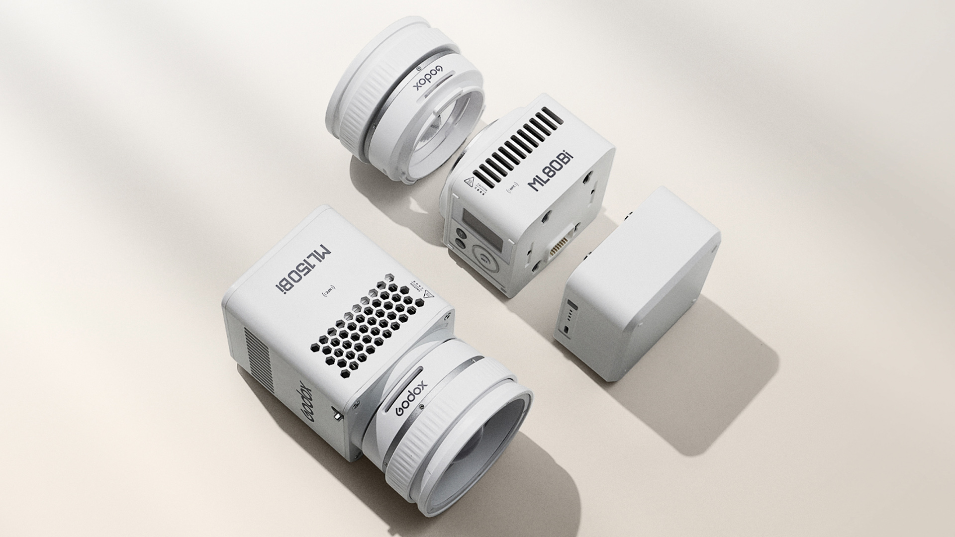
Godox is introducing two compact bi-color LED lights, the ML80Bi and ML150Bi, to their lineup of video lighting tools. Both lights use a modular system that adapts to different power and accessory setups, making them versatile and practical for…

Samsung is turning up the heat this summer with its exciting Summer Is On Us campaign – a celebration of big screens, bold entertainment, and even bigger rewards. From 20 October 2025 to 25…