Powered by Samsung’s Crystal Architecture™, the R20 delivers superior image clarity, contrast, and diagnostic accuracy across a wide range of clinical applications
Equipped with AI-driven automation tools and an ergonomic design, the R20 enhances workflow efficiency and elevates the clinician experience
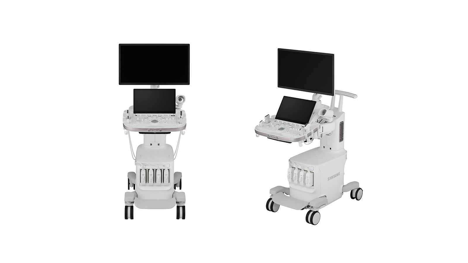
Samsung, India’s largest consumer electronics brand, today announced the launch of its super-premium, next-generation R20 ultrasound system for general imaging. The R20 represents a major leap forward in general imaging, combining advanced artificial intelligence tools, superior image clarity, and an ergonomic design focused on clinician comfort and efficiency.
Built on Samsung’s state-of-the-art Crystal Architecture™, the R20 delivers exceptional image uniformity, resolution, and penetration across a wide range of general imaging applications. Its next-generation imaging engine, powerful GPU, and ultra-high-definition OLED monitor provide clinicians with remarkable visualization and diagnostic confidence in every scan.
The R20 is equipped with a comprehensive suite of AI-powered clinical and workflow enhancement tools that streamline complex procedures and automate repetitive tasks. Key technologies include:
- Live LiverAssist – Detects a suspicious focal lesion during live ultrasound scan
- Live BreastAssist – Real-time detection of Breast lesions with BIRADS Classification and reporting.
- Auto measurement tools– AI-based automatic detection, measurement of internal structures with guided reporting for high consistency and maximum throughput
- Deep USFF– AI Based Deep Ultrasound Fat Fraction quantification with proven high correlation to the gold standard, i.e., MRI PDFF
With its superior imaging architecture, the R20 delivers remarkable performance across a wide spectrum of clinical applications — including abdomen, thyroid, musculoskeletal, vascular, breast, obstetrics, gynaecology, and urology imaging. Enhanced Doppler sensitivity and colour flow visualization allow clinicians to detect subtle vascular structures and pathologies with greater precision and confidence. This versatility ensures that healthcare professionals can achieve consistent, high-quality diagnostic results across diverse patient profiles.
“The R20 embodies Samsung’s commitment to advancing healthcare through intelligent innovation. With AI at its core and a focus on both image excellence and clinician comfort, the R20 is a paradigm shift in ultrasound technology helping doctors ensure detection of lesions during live scanning,” said Atantra Das Gupta, Head of HME Business, Samsung India.
Beyond its imaging capabilities, the R20 emphasizes user comfort and operational excellence. Designed with ergonomics in mind, it features lightweight transducer cables, an intuitive touch interface, and customizable system configurations to meet varied clinical needs. The system’s refined design minimizes strain and fatigue, enabling clinicians to focus on what matters most — their patients.
With the launch of the R20, Samsung reaffirms its commitment to shaping the future of healthcare technology. Combining AI-driven intelligence, superior imaging performance, the R20 is set to transform the landscape of general imaging, and a design that puts the clinicians and the patient at the centre of care.
For more information about Samsung R20, please visit: https://samsunghealthcare.com/en