Tokyo, November 25, 2025 – Mitsubishi Heavy Industries, Ltd. (MHI) has been awarded the engineering, procurement, and construction (EPC) contract for a Cyclo Olefin Polymer (COP)(Note) production plant planned by Zeon Corporation (hereinafter “Zeon”) in Shunan City, Yamaguchi Prefecture. The plant is scheduled for completion in the first half of fiscal 2028.
Leveraging its extensive experience in chemical plant design and construction, MHI will deliver process design, procurement of major equipment, and installation of plant machinery. The architectural and civil engineering works will be undertaken by Fujita Corporation.
MHI’s proposal was highly evaluated based on its abundant knowledge and expertise in high-performance chemicals.
MHI is focusing not only on conventional plants such as methanol and ammonia but also on high-performance chemicals including COP. This contract further strengthens MHI’s presence in the domestic and global chemical manufacturing plant markets.
MHI remains committed to contributing to the advancement of global industry and the realization of a sustainable society through initiatives and products aligned with societal and environmental needs.
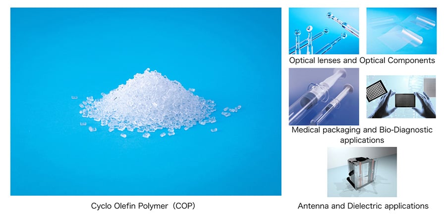
- Cyclo Olefin Polymer (COP) is a high-performance chemical characterized by excellent optical properties, low moisture absorption, and extremely low impurity levels. Demand is expected to grow in optical, medical, and semiconductor applications.