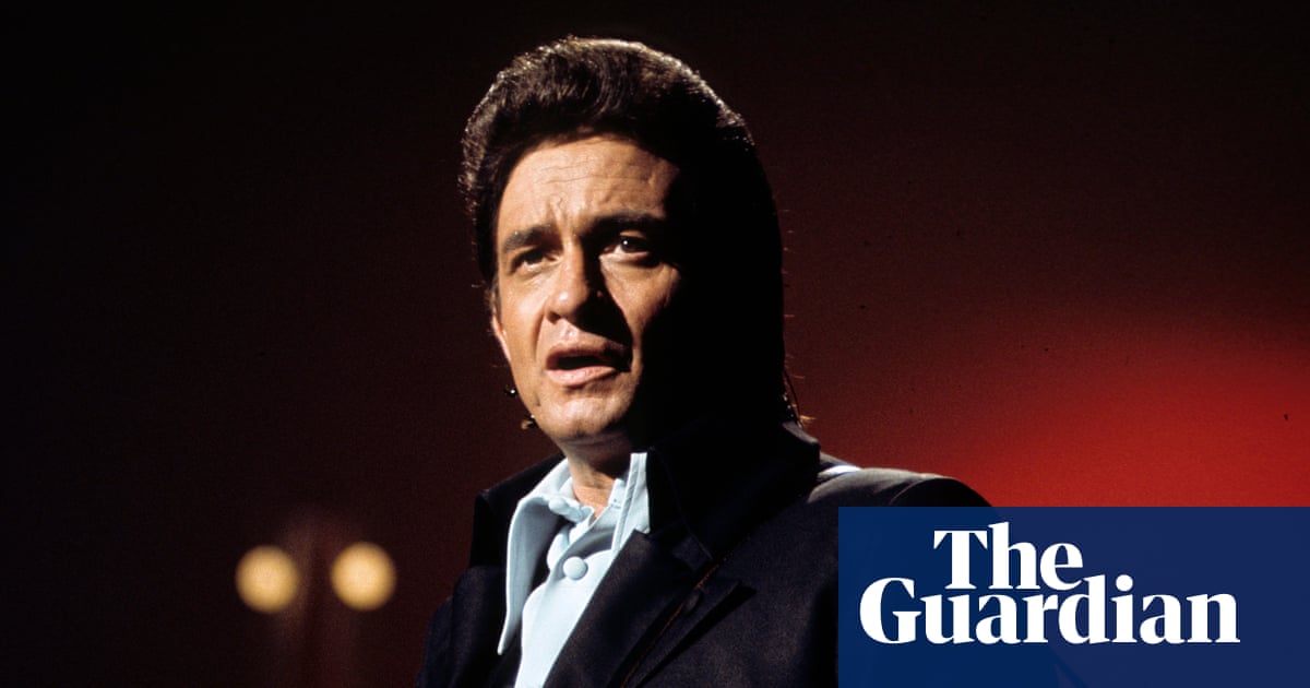
Introduction
Neuromyelitis optica spectrum disorder (NMOSD) is a rare autoimmune neurological condition which primarily manifests as recurrent optic neuritis and myelitis, with less frequent involvement of cerebral hemisphere, brainstem, and diencephalon.1,2 Repeated attacks often result in permanent neurological deficits, including blindness and paralysis.3,4 Approximately 75% of NMOSD cases harbor pathogenic autoantibodies against aquaporin-4 (AQP4), which serve as a primary effector and a diagnostic biomarker of NMOSD.5–7 The Binding of AQP4-IgG to AQP4 on astrocytes triggers classical complement activation, and antibody-dependent cellular cytotoxicity, resulting in astrocytic injury, neuronal loss, and demyelination.8,9 Additionally, B cells contribute to NMOSD pathophysiology by activating autoimmune T cells through antigen presentation, driving plasma and plasmablasts cells producing pathogenic AQP4-IgG, and secreting pro-inflammatory cytokines.10,11 Improved knowledge of the pathogenic mechanisms of NMOSD has facilitated the development of targeted therapies.
Inebilizumab, a monoclonal antibody targeting and depleting CD19-expressing B lymphocytes, was first approved globally for use in adults with AQP4-IgG seropositive NMOSD in 2020.12 Inebilizumab has been shown excellent effectiveness in reducing the risk of attack and preventing disability progression assessed by expanded disability status scales (EDSS) in NMOSD.1 Moreover, greater reductions in NMOSD-related hospitalizations and cumulative MRI activities were observed with inebilizumab than with placebo.13,14 Beyond NMOSD, its wide B-cell depletion may benefit other neuroimmunological conditions, such as steroid-refractory neurologic immune-related adverse events and autoimmune encephalitis, which often require prolonged immunosuppression.15–17 Regarding safety, inebilizumab exhibited favorable safety in clinical trials, with most mild to moderate side effects, such as urinary tract infections, arthralgia and infusion-related reactions.18 Current evidence suggests that while anti-CD20 antibodies alone may not require routine PJP prophylaxis, the coexistence of additional risk factors (eg, high-dose corticosteroids, absolute lymphopenia, or concurrent immunosuppressants) warrants preventive measures.19 Although inebilizumab’s broader CD19 targeting theoretically poses higher infection risk than CD20-specific agents,20 no PJP cases have been reported. Here, we presented a case of an NMOSD patient that developed severe respiratory failure after inebilizumab therapy and was finally diagnosed with PJP. In addition, multiple PJP cases in patients with autoimmune disorders of the central nervous system (CNS) were summarized.
Case Presentation
Initial NMOSD Diagnosis
A 41-year-old woman with NMOSD was admitted to our hospital who complained of intermittent fever and a mild cough. She denied recent travel, previous asthma, tuberculosis, tumors, and poor working or living situations. On March 8th, 2024, the patient presented skin itchiness and hyperalgesia in the right upper limb in the absence of identifiable triggers. Over time, the scratched skin developed numbness, spreading from the fingers to the right shoulder. In April, contrast-enhanced magnetic resonance imaging (MRI) of the cervical spine revealed edema in C2-C5 of the cervical spinal cord, while cranial MRI showed no abnormalities. In May, the patient experienced worsening numbness and cramps, with a new onset of numbness in her right leg. Contrast-enhanced thoracic and lumbar MRI revealed disc bulging at the L3/4, L4/5, and L5/S1 levels. The patient underwent antibody testing, confirming AQP4-IgG-seropositivity (1:32). Based on her clinical symptoms, laboratory results, and radiological features, the patient was diagnosed with NMOSD.
B-Cell Depletion and Response
Methylprednisolone was administered at 1000 mg for 3 days, followed by gradually tapered doses to 80 mg and maintained. Inebilizumab (300 mg) was administered on June 6th and 21st, respectively, after which the patient’s CD19+ B cells dropped from 11.04% to 0.02%, while CD20+ B cells decreased from 11.20% to 0.39% of the total lymphocytes. At the time of discharge on June 28th, her symptoms of numbness and cramps had significantly diminished with AQP4-IgG titer 1:10, and her EDSS score decreased from 2.5 to 1.5 points.
Onset of Respiratory Symptoms and Diagnostic Workup
However, the patient developed intermittent fever (peaking at 38.8°C) and headache since July 25th, with symptomatic fluctuations following ibuprofen administration. Upon admission on August 12th, the physical examination found an afebrile status (36.6°C) and fine moist rales in her lower lung lobes. An arterial blood gas analysis showed partial oxygen pressure (pO2) of 87 mmHg and partial pressure of carbon dioxide (pCO2) of 29 mmHg. Laboratory tests revealed a high neutrophil count (7.32*109/L) and C-reactive protein (CRP) level (37.73 mg/L), a decreased lymphocyte count at 0.66*109/L, and a low-level CD4 count of 324 cells/L. A rapid influenza virus antigen tested negative. The chest computed tomography (CT) revealed bilateral ground glass opacity with linear and patchy shadows (Figure 1). Empirical treatment with ceftriaxone and prednisone acetate (40 mg/day) was administrated. On the second day of hospitalization, the patient developed abrupt shortness of breath and her pulse oxygen saturation decreased to 87%. Due to hypoxemia (pO2 59 mmHg, pCO2 38 mmHg), she was given high-flow oxygen with a mask and atomization inhalation and was diagnosed with acute respiratory failure. To identify etiological factors, the patient underwent a series of pathogen tests (Table 1). Electric bronchoscopy was performed and bronchoalveolar lavage fluid (BALF) was collected, which was analyzed through culture, smear, galactomannan test, and metagenome next-generation sequencing (mNGS). On the third day, her axillary temperature rose to 38.7°C. Laboratory tests showed (1,3)-beta-D-glucan(G-Test) of 1378. 40 pg/mL. Additionally, mNGS revealed Pneumocystis jirovecii in BALF with reads per million (RPM) of 45776 and cytomegalovirus with 349 RPM, while no other pathogenic microorganism was detected (Table 1).
Table 1 Pathogen Examination and Results
|
![]() |
Figure 1 Chest CT imaging of the patient. (A–D) On August 12, upon admission, chest CT revealed ground-glass opacities in both lungs, with linear and patchy opacities of increased density (indicated by white arrows). (a–d) Repeat chest CT on August 20 demonstrated significant resolution of the bilateral lung lesions compared to the previous scan.
|
PJP Confirmation and Treatment
Accordingly, a diagnosis of PJP was confirmed via BALF mNGS and the patient was prescribed oral trimethoprim- sulfamethoxazole (TMP-SMX) 240 mg/1200 mg every 6 hours, thymalfasin to boost immunity, and methylprednisolone (40 mg for 5 days, then decreased to 35 mg). On August 24th, the patient was free from cough and breathlessness. A repeat chest CT showed an obvious decrease in exudation (Figure 1a–d). In addition, laboratory tests revealed normal CRP and decreased G-Test level (526.50 pg/mL). She was discharged and advised for an additional four cycles of PJP prophylaxis (160 mg TMP/800 mg SMZ daily). During the two-month follow-up, the patient remained asymptomatic for respiratory symptoms. In September 2024, comparative analysis showed negative anti-AQP4 antibody, undetectable CD19 cells (0 cells/μL), and a stable EDSS score of 1.5. In October, the G-test was evaluated within normal limits.
Discussion
PJP Clinical Characteristics in Immunocompromised Hosts
PJP, an opportunistic fungal infection, remains a serious threat to the immunocompromised population, especially in human immunodeficiency virus (HIV) infected individuals.21 The incidence of PJP in non-HIV-infected patients has been increasing in recent decades for the widespread use of immunosuppressive agents.22–26 Patients with PJP classically present with fever, non-productive cough and dyspnea accompanied by interstitial opacities on chest imaging.27 Nevertheless, differences exist between non-HIV and HIV-infected patients in terms of presentation, clinical progression, and outcomes. Specifically, non-HIV PJP usually exhibits more severe manifestations, evolves more rapidly, and carries a higher mortality.28–31 Early treatment for PJP is of paramount significance, for a delay of 4 days in treatment initiation has been shown to accelerate the time to death by 6.75-fold.30 Therefore, prompt and thorough examination of suspected PJP cases is crucial. To figure out the pathogen, we conducted a series of etiological tests, and eventually mNGS confirmed the diagnosis of PJP. Timely administration of anti-pneumocystis drugs prompted rapid improvement in the patient’s condition.
B Cell Depletion and PJP Risk
PJP is associated with low CD4+ T-cell counts, HIV infection, solid organ or stem cell transplantation, malignancies, and the use of chemotherapeutic or immunosuppressive agents.32 A decline in CD4+ T cell counts below 200 cells/μL renders highly susceptible to PJP, serving as a determinant for the initiation of prophylaxis against opportunistic infections.33–36 However, in this case, we conducted simultaneous testing of CD4+ and CD3+T cell count, ruling out PJP due to the reactive activation of T cells. Furthermore, there was no evidence of HIV infection or malignancies, and the patient had not received other immunosuppressive drugs known to cause PJP. Given the initiation of inebilizumab and corticosteroid treatment, drug-induced PJP was considered as the most probable cause of the symptoms. Based on the Naranjo Adverse Drug Reaction Scale, the probability of inebilizumab-induced PJP in the case is identified as probable (Supplementary Table 1). To the best of our knowledge, this is the first reported PJP case in an NMOSD patient following the use of inebilizumab and corticosteroids.
Previous studies have indeed demonstrated the critical contribution of T lymphocytes response against pneumocystis, wherein T cells mediate clearance of pathogens through the recruitment and activation of effector cells.37 The increasing utilization of B cell-targeted therapies in autoimmune diseases and hematological malignancies has brought increasing attention to the role of B lymphocytes in pulmonary defense against Pneumocystis. By producing Pneumocystis-specific IgM antibodies which target fungal cell wall carbohydrates, B cells enhance pathogen opsonization, complement activation, and independent phagocytosis, thereby promoting the clearance of microbial pathogens.38 This protective role is evidenced in B-cell-deficient models, which exhibit progressively increasing Pneumocystis burden and delayed pathogen elimination.39 Clinical relevance is underscored by data from CD40 ligand knockout mice, where decreased CD19+ B cells correlated with heightened susceptibility to severe PJP, contrasting with immunocompetent mice that mount robust responses and effectively resolve infection.40 Further supporting this, clinical observations identify decreased CD19+ B-cell counts as an independent risk factor for PJP.41 Moreover, B cells contribute to post-infection immune reconstitution, facilitating bone marrow lymphocyte repopulation after pneumocystis lung infection.42 Clinically, B cell depletion with rituximab predisposes patients to both PJP and subsequent severe lung injury.43 Collectively, B cells are indispensable for effective anti-Pneumocystis, acting through antibody-dependent mechanisms and immunomodulatory pathways. Their deficiency not only impairs pathogen clearance but also disrupts immune homeostasis, exacerbating PJP severity. These insights advocate for further exploration of B cell-targeted strategies to augment host defense or modulate dysregulated inflammation in PJP.
Corticosteroids in PJP: Dose–Response Controversies and Adjunctive Therapy Dilemmas
Corticosteroids remain the most commonly implicated pharmacological risk factor for PJP development.44 In a retrospective analysis involving 116 HIV-negative cases, 90.5% had received corticosteroids systemically within one month before PJP diagnosis,45 corresponding with our review (Table 2). However, evidence regarding the dose of corticosteroids associated with PJP risk in non-HIV patients remains highly controversial. In a nationwide retrospective study of patients with autoimmune rheumatic diseases, the use of glucocorticoid above 10 mg daily was identified as conferring the highest PJP risk.46 Another research suggested threshold effects at more than 20 mg prednisone monotherapy, though the small sample size (n=21) may limit its generalizability.47 Moreover, a dose-dependent hierarchical pattern was observed with median glucocorticoid doses of the PJP group reaching 38.7 mg/day,48 consistent with several studies.49–51 Nevertheless, this association appears context-dependent, as low-dose prednisone (20–30 mg/day) did not significantly increase PJP risk in patients receiving concurrent rituximab.31 These inconsistencies may reflect differences in study populations and treatment regimens. In conclusion, current evidence supports a dose–response relationship, but the precise risk threshold may vary based on underlying immune status and combination therapies, underscoring the need for individualized risk assessment when considering PJP prophylaxis.
![]() |
Table 2 Overview of Pneumocystis jirovecii Pneumonia on Autoimmune CNS Disorders
|
Controversy over the appropriateness of adjunctive corticosteroid therapy to PJP outcomes persists, and the optimal dosage remains inadequately explored.60 A prospective observational study including 49 intensive care units (158 cases) demonstrated that adjunctive corticosteroids increased mortality rate in non-HIV patients, but found no obvious association in HIV individuals.30 In contrast, a multicentre retrospective cohort of 139 non-HIV PJP patients indicated no significant differences between high-dose pulse methylprednisolone (500 to 1000 mg/day) and moderate-dose corticosteroid (<500 mg/day) in 30- or 180-day mortality, even in the subgroup analysis of respiratory failure cases.61 Surprisingly, a large-scale meta-analysis of 16 retrospective cohorts (2518 PJP cases) showed that low-dose corticosteroids (1 mg/kg/day prednisone equivalent) were linked to higher mortality, except in severe acute respiratory failure (PaO2<60mmHg), where corticosteroids were related to better clinical outcomes and decreased mortality.60 Conversely, others reported no significant survival benefit with adjunctive corticosteroids in moderate-to-severe PJP, irrespective of recent corticosteroid use.62 After discussion, adjunctive corticosteroid therapy was ultimately administered to the patient to prevent irreversible neurological deficits from potential subsequent attacks induced by opportunistic infections.
PJP in CNS Autoimmune Disorders
We conducted a comprehensive literature review of PJP patients with CNS autoimmune diseases across the PubMed database, spanning publications from its inception to 1 February 2025. The search strategy was provided in the supplementary file. A total of 12 patients (including one from our study) were included in the statistical analysis (Table 2). The median age of the patients was 32 years, ranging from 0.92 to 82 years. Despite higher prevalence among female NMOSD and multiple sclerosis (MS) patients, our retrospective analysis revealed a balanced gender distribution in PJP, potentially due to a small sample size introducing bias. The clinical presentation of PJP in this review was characterized by dyspnea (66.7%), cough (58.3%), and fever (41.7%), stressing the imperative to maintain a high index of suspicion for agnogenic respiratory symptoms in CNS autoimmune disorders. Of note, 58.3% of patients had undergone B-cell depleting therapies with PJP onset at a median of 8 weeks (range from 5 to 12 weeks) after the last treatment, indicating a potential synergistic risk between these immunosuppressive modalities possibly mediated by lymphopenia (Patients 2, 9, and 12). Diagnostically, the low sensitivity of sputum smear microscopy (8.3% in this study) for PJP correlates with typically scant sputum production. Six patients (50%) were diagnosed through BALF analysis, which demonstrates superior diagnostic efficacy of early BALF sampling for prompt diagnosis of PJP. It is necessary to acquire BALF when clinical suspicion persists despite negative sputum etiological tests. Currently, the emerging utility of mNGS further facilitates a wide detection range, particularly benefits hosts with unidentified etiologies, immunocompromised status, or infection caused by atypical or culture-negative pathogens.
Prophylaxis Challenges and Recommendations
Providing appropriate PJP prophylaxis treatments to immuno-compromised patients is crucial to reduce mortality and improve clinical outcomes.63 Largely based on retrospective studies involving oncology and transplant patients, a widely cited recommendation is to initiate PJP prophylaxis when receiving ≥20 mg prednisone above 2 to 4 weeks.45,64 However, the risk-benefit assessment becomes more complex in the context of CNS autoimmune diseases, such as NMOSD and MS. These patients are often managed with high-dose glucocorticoids and additional immunomodulators, yet the low incidence of PJP and non-negligible adverse effects of prophylaxis regimens have led to a lack of consensus on routine prevention.31,64 Notably, in our review of CNS autoimmune disease cases, none of the patients had received prophylaxis for PJP (Table 2), indicating that the absence of prophylactic measures may serve as a significant risk factor. Given the current gaps in evidence, large-scale prospective researches are urgently needed to evaluate the incidence of PJP in autoimmune CNS disorders, optimal thresholds, and long-term safety profiles of preventive regimens. Clinicians should weigh individual risk factors, such as cumulative immunosuppression, lymphopenia, and comorbidities when considering PJP prophylaxis in this population. A tailored approach, rather than universal prophylaxis, may offer the best balance between efficacy and safety.
Limitations
Our retrospective analysis has several limitations that warrant consideration. First, our review was constrained by small sample sizes and incomplete data, such as lymphocyte counts and B-cell depleting therapy duration, compromising the accuracy and generalizability of our findings. Moreover, heterogeneity in immunosuppressive regimens and baseline comorbidities among patients could introduce confounding factors, potentially obscuring real relationships between specific risk factors and PJP development. To better elucidate the risk of PJP in autoimmune CNS disorders, future research should employ prospective designs with larger patient cohorts and comprehensive documentation of immune profiles and treatment histories.
Conclusion
PJP is rare in autoimmune CNS disorders but may progress rapidly. Clinicians should maintain heightened vigilance for PJP and promptly obtain BALF for microbiological confirmation to facilitate early diagnosis and timely therapeutic intervention. Our report demonstrates the first confirmed NMOSD case of PJP due to maintenance therapy with inebilizumab and corticosteroids, highlighting the emerging safety concern of this highly efficacious treatment. The diagnosis of PJP in this case was unexpected, and we attributed her susceptibility to deep B cell depletion response to inebilizumab and corticosteroid therapy. Our finding supported previous research indicating the critical role of B lymphocytes in the protective immune response against PJP. The consistent temporal relationship between treatment initiation and PJP onset emphasizes the need for vigilant monitoring and proactive prophylactic strategies to mitigate this risk. Prospective multicenter studies and registries are needed to quantify risk and guide prophylaxis duration. Clinicians should consider individual risk assessment for PJP prophylaxis in NMOSD patients treated with B-cell depleting agents.
Abbreviations
NMOSD, neuromyelitis optica spectrum disorder; PJP, Pneumocystis jirovecii pneumonia; AQP4, aquaporin-4; EDSS, expanded disability status scales; CNS, central nervous system; MRI, magnetic resonance imaging; pO2, partial oxygen pressure; pCO2, pressure of carbon dioxide; CRP, C-reactive protein; CT, computed tomography; BALF, bronchoalveolar lavage fluid; mNGS, metagenome next-generation sequencing; G-Test, (1,3)-beta-D-glucan; TMP-SMX, trimethoprim-sulfamethoxazole; HIV, human immunodeficiency virus.
Data Sharing Statement
Data will be provided by the corresponding author upon reasonable request.
Ethics Approval and Consent to Participate
The ethics committee of Shenzhen Traditional Chinese Medicine Hospital confirmed that no separate institutional approval was required for publishing anonymized case data under local regulations.
Consent Statement
Written informed consent was obtained from the patient for the publication of any potentially identifiable images or data included in this case report prior to inclusion.
Acknowledgments
The authors thank all the clinical staff who contributed to the study.
Funding
This work was supported by the Sanming Project of Medicine in Shenzhen [grant numbers SZZYSM202111011]; “3030 project” of Clinical Research Program in Shenzhen Traditional Chinese Medicine Hospital in 2021 [grant numbers G3030202132].
Disclosure
The authors report no conflicts of interest in this work.
References
1. Hor JY, Asgari N, Nakashima I, et al. Epidemiology of neuromyelitis optica spectrum disorder and its prevalence and incidence worldwide. Front Neurol. 2020;11:501. doi:10.3389/fneur.2020.00501
2. Cree B, Bennett JL, Kim HJ, et al. Inebilizumab for the treatment of neuromyelitis optica spectrum disorder (N-MOmentum): a double-blind, randomised placebo-controlled Phase 2/3 trial. Lancet. 2019;394(10206):1352–1363. doi:10.1016/S0140-6736(19)31817-3
3. Wingerchuk DM, Banwell B, Bennett JL, et al. International consensus diagnostic criteria for neuromyelitis optica spectrum disorders. Neurology. 2015;85(2):177–189. doi:10.1212/WNL.0000000000001729
4. Asseyer S, Schmidt F, Chien C, et al. Pain in AQP4-IgG-positive and MOG-IgG-positive neuromyelitis optica spectrum disorders. Mult Scler J Exp Transl Clin. 2018;4(3):1841750644.
5. Lucchinetti CF, Mandler RN, McGavern D, et al. A role for humoral mechanisms in the pathogenesis of Devic’s neuromyelitis optica. Brain. 2002;125(Pt 7):1450–1461. doi:10.1093/brain/awf151
6. Waters PJ, Pittock SJ, Bennett JL, Jarius S, Weinshenker BG, Wingerchuk DM. Evaluation of aquaporin-4 antibody assays. Clin Exp Neuroimmunol. 2014;5(3):290–303. doi:10.1111/cen3.12107
7. Lennon VA, Kryzer TJ, Pittock SJ, Verkman AS, Hinson SR. IgG marker of optic-spinal multiple sclerosis binds to the aquaporin-4 water channel. J Exp Med. 2005;202(4):473–477. doi:10.1084/jem.20050304
8. Nishiyama S, Misu T, Nuriya M, et al. Complement-dependent and -independent aquaporin 4-antibody-mediated cytotoxicity in human astrocytes: pathogenetic implications in neuromyelitis optica. Biochem Biophys Rep. 2016;7:45–51. doi:10.1016/j.bbrep.2016.05.012
9. Roemer SF, Parisi JE, Lennon VA, et al. Pattern-specific loss of aquaporin-4 immunoreactivity distinguishes neuromyelitis optica from multiple sclerosis. Brain. 2007;130(Pt 5):1194–1205. doi:10.1093/brain/awl371
10. Bennett JL, O’Connor KC, Bar-Or A, et al. B lymphocytes in neuromyelitis optica. Neurol Neuroimmunol Neuroinflamm. 2015;2(3):e104. doi:10.1212/NXI.0000000000000104
11. Hausser-Kinzel S, Weber MS. The role of B cells and antibodies in multiple sclerosis, neuromyelitis optica, and related disorders. Front Immunol. 2019;10:201. doi:10.3389/fimmu.2019.00201
12. Frampton JE. Inebilizumab: first approval. Drugs. 2020;80(12):1259–1264. doi:10.1007/s40265-020-01370-4
13. Marignier R, Bennett JL, Kim HJ, et al. Disability outcomes in the N-MOMENTUM trial of inebilizumab in neuromyelitis optica spectrum disorder. Neurol Neuroimmunol Neuroinflamm. 2021;8(3). doi:10.1212/NXI.0000000000000978
14. Cree B, Kim HJ, Weinshenker BG, et al. Safety and efficacy of inebilizumab for the treatment of neuromyelitis optica spectrum disorder: end-of-study results from the open-label period of the N-MOmentum trial. Lancet Neurol. 2024;23(6):588–602. doi:10.1016/S1474-4422(24)00077-2
15. Rossi S, Farina A, Malvaso A, et al. Clinical course of neurologic adverse events associated with immune checkpoint inhibitors: focus on chronic toxicities. Neurol Neuroimmunol Neuroinflamm. 2024;11(6):e200314. doi:10.1212/NXI.0000000000200314
16. Antonio M, Cerne D, Sara B, et al. Retrograde amnesia in LGI1 and CASPR2 limbic encephalitis: two case reports and a systematic literature review. Eur J Neurol. 2025;32(3):e70113. doi:10.1111/ene.70113
17. Malvaso A, Giglio P, Diamanti L, et al. Unravelling the acute, chronic and steroid-refractory management of high-grade neurological immune-related adverse events: a call to action. Brain Sci. 2024;14(8):764. doi:10.3390/brainsci14080764
18. Nie T, Blair HA. Inebilizumab: a review in neuromyelitis optica spectrum disorder. CNS Drugs. 2022;36(10):1133–1141. doi:10.1007/s40263-022-00949-7
19. Zhou S, Aitken SL. Prophylaxis against pneumocystis jirovecii pneumonia in adults. JAMA. 2023;330(2):182–183. doi:10.1001/jama.2023.9844
20. Forsthuber TG, Cimbora DM, Ratchford JN, Katz E, Stuve O. B cell-based therapies in CNS autoimmunity: differentiating CD19 and CD20 as therapeutic targets. Ther Adv Neurol Disord. 2018;11:1276983265. doi:10.1177/1756286418761697
21. Thomas CJ, Limper AH. Pneumocystis pneumonia. N Engl J Med. 2004;350(24):2487–2498. doi:10.1056/NEJMra032588
22. Pereira-Diaz E, Moreno-Verdejo F, De la horra C, Guerrero JA, Calderon EJ, Medrano FJ. Changing trends in the epidemiology and risk factors of pneumocystis pneumonia in Spain. Front Public Health. 2019;7:275. doi:10.3389/fpubh.2019.00275
23. Gronseth S, Rogne T, Hannula R, Asvold BO, Afset JE, Damas JK. Epidemiological and clinical characteristics of immunocompromised patients infected with Pneumocystis jirovecii in a twelve-year retrospective study from Norway. BMC Infect Dis. 2021;21(1):659. doi:10.1186/s12879-021-06144-1
24. Kolbrink B, Scheikholeslami-Sabzewari J, Borzikowsky C, et al. Evolving epidemiology of pneumocystis pneumonia: findings from a longitudinal population-based study and a retrospective multi-center study in Germany. Lancet Reg Health Eur. 2022;18:100400. doi:10.1016/j.lanepe.2022.100400
25. Wu HY, Wu KS, Huang YL, et al. Identifying predictors for bacterial and fungal coinfection on chest computed tomography in patients with Pneumocystis pneumonia. J Microbiol Immunol Infect. 2021;54(4):701–709. doi:10.1016/j.jmii.2020.06.007
26. Pates K, Periselneris J, Russell MD, Mehra V, Schelenz S, Galloway JB. Rising incidence of Pneumocystis pneumonia: a population-level descriptive ecological study in England. J Infect. 2023;86(4):385–390. doi:10.1016/j.jinf.2023.02.014
27. Wilkin A, Feinberg J. Pneumocystis carinii pneumonia: a clinical review. Am Fam Physician. 1999;60(6):
28. Salzer H, Schafer G, Hoenigl M, et al. Clinical, diagnostic, and treatment disparities between HIV-infected and non-HIV-infected immunocompromised patients with pneumocystis jirovecii pneumonia. Respiration. 2018;96(1):52–65. doi:10.1159/000487713
29. Cordonnier C, Cesaro S, Maschmeyer G, et al. Pneumocystis jirovecii pneumonia: still a concern in patients with haematological malignancies and stem cell transplant recipients. J Antimicrob Chemother. 2016;71(9):2379–2385. doi:10.1093/jac/dkw155
30. Kamel T, Janssen-Langenstein R, Quelven Q, et al. Pneumocystis pneumonia in intensive care: clinical spectrum, prophylaxis patterns, antibiotic treatment delay impact, and role of corticosteroids. A French multicentre prospective cohort study. Intensive Care Med. 2024;50(8):1228–1239. doi:10.1007/s00134-024-07489-2
31. Park JW, Curtis JR, Jun KI, et al. Primary prophylaxis for pneumocystis jirovecii pneumonia in patients receiving rituximab. Chest. 2022;161(5):1201–1210. doi:10.1016/j.chest.2021.11.007
32. Pike-Lee T, Syed S, Willis MA, Li Y. Pneumocystis jirovecii pneumonia in neurologic disorders: is prophylaxis necessary? Neurol Clin Pract. 2021;11(3):242–248. doi:10.1212/CPJ.0000000000000923
33. Evernden C, Dowhan M, Dabas R, et al. High incidence of Pneumocystis jirovecii pneumonia in allogeneic hematopoietic cell transplant recipients in the modern era. Cytotherapy. 2020;22(1):27–34. doi:10.1016/j.jcyt.2019.11.002
34. Anyimadu H, Pingili C, Sivapalan V, Hirsch-Moverman Y, Mannheimer S. The impact of absolute CD4 count and percentage discordance on Pneumocystis jirovecii pneumonia prophylaxis in HIV-infected patients. J Int Assoc Provid AIDS Care. 2018;17:1499741215. doi:10.1177/2325958218759199
35. Phair J, Munoz A, Detels R, Kaslow R, Rinaldo C, Saah A. The risk of Pneumocystis carinii pneumonia among men infected with human immunodeficiency virus type 1. multicenter AIDS cohort study group. N Engl J Med. 1990;322(3):161–165. doi:10.1056/NEJM199001183220304
36. Messiaen PE, Cuyx S, Dejagere T, van der Hilst JC. The role of CD4 cell count as discriminatory measure to guide chemoprophylaxis against Pneumocystis jirovecii pneumonia in human immunodeficiency virus-negative immunocompromised patients: a systematic review. Transpl Infect Dis. 2017;19(2). doi:10.1111/tid.12651
37. Morris A, Norris KA. Colonization by Pneumocystis jirovecii and its role in disease. Clin Microbiol Rev. 2012;25(2):297–317. doi:10.1128/CMR.00013-12
38. Rapaka RR, Ricks DM, Alcorn JF, et al. Conserved natural IgM antibodies mediate innate and adaptive immunity against the opportunistic fungus Pneumocystis murina. J Exp Med. 2010;207(13):2907–2919. doi:10.1084/jem.20100034
39. Rong HM, Li T, Zhang C, et al. IL-10-producing B cells regulate Th1/Th17-cell immune responses in Pneumocystis pneumonia. Am J Physiol Lung Cell Mol Physiol. 2019;316(1):L291–L301. doi:10.1152/ajplung.00210.2018
40. Hernandez-Novoa B, Bishop L, Logun C, et al. Immune responses to Pneumocystis murina are robust in healthy mice but largely absent in CD40 ligand-deficient mice. J Leukoc Biol. 2008;84(2):420–430. doi:10.1189/jlb.1207816
41. Li XL, Liu ZX, Liu ZJ, et al. Pneumocystis pneumonia in liver transplant recipients. Am J Transl Res. 2021;13(12):13981–13992.
42. Hoyt TR, Dobrinen E, Kochetkova I, Meissner N. B cells modulate systemic responses to Pneumocystis murina lung infection and protect on-demand hematopoiesis via T cell-independent innate mechanisms when type I interferon signaling is absent. Infect Immun. 2015;83(2):743–758. doi:10.1128/IAI.02639-14
43. Martin-Garrido I, Carmona EM, Specks U, Limper AH. Pneumocystis pneumonia in patients treated with rituximab. Chest. 2013;144(1):258–265. doi:10.1378/chest.12-0477
44. Ghembaza A, Vautier M, Cacoub P, Pourcher V, Saadoun D. Risk factors and prevention of Pneumocystis jirovecii pneumonia in patients with autoimmune and inflammatory diseases. Chest. 2020;158(6):2323–2332. doi:10.1016/j.chest.2020.05.558
45. Yale SH, Limper AH. Pneumocystis carinii pneumonia in patients without acquired immunodeficiency syndrome: associated illness and prior corticosteroid therapy. Mayo Clin Proc. 1996;71(1):5–13. doi:10.4065/71.1.5
46. Hsu HC, Chang YS, Hou TY, et al. Pneumocystis jirovecii pneumonia in autoimmune rheumatic diseases: a nationwide population-based study. Clin Rheumatol. 2021;40(9):3755–3763. doi:10.1007/s10067-021-05660-4
47. Mecoli CA, Saylor D, Gelber AC, Christopher-Stine L. Pneumocystis jiroveci pneumonia in rheumatic disease: a 20-year single-centre experience. Clin Exp Rheumatol. 2017;35(4):671–673.
48. Zhao Z, Huang Y, Ming B, Zhong J, Dong L. Characterization and associated risk factors of Pneumocystis jirovecii pneumonia in patients with AIRD: a retrospective study. Rheumatology. 2022;61(9):3766–3776. doi:10.1093/rheumatology/keab941
49. Youssef J, Novosad SA, Winthrop KL. Infection risk and safety of corticosteroid use. Rheum Dis Clin North Am. 2016;42(1):157–176. doi:10.1016/j.rdc.2015.08.004
50. Winthrop KL, Baddley JW. Pneumocystis and glucocorticoid use: to prophylax or not to prophylax (and when?); that is the question. Ann Rheum Dis. 2018;77(5):631–633. doi:10.1136/annrheumdis-2017-212588
51. Park JW, Curtis JR, Moon J, Song YW, Kim S, Lee EB. Prophylactic effect of trimethoprim-sulfamethoxazole for pneumocystis pneumonia in patients with rheumatic diseases exposed to prolonged high-dose glucocorticoids. Ann Rheum Dis. 2018;77(5):644–649. doi:10.1136/annrheumdis-2017-211796
52. Garcia-Moreno J, Igartua LJ, Montes RM. Pneumocystis Jirovecii pneumonia in a patient with anti-N-methyl-D-aspartate receptor Postherpetic Encephalitis. Pediatr Infect Dis J. 2016;35(7):816–817. doi:10.1097/INF.0000000000001165
53. Yann K, Jackson F, Sharaf N, et al. Acute respiratory distress syndrome following alemtuzumab therapy for relapsing multiple sclerosis. Mult Scler Relat Disord. 2017;14:1–3. doi:10.1016/j.msard.2017.03.001
54. Lau AY, Lui G, Chan KP, Au C, Mok V, Ziemssen T. Pneumocystis pneumonia in a patient treated with alemtuzumab for relapsing multiple sclerosis. Mult Scler Relat Disord. 2020;38:101503. doi:10.1016/j.msard.2019.101503
55. Sadeghi HZM, Mehdinezhad H, Mehraeen R, Golparvar AM, Tavakoli PA. Pneumocystis jirovecii pneumonia complicated a case of SARS-CoV-2 infection and multiple sclerosis after treatment with rituximab. Clin Case Rep. 2023;11(6):e7455. doi:10.1002/ccr3.7455
56. Nakano H, Motoyama R, Tanaka K, Tanaka M. A case of neuromyelitis optica with varicella zoster virus meningitis during mitoxantrone treatment. Rinsho Shinkeigaku. 2011;51(9):703–705. doi:10.5692/clinicalneurol.51.703
57. Zhou J, Zhang Y, Jin Y, et al. Pneumocystis pneumonia during Rituximab treatment in children with autoimmune nerve system diseases. J Neuroimmune Pharmacol. 2018;13(3):277–278. doi:10.1007/s11481-018-9791-8
58. Liu L, Huang Y, Shu S, Zhou H, Fang F, Liu X. Nocardia farcinica pneumonia complicated by pneumocystis jiroveci infection in children with Neuromyelitis optica spectrum disorders: a case report and literature review. Ital J Pediatr. 2024;50(1):255. doi:10.1186/s13052-024-01827-2
59. Baulier G, Issa N, Gabriel F, Accoceberry I, Camou F, Duffau P. Guidelines for prophylaxis of Pneumocystis pneumonia cannot rely solely on CD4-cell count in autoimmune and inflammatory diseases. Clin Exp Rheumatol. 2018;36(3):490–493.
60. Ding L, Huang H, Wang H, He H. Adjunctive corticosteroids may be associated with better outcome for non-HIV Pneumocystis pneumonia with respiratory failure: a systemic review and meta-analysis of observational studies. Ann Intensive Care. 2020;10(1):34. doi:10.1186/s13613-020-00649-9
61. Morimoto Y, Matsui H, Fujioka H, et al. Effectiveness of pulse methylprednisolone in patients with non-human immunodeficiency virus pneumocystis pneumonia: a multicentre, retrospective registry-based cohort study. BMC Infect Dis. 2024;24(1):1233. doi:10.1186/s12879-024-10151-3
62. Moon SM, Kim T, Sung H, et al. Outcomes of moderate-to-severe Pneumocystis pneumonia treated with adjunctive steroid in non-HIV-infected patients. Antimicrob Agents Chemother. 2011;55(10):4613–4618. doi:10.1128/AAC.00669-11
63. Gaborit BJ, Tessoulin B, Lavergne RA, et al. Outcome and prognostic factors of Pneumocystis jirovecii pneumonia in immunocompromised adults: a prospective observational study. Ann Intensive Care. 2019;9(1):131. doi:10.1186/s13613-019-0604-x
64. Schmajuk G, Jafri K, Evans M, et al. Pneumocystis jirovecii pneumonia (PJP) prophylaxis patterns among patients with rheumatic diseases receiving high-risk immunosuppressant drugs. Semin Arthritis Rheum. 2019;48(6):1087–1092. doi:10.1016/j.semarthrit.2018.10.018