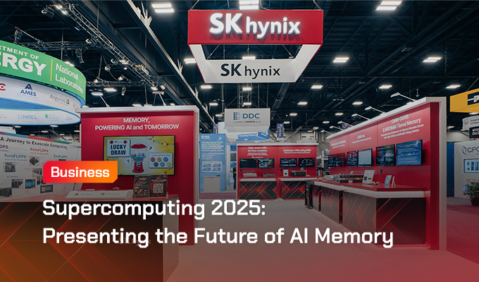
Prolonged dengue viremia with predominantly ocular manifestations in a lung transplant recipient | BMC Infectious Diseases

A 40-year-old Caucasian woman was referred to the ophthalmology clinic by her optometrist with a two-day history of bilateral scotomata and rapidly deteriorating visual acuity, alongside a four-day history of bifrontal and retro-orbital headache. She further reported mild photophobia and orbital fullness, but denied diplopia, flashes, amaurosis, pain with eye movements, neck stiffness and phonophobia. She had no history of prior ocular pathology but had had a bilateral lung transplant for cystic fibrosis 15 years prior and was stable on tacrolimus 1.5 mg twice-daily (target trough level 6–8 microg/L), mycophenolate mofetil 1000 mg twice-daily and prednisolone 5 mg daily. She had never experienced rejection episodes but had one episode of cytomegalovirus (CMV) colitis seven years prior. She had recently returned from a two-week trip to Bali, Indonesia. While overseas, she experienced a febrile illness featuring a retro-orbital headache and generalized arthralgia that self-resolved after two days. She departed Bali three days after this febrile illness started (D + 3, where D + 0 is the first day of the febrile illness). The presenting headache started ten days after the febrile illness began (D + 10), the visual symptoms on D + 12, and hospital presentation was on D + 14. She had not travelled elsewhere recently and had no sick contacts. A timeline of key events is shown in Fig. 1.

Fig. 1

Timeline of key clinical and ocular findings, investigation results and treatment given

There was no nausea, vomiting, abdominal pain, myalgia, arthralgia or rash at the time of admission. Vital signs, blood counts, haematocrit, electrolytes, renal and liver function tests were normal. Visual acuity (VA) was 6/30 on the right and 6/9.5 on the left on admission, which worsened to counting fingers on the right and 6/60 on the left the next day. Fundoscopy (Fig 2 A-B) showed bilateral cystoid macular oedema (CMO), nasal optic disc swelling, intra-retinal haemorrhages and chorioretinal atrophy. Ocular coherence tomography (OCT) (Fig 2 C-F) showed bilateral CMO with central macular thickness (CMT) of 305 micrometres on the right, and 310 micrometres on the left. Fundus fluorescein angiography showed bilateral retinal vasculitis (phlebitis more so than arteritis) and non-specific diffuse hyperfluorescent white lesions. Bilateral aqueous humour aspiration followed by intravitreal foscarnet injections were performed to investigate for and pre-emptively treat CMV retinitis given the history of CMV disease, but all samples were negative on polymerase chain reaction (PCR) for CMV, herpes simplex virus 1 and 2. Alphavirus IgM and IgG were sent to investigate for chikungunya, which returned negative.

Fig. 2
figure 2

Ocular imaging of the eye obtained on D+14 of illness. A and B: Colour fundus photography of the left and right eye respectively showing scattered macular retinitis (arrows). CF: Ocular coherence tomography of the left (C and D) and right (E and F) eyes through the fovea showing macular oedema (asterisks) and retinitis (dashed arrows)

Dengue virus (DENV) serology performed on D + 15 returned as NS1 positive, IgM positive and IgG negative. Given these results, serum, urine and the bilateral aqueous humour samples were sent to a reference laboratory for DENV PCR and confirmatory flavivirus serology. Prednisolone 50 mg daily was commenced on D + 15 for the ocular inflammation, but low-grade fevers, headaches and arthralgias resembling the initial illness occurred, and the dose was returned to 5 mg daily on D + 18. CMT peaked at this time (right eye (RE) 892 micrometres, left eye (LE) 703 micrometres). The PCR results of serum, urine and aqueous humour returned on D + 23; all were positive for DENV serotype 1 (DEN-1), and a Luminex-based microsphere immunoassay [5] detected IgM to DEN-1. To aid clearance of DENV, tacrolimus dosing was titrated to a lower target trough level of 4–5 microg/L, and mycophenolate mofetil was reduced to 500 mg twice-daily, without complications. VA and CMO improved on serial eye examinations. On D + 36, while VA and CMO continued to show improvement (RE VA 6/48 and CMT 406 micrometres, LE VA 6/38, CMT 397 micrometres), new retinal haemorrhages with increased intraocular pressure were noted on examination, prompting reinitiation of prednisolone 50 mg, this time without adverse effects. PCR of vitreous fluid from the right eye at this time was negative for CMV, but positive for DENV. VA and CMO continued to improve, and tapering of the prednisolone dose was commenced. Further serum samples were sent for DENV PCR on D + 32, D + 43, D + 49, D + 69 and D + 89; DEN-1 was detected on the samples from D + 32 and D + 49, but not on the samples from D + 43, D + 69 and D + 89. The patient was able to resume driving on D + 84, VA being 6/12 on the right, 6/15 with pinhole correction and 6/38 without on the left and 6/7.5 with both eyes open at that time. The prednisolone dose was 7.5 mg daily by D + 91. Tacrolimus and mycophenolate mofetil doses were returned to their original prescribed regimen (prior to dengue onset) by D + 98. The left eye CMO resolved by D + 111. The patient resumed part-time work on D + 126 whilst continuing prednisolone 7.5 mg daily and prednisolone eye drops for fluctuating symptomatic right-sided CMO, manifesting as scotoma and blurred vision. DENV serology performed on D + 182 showed NS1 negative, IgM positive and IgG negative, but subsequent serology on D + 319 demonstrated seroconversion with NS1 negative, IgM positive and IgG positive.

Continue Reading