 PA Media
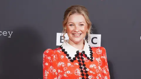
Gavin & Stacey star Joanna Page has said she was told at drama school that her performances were poor because she was Welsh.
Page, who played Stacey…
 PA Media
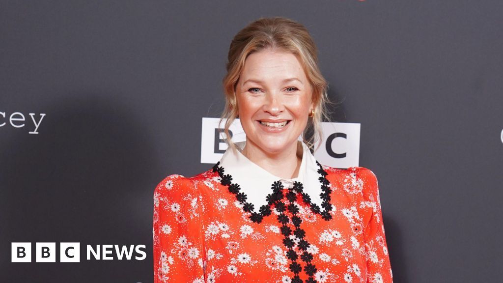
Gavin & Stacey star Joanna Page has said she was told at drama school that her performances were poor because she was Welsh.
Page, who played Stacey…