Harry Low,Londonand
Helen Drew,London political reporter
 BBC
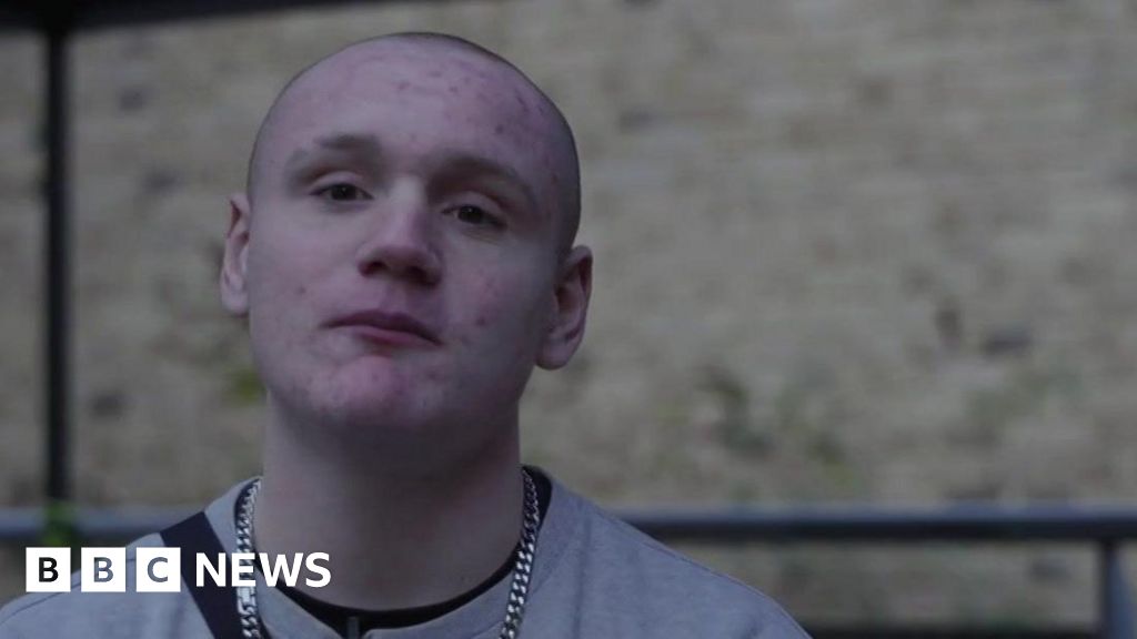
BBC“It keeps me busy, keeps me out of trouble, keeps me doing something and with the work we do as well, it gives me a…
Harry Low,Londonand
Helen Drew,London political reporter
BBC“It keeps me busy, keeps me out of trouble, keeps me doing something and with the work we do as well, it gives me a…