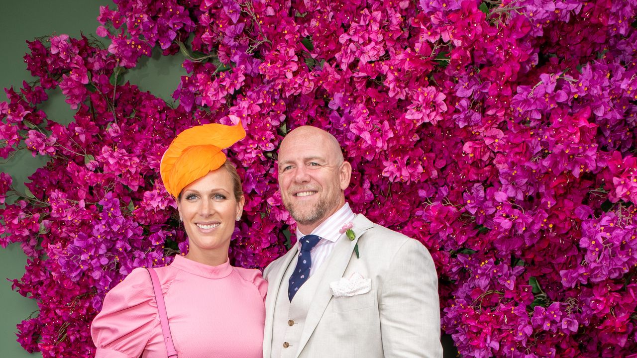
Macrophage-targeted nanomedicine for the treatment of atherosclerosis

Introduction

Atherosclerosis (AS) is a chronic and progressive disease1 characterized by subtle changes in the vascular walls that often precede the onset of organ damage due to vascular narrowing.2 In recent years, shifts in the demographic patterns of AS have been observed, notably a decrease in the age of onset and an increase in the prevalence among women.3

Table 1 Comparative Analysis of Nanocarrier Platforms for Atherosclerosis Therapy

Statins, widely recognized for their potent lipid-lowering efficacy, remain the cornerstone of AS management in both primary and secondary cardiovascular prevention. Robust clinical research4,5 underscores their ability to stabilize plaques and reduce cardiovascular events by modulating lipid profiles. However, their therapeutic utility is constrained by dose-dependent adverse effects, including hepatotoxicity and myopathy,6 which necessitate periodic monitoring of hepatic enzymes and creatine kinase levels. In recent years, proprotein convertase subtilisin/kexin type 9 (PCSK9) inhibitors have emerged as a transformative adjunctive therapy, demonstrating unparalleled efficacy in lowering low-density lipoprotein cholesterol (LDL-C) levels through targeted degradation of hepatic LDL receptors.7,8 Despite their significant promise, logistical challenges such as high costs and the need for subcutaneous administration currently limit their accessibility in routine clinical practice.9 Therapeutic strategies for advanced AS complications, notably thrombotic events, incorporate antiplatelet agents (eg, aspirin) to mitigate platelet aggregation. Long-term antiplatelet regimens, while effective in secondary prevention, carry an elevated risk of hemorrhagic complications,10,11 underscoring the need for risk stratification during patient selection.

In recent years, the application of nanotechnology in treating various diseases has grown exponentially.12,13 Compared with traditional drug formulations, nanomedicines can achieve precise localization and targeted delivery to lesion sites by modifying specific targeting molecules, such as antibodies and peptides.14 Furthermore, nanotechnology can potentially enhance drug solubility, stability, and bioavailability.15 Nanomedicines also enable responsive drug release,16 enhancing therapeutic efficacy and reducing side effects associated with premature release. As research continues to deepen our understanding of the pathophysiological mechanisms of macrophages in AS and as nanotechnology undergoes ongoing innovation, nanomedicines are expected to play a more significant role in the treatment of AS, providing new strategies and methods for the prevention and treatment of cardiovascular and cerebrovascular diseases.

In this review, we explore advancements in research on macrophage-targeted nanomedicines for the treatment of AS. The dual role and plasticity of macrophages in AS are initially examined. Subsequently, methodologies for targeting plaque macrophages with nanodrugs are investigated, and strategies for achieving controlled drug release through the manipulation of internal or external environmental cues are discussed. Moreover, the latest developments in the application of macrophage-associated biomimetic nanodrugs for the management of AS are reviewed. Finally, the challenges faced by nanomedicines in the clinical translation of AS treatment are analyzed, and a perspective on their future development is provided.

Dual Role of Macrophages in Atherosclerosis

Revisiting Macrophages: Pathogenic Agents and Therapeutic Targets

The pathological hallmark of AS is the accumulation of lipids and abnormal inflammatory responses within the arterial wall, with macrophages playing a crucial role in the development of AS and the formation of foam cells, which constitute more than 80% of the cellular components of plaques.17 During the initial stages of AS, hyperlipidemia or long-term hypertension leads to endothelial cell damage,18 allowing apolipoprotein B-containing lipoproteins (ApoB-LPs), such as LDL, to enter the arterial intima. Under the influence of proteoglycans, LDL is oxidized to form ox-LDL, which accumulates beneath the endothelium.19 Oxidized LDL (Ox-LDL) is a key trigger for early inflammatory responses. It activates the overlying endothelial cells, prompting them to secrete chemokines that recruit monocytes from the bloodstream into the subintimal layer.20 Monocytes differentiate into macrophages under the influence of macrophage-colony stimulating factor (M-CSF), with the upregulation of scavenger receptor A1 (SR-A1) and CD-36,21 enabling them to recognize and internalize ox-LDL. The engulfment of ox-LDL transforms macrophages into foam cells, which are laden with lipids.22 Foam cells, in turn, secrete various cytokines and inflammatory mediators, including tumor necrosis factor-alpha (TNF-α) and interleukin-6 (IL-6).23 These molecules stimulate the upregulation of adhesion molecules on endothelial cells, such as intercellular adhesion molecule-1 (ICAM-1) and vascular cell adhesion molecule-1 (VCAM-1). VCAM-1 can be recognized by integrins α4 and β1 on the macrophage membrane, thereby recruiting more macrophages into the inflammatory area.24 These macrophages then transform into foam cells, creating a vicious cycle that intensifies the inflammatory response and the progression of atherosclerotic lesions.

In advanced lesions, macrophages may undergo apoptosis through various mechanisms, including growth factor deprivation,19 oxidative stress, and endoplasmic reticulum stress.25 Furthermore, due to impairments in phagocytic clearance mechanisms,26 apoptotic macrophages are not effectively removed, leading to the accumulation of cellular debris and lipids within the plaque and the formation of a necrotic core. The formation of a necrotic core further exacerbates the inflammatory response and increases the risk of plaque rupture. Moreover, macrophages secrete substances such as matrix metalloproteinases (MMPs),27 which degrade the extracellular matrix, thereby compromising the structural integrity of the fibrous cap and increasing the plaque more susceptibility to rupture. Upon plaque rupture, the exposure of subendothelial components activates the coagulation system, leading to platelet aggregation and thrombus formation, triggering acute cardiovascular and cerebrovascular events.

In summary, macrophages are significant pathogenic factors contributing to the progression of AS and are highly promising targets for disease treatment. Future in-depth research into the complex mechanisms of macrophages in AS is expected to yield more innovative and effective therapeutic strategies. These advancements have the potential to offer new hope for patients with AS and to drive significant advancements in AS treatment.

Plasticity of Macrophages and Their Transformation in the Progression of Atherosclerosis

Macrophages exhibit a high degree of plasticity, enabling them to adopt diverse phenotypic characteristics under various microenvironmental stimuli. Classically activated macrophages (M1) and alternatively activated macrophages (M2) are the two most common phenotypes of macrophages. M1 macrophages, which are typically induced by cytokines such as lipopolysaccharide (LPS) and interferon-γ (IFN-γ),28 activate key signaling pathways, including nuclear factor-κB (NF-κB) and STAT1 (signal transducer and activator of transcription 1). This activation leads to the secretion of substantial quantities of inflammatory mediators, including IL-1β, IL-6, reactive oxygen species (ROS), and nitric oxide (NO).29 These mediators play critical roles in driving inflammatory responses, recruiting additional immune cells to the injury site, and contributing to the expansion of plaque volume. Consequently, the fibrous cap becomes thinner, plaque stability is significantly compromised, and the risk of acute cardiovascular and cerebrovascular events is markedly elevated.

In contrast, under the influence of anti-inflammatory cytokines such as IL-4 and IL-13,30 certain macrophages polarize toward the M2 phenotype. M2 macrophages highly express IL-10 while downregulating IL-12 and IL-23.31 These cells primarily focus on anti-inflammatory functions, suppress inflammatory cascades, and participate in tissue repair and vascular regeneration. By stabilizing plaque structure and improving the local vascular environment, M2 macrophages offer a potential therapeutic opportunity for alleviating AS lesions. Studies have shown that M1 macrophages are predominantly found in late-stage unstable plaques, whereas M2 macrophages are more highly expressed in stable plaque regions,32 indicating that the balance between M1 and M2 macrophages is closely related to plaque outcomes. Therefore, promoting the polarization of M1 to M2 macrophages has become a promising strategy for treating AS.

In addition, various intermediate phenotypes and unique subtypes of macrophages are present within plaques, such as the Mox macrophage population, M(Hb) and Mhem populations, M4 macrophages, and IL-17A-stimulated macrophages.33 Among these, Mox macrophages, which are induced by oxidized phospholipids, are widely distributed in plaques and account for 30% of plaque macrophages.34 The role of Mox macrophages in AS has yet to be fully explored. Zhai et al reported that nanoparticles (NPs) with intermediate hydrophobicity (C4NP) could induce macrophage polarization toward the Mox phenotype by activating the Nrf2 and HO-1 signaling pathways,35 which helps to further understand the complex role of macrophages in the occurrence and development of AS and provides new clues for exploring the pathogenesis of AS (Figure 1).

Figure 1 Macrophage Polarization Phenotypes and Their Roles in AS. During the development and progression of atherosclerosis, the recruitment, adhesion, and migration of monocytes, as well as the different phenotypes of macrophage polarization. Created by Figdraw.

Macrophage-Targeted Nanomedicines

Nanocarrier Platforms: Classification and Design Principles

The efficacy of macrophage-targeted nanomedicines heavily relies on the rational design of nanocarrier platforms. Based on material composition and functional mechanisms, current nanocarriers can be categorized into four major classes (Table 1). The structure of the nanomedicine delivery system is illustrated in Figure 2.

Figure 2 Schematic representation of the drug delivery nanosystem. (a) Targeting macrophage-specific ligands. (b) Cargoes exerting therapeutic effects. (c) Nanomaterials commonly used for the treatment of atherosclerosis. (d) The types of biomimetic nanomaterials currently in use. Created by Figdraw.

Organic Nanocarriers

Lipid-Based Nanoparticles (LNPs)

Lipid-based nanoparticles (LNPs), encompassing liposomes and solid lipid nanoparticles (SLNs), are structurally characterized by spherical vesicles formed from a phospholipid bilayer (liposomes) or a solid lipid core (SLNs).36 To enhance macrophage-specific targeting, these nanoparticles are often surface-functionalized with ligands such as mannose37 or CD36 antibodies,38 which facilitate receptor-mediated uptake via scavenger receptors (eg, SR-A1 or CD36) expressed on macrophages. Liposomes, for instance, can be loaded with docosahexaenoic acid, a bioactive lipid that polarizes macrophages toward the anti-inflammatory M2 phenotype,39 thereby reducing plaque inflammation and promoting cholesterol efflux from foam cells. SLNs, on the other hand, excel in encapsulating hydrophobic therapeutics, such as simvastatin,40 enabling sustained drug release within the plaque microenvironment to inhibit macrophage-driven necrotic core expansion.

Polymeric Micelles

Polymeric micelles, self-assembled from amphiphilic block copolymers like poly(lactic-co-glycolic acid) (PLGA) and polyethylene glycol (PEG), form a hydrophobic core (for drug encapsulation) and hydrophilic shell that enhances stability and prolongs circulation by minimizing opsonization and renal clearance. To target macrophages, micelles are functionalized with hyaluronic acid (HA), which binds CD44 receptors on pro-inflammatory macrophages in plaques. HA-PLGA micelles co-deliver anti-inflammatory agents (eg, rapamycin) and ROS scavengers (eg, SOD mimetics), synergistically reducing oxidative stress and stabilizing plaques. Their stimuli-responsive design—activated by plaque-specific acidic pH or ROS41—enables controlled drug release at the lesion site, minimizing systemic toxicity.

Dendrimers

Dendrimers are hyperbranched, monodisperse polymers with precise radial architecture and surface functionality. Kou et al demonstrated that polyamidoamine (PAMAM) dendrimers exhibit a hollow architecture and surface amine groups modifiable with PEG-mannose, enabling macrophage-specific targeting via mannose receptors. By employing flash nanoprecipitation with a custom multi-inlet vortex mixer, scalable synthesis (9.6 L/h) of cross-linked NPs was achieved with unprecedented drug-loading capacity (37%) and encapsulation efficiency (76%). The PEGylation reduced surface charge (+17.3 mV to +4.1 mV), minimizing cytotoxicity in RAW 264.7 macrophages and HUVECs.42

Inorganic Nanocarriers

Mesoporous Silica Nanoparticles (MSNs)

MSNs are characterized by a high-surface-area silica framework with radially ordered mesopores, enabling efficient encapsulation of therapeutic agents.43 These nanoparticles are functionalized with stimuli-responsive linkers, such as thioketal bonds, to achieve ROS-triggered drug release within atherosclerotic plaques.44 Their engineered near-infrared fluorescence properties (eg, via ICG loading) further support real-time tracking of drug delivery dynamics.45

Gold Nanoparticles (AuNPs)

Gold nanoparticles, featuring tunable surface plasmon resonance properties, adopt either spherical or rod-shaped morphologies to optimize light-to-heat conversion efficiency. Under near-infrared irradiation, AuNPs generate localized hyperthermia, inducing photothermal ablation46 of pro-inflammatory macrophages and reducing necrotic core expansion. Concurrently, their strong photoacoustic signals enable high-resolution imaging of plaque morphology.47

Superparamagnetic Iron Oxide Nanoparticles (SPIONs)

SPIONs consist of iron oxide cores coated with dextran or PEG, which ensures colloidal stability and reduces immunogenicity. Functionalization with anti-CCR2 antibodies facilitates targeted binding to inflammatory monocytes expressing CCR2 receptors, thereby directing nanoparticles to the atherosclerotic lesions.48 Magnetic guidance further improves their accumulation in plaque microenvironments.

Biomimetic Nanocarriers

Cell Membrane-Coated Nanoparticles (CMC@NPs)

CMC@NPs are synthesized via extrusion, ultrasonication, or microfluidics, balancing yield, stability, and scalability. These membranes include red blood cells (RBCs), which confer immune evasion via CD47-mediated “don’t eat me” signals to prolong circulation (>72 h);49 leukocytes (eg, neutrophils, macrophages) that inherit chemotactic receptors (CCR2, CXCR4) for active homing to inflammatory or tumor sites;50 platelets rich in adhesion molecules (P-selectin) for targeting injured vasculature;51 cancer cell membranes enabling homotypic tumor targeting via surface biomarkers;52 stem cell membranes with inherent tropism for diseased tissues;53 bacterial outer membranes carrying pathogen-associated molecular patterns for immune modulation;54 and hybrid membranes (eg, RBC-platelet fusions55) that integrate multifunctional properties. The membrane coating strategy offers inherent advantages, including immune evasion, ligand-receptor-driven targeting, enhanced biocompatibility, and protection of encapsulated payloads from enzymatic degradation.

Reconstituted High-Density Lipoprotein (rHDL)

rHDL nanoparticles are engineered by complexing apoA-I mimetic peptides with phospholipids,56 forming discoidal or spherical nanostructures that mimic endogenous HDL. rHDL can be functionalized with hydrophobic drugs (eg, statins57) to achieve dual therapeutic effects: promoting cholesterol efflux via ATP-binding cassette transporter A1 (ABCA1) and suppressing plaque inflammation.57

Exosome-Based Therapeutic Systems

Exosomes, natural extracellular vesicles (30–150 nm) secreted by stem cells or macrophages,58 are typically engineered to deliver immunomodulatory cargo (eg, anti-miR-155,59 IL-10 mRNA60) for cardiovascular therapy. Macrophage-derived exosomes polarize pro-inflammatory M1 macrophages toward anti-inflammatory M2 phenotypes via miRNA transfer, stabilizing atherosclerotic plaques in preclinical models.61 Their innate biocompatibility, low immunogenicity, and ability to cross biological barriers make them superior to synthetic carriers for sustained drug delivery. However, challenges include low yield during ultracentrifugation-based isolation, variability in cargo loading, and insufficient targeting precision.58

Emerging Hybrid Systems

Emerging hybrid nanosystems represent a transformative approach in cardiovascular therapeutics, particularly for AS, by synergistically integrating organic, inorganic, and biomimetic components to address multifactorial disease mechanisms. Macrophage-lipoprotein hybrid nanovesicles (MLP-NVs), engineered by fusing anti-inflammatory M2 macrophage membranes with lipidated peptides,62 demonstrate dual functionality: they suppress inflammation and enhance cholesterol efflux. Other hybrid platforms, such as shear-responsive nanogels63 and phototherapeutic nanosystems, further illustrate the versatility of this approach. Collectively, these hybrid systems overcome traditional limitations, such as poor targeting and systemic toxicity, by combining biomimetic tropism, microenvironment-responsive drug release, and multifunctional synergy, positioning them as promising candidates for precision medicine in cardiovascular diseases.

Passive Targeting

The design of nanomedicines necessitates not only precise targeting specificity to the diseased site but also stability within the physiological environment. Figure 3 illustrates the systemic uptake, plaque accumulation, and therapeutic action of nanomedicine.

Figure 3 Journey of nanomedicines to macrophages. (a) Due to increased vascular permeability, nanomedicines accumulate at atherosclerotic plaque sites. (b) After entering the plaque area, macrophages recognize and phagocytose nanomedicines through passive or active targeting. Under the stimulation of the plaque microenvironment or external environments, such as laser and ultrasound, they release the loaded drugs, acting on macrophages to exert therapeutic effects on atherosclerosis. Created by Figdraw.

Leveraging the intrinsic properties of these nanocarriers, passive targeting strategies exploit macrophage phagocytosis64 and pathological enhanced permeability and retention (EPR) effects to localize therapeutics within plaques.65,66 These NPs are subsequently internalized by macrophages via non-specific phagocytosis, with the majority being transported to lysosomes for degradation or further processing (Table 2).

Table 2 Passive Targeting of Macrophages for the Treatment of Atherosclerosis

The selection of nanomaterials plays a crucial role in the treatment of AS. Among the commonly used nanomaterials are polymeric nanomaterials, metallic nanomaterials, liposomes, dendrimers, and biological nanomaterials. Notably, the US Food and Drug Administration (FDA) has approved two polymeric materials, PLA and PLGA, for their extensive application in creating degradable high-molecular-weight nano-drug carriers.83 Giacalone et al encapsulated RSG in PLA-PEG NPs to evaluate its uptake by RAW264.7 cells compared with that of free radiolabeled rosiglitazone (RSG). These findings revealed that nanoparticle-mediated RSG uptake was markedly greater than that of free RSG.81 Similarly, Sun et al utilized PLGA to encapsulate pitavastatin (P-NP) and reported that, in ApoE−/− mice with AS, the P-NP group presented a significantly lower plaque area (15.7±1.08%) than did the intravenous (26.9±1.55%) and oral (17.3±0.56%) pitavastatin groups.79 These results substantiate the efficacy of employing nanomaterials as drug delivery vehicles, which can increase drug concentrations within the pathological region, extend the therapeutic duration of the medication, and thereby enhance treatment efficacy. In atherosclerosis, macrophages ingest ox-LDL and other materials, resulting in lysosomal accumulation of free cholesterol and cholesteryl esters, which impairs lysosomal function and disrupts acidic pH maintenance.84,85 As acidic NPs, PLA and PLGA can undergo hydrolysis to produce glycolic and/or lactic acid, directly restoring lysosomal acidity and function.74 Zhang et al assessed the capacity of PLA and PLGA to ameliorate macrophage lysosomal dysfunction through both in vivo and in vitro studies. Experiments in cultured macrophages revealed that PLGA NPs were particularly effective at targeting and acidifying lysosomes, whereas PLA did not affect acidification. In ApoE−/− mice fed a Western diet, treatment with PLGA NPs reduced the complexity of aortic root lesions, as evidenced by decreased cell apoptosis, reduced necrotic core formation, diminished cytotoxic protein aggregation, and increased fibrous cap formation.74 These results underscore the therapeutic potential of acidic NPs, especially those based on PLGA, in addressing macrophage lysosomal dysfunction associated with AS.

In addition to conventional carrier-based delivery systems, carrier-free drug self-delivery systems have emerged in recent years as promising strategies for achieving theranostic nanoplatforms for AS. Chen et al developed an indole molecule-templated self-assembly method to prepare probucol (PB) nanomedicines tailored for atherosclerotic theranostics. This PB-based nanomedicine not only neutralized ROS but also exhibited anti-inflammatory properties and inhibited macrophage foam cell formation. The theranostic functionality was enhanced by the ability of indole molecules to be used for fluorescence imaging.71 Wu et al designed a study focusing on tannic acid-poloxamer self-assembled nanoparticles (TPNPs) for the treatment of advanced AS. In vitro experiments demonstrated that TPNPs could effectively inhibit inflammatory responses and modulate macrophage polarization. In vivo studies further demonstrated that TPNPs could reduce the production of ROS and promote the polarization of M2 macrophages, leading to significant alleviation of advanced atherosclerotic plaques. Given their advantages, including low cytotoxicity, straightforward synthesis, and multifunctionality, TPNPs hold great promise as an intervention platform not only for AS but also for other inflammation-related diseases32 (Figure 4).

Figure 4 Passive Targeting strategies for the treatment of Atherosclerosis. (a) Schematic illustration of the synthesis procedure and the anti-atherosclerotic impacts for TPNP. Reprinted from Wu H, Sheng J, Wang Z, et al. Tannic acid-poloxamer self-assembled nanoparticles for advanced atherosclerosis therapy by regulation of macrophage polarization. J Mat Chem B. 2024;12:4708–4716. doi:10.1039/D3TB01157G. Copyright 2024, Royal Society of Chemistry.32 Copyright 2024, Royal Society of Chemistry. (b) Preparation of PB nanoparticles templated by indole molecules via hydrophobic interactions (IPNP or IRPNP) to serve as a smart theranostic agent for atherosclerosis, administered i.v. to eliminate ROS, reduce inflammatory cytokine expression, and enable FL imaging. Reprinted from Chen F. Theranostics of atherosclerosis by the indole molecule-templated self-assembly of probucol nanoparticles. J Mater Chem B. 2021;9:4134–42. Copyright 2021, Royal Society of Chemistry.71 Copyright 2021, Royal Society of Chemistry. (c) Overview of how acidic nanoparticles contribute to stabilizing atherosclerotic plaques. Reprinted from Zhang X, Misra SK, Moitra P, et al. Use of acidic nanoparticles to rescue macrophage lysosomal dysfunction in atherosclerosis. Autophagy. 2023;19(3):886–903. doi:10.1080/15548627.2022.2108252. Copyright 2022, Taylor & Francis.74

Active Targeting

Scavenger Receptors

Scavenger receptors are prominently overexpressed on the surface of activated macrophages. During the pathological progression of atherosclerosis in its early stages, the overexpression of class A scavenger receptors (SR-A) on macrophages is particularly significant.17 Leveraging this observation, Bai et al designed microRNA-146a-coated superparamagnetic iron oxide NPs (miR-146a-SPIONs).86 The outer layer of these NPs consists of miR-146a oligonucleotides, which naturally bind to SR-A receptors on macrophages and endothelial cells within atherosclerotic plaques, promoting the internalization of NPs by cells without the need for additional targeting ligands, thus achieving targeted drug delivery. The in vivo distribution of miR-146a-SPIONs was rigorously validated via various experimental techniques, including inductively coupled plasma mass spectrometry (ICP-MS), fluorescence imaging, and flow cytometry. The results indicated that the accumulation of miR-146a-SPIONs in the aorta and heart was significantly higher compared to PEG-SPIONs. Moreover, miR-146a-SPIONs exhibited stronger binding to macrophages and endothelial cells in the aorta, particularly to cells rich in SR-A, with the liver also being one of the main accumulation sites. These findings confirmed the ability of miR-146a to actively target macrophage scavenger receptors. When miR-146a-SPIONs were administered to ApoE−/− mice fed a high-cholesterol diet, qRT-PCR analysis revealed that miR-146a-SPIONs significantly reduced the expression of genes associated with the NF-κB pathway in the aorta, demonstrating their anti-inflammatory effects. In addition, miR-146a expression in the aorta was increased, confirming its gene regulatory action in vivo. Further evidence from Oil Red O staining indicated that miR-146a-SPIONs have therapeutic potential in improving atherosclerotic plaques. CD36, a membrane glycoprotein and member of the class B scavenger receptor family, is expressed on macrophages and plays a role in promoting foam cell formation upon the uptake of ox-LDL.87 Wang et al engineered a nanomedicine based on composite mesoporous silica NPs (CMSNs) that incorporated the SIRT1 activator SRT1720 and targeted macrophages with high CD36 expression via anti-CD36 antibodies to increase therapeutic efficacy. Both in vitro and in vivo studies confirmed the excellent targeting ability of CMSN@SRT@Anti. Notably, coculture experiments involving RAW264.7 cells and ox-LDL-stimulated RAW264.7 cells with CMSN@SRT@Anti revealed stronger fluorescence in ox-LDL-stimulated cells, potentially due to increased CD36 expression after ox-LDL treatment.88 These experiments, which spanned the molecular, cellular, and whole-animal levels, comprehensively validated the targeting specificity of CMSN@SRT@Anti. In addition to anti-CD36 antibodies, oxidized phospholipids can serve as ligands targeting CD36. Craparo et al selected KOdia-PC as a key component for targeting CD36 and validated its effective targeting properties through in vitro cellular experiments89 (Figure 5).

Figure 5 Active targeting Strategies for the treatment of atherosclerosis. (a) IBR@PDA-PEG-AntiCD47 facilitates atherosclerosis treatment by regulating B cells and macrophages. Reprinted from Wang H, Zhao R, Peng L, et al. A dual-function CD47-targeting nano-drug delivery system used to regulate immune and anti-inflammatory activities in the treatment of atherosclerosis. Adv Healthcare Mater. 2024;13:e2400752. doi:10.1002/adhm.202400752. Copyright 2024, Wiley-VCH.90 (b) SR-A-Ce6NB-driven SDT facilitates the transition of macrophages from the M1 to M2 phenotype, achieving both anti-inflammatory and anti-atherosclerotic effects. Reprinted from Chen Y, Wang H, Pan J, et al. Macrophage-targeted ultrasound nanobubbles for highly efficient sonodynamic therapy of atherosclerotic plaques by modulating M1-to-M2 polarization. Atherosclerosis. 2024;389:117423. doi:10.1016/j.atherosclerosis.2023.117423. Copyright 2024, Elsevier.91 (c) Schematic illustration of HNSs. Reprinted from Cao Z, Yuan G, Zeng L, et al. Macrophage-targeted sonodynamic/photothermal synergistic therapy for preventing atherosclerotic plaque progression using CuS/TiO(2) heterostructured nanosheets. ACS nano. 2022;16:10608–10622. doi:10.1021/acsnano.2c02177. Copyright 2022, American Chemical Society.14

Mannose Receptor

Macrophages harbor various carbohydrate receptors, including the macrophage galactose-binding lectin (MGL, CD301) and the mannose receptor (MR, CD206).92 Chen et al investigated the targeting potential of macrophages by decorating NPs with mannose, galactose, and dextran. They co-cultured Cy5.5-labeled NPs with RAW 264.7 macrophages and evaluated the percentage of Cy5.5-positive cells and mean fluorescence intensity (MFI) using flow cytometry. These findings revealed that NPs modified with mannose and dextran were more effective at increasing the transfection efficiency, with mannose-modified NPs being particularly effective at targeting macrophages.64 He et al employed a refined mannose-decorated dendrimeric nanoparticle system for targeted macrophage therapy, achieving dual delivery of SR-A siRNA to lower LDL uptake and LXR-L to increase cholesterol efflux.93 In vivo experiments using LDLR-deficient mice demonstrated that the treatment specifically reduced SR-A expression and increased ABCA1 and ABCG1 levels within atherosclerotic plaques in the aortic arch without affecting other tissues. The treatment also resulted in reduced plasma triglycerides and a significant decrease in plaque area and cholesterol levels in the aortic arch compared with those in the control group. Current evidence suggests that MRs are predominantly expressed in M2 macrophages, whereas their expression is either minimal or suppressed in M1 macrophages.94 In addition, Chen et al developed a novel and highly selective MR-targeting ligand and explored its potential for selectively targeting M2 macrophages for nanoparticle delivery. Confocal microscopy analysis revealed significantly greater mannose receptor targeting ligand (MRTL) uptake in M2 macrophages than in resting and M1 macrophages, with 2.4-fold and 11.8-fold increases, respectively. This research confirms the viability of MRTL-mediated M2 targeting, paving the way for in vivo studies.95

CD44

A substantial body of research indicates that the transmembrane glycoprotein CD44 is upregulated in activated macrophages and has a high affinity for HA.96 −,-Epicatechin gallate (ECG) has been shown to inhibit macrophage foam cell formation and suppress the abnormal proliferation of vascular smooth muscle cells (VSMCs), suggesting its potential as an anti-atherosclerotic agent.97 However, its poor biostability and rapid metabolism have limited its clinical application.98 To address these challenges, Yu et al leveraged the inclusion capacity and functional modification properties of β-cyclodextrin (β-CD) to design and synthesize pH-responsive acetalized β-CD (PH-CD) and CD44-targeted HA-cyclodextrin (HA-CD) dual-carrier nano-drug delivery systems,99 which encapsulate ECG to increase its stability and achieve targeted delivery for the treatment of AS. In vitro experiments using ox-LDL-induced macrophages demonstrated that, compared with free ECG, drug-loaded NPs significantly suppressed the expression of TNF-α, IL-6, and ROS in macrophages. In addition, the NPs altered the cell cycle distribution, reduced apoptosis, promoted macrophage migration, and decreased the expression of ox-LDL receptor proteins. In an ApoE−/− mouse model of AS induced by a high-fat diet, treatment with ECG-NPs improved blood lipid levels, reduced hepatic lipid accumulation, and regulated the expression of cholesterol metabolism-related genes. Notably, dual-NPs were more effective than free ECG in regulating blood lipid abnormalities and hepatic lipid accumulation while also reducing the expression of serum inflammatory mediators. Both in vitro and in vivo experiments confirmed that this nano-platform exhibits excellent biosafety.

Stabilin-2 Receptors

In atherosclerotic lesions, the expression of stabilin-2 on the surface of macrophages is significantly upregulated, positioning stabilin-2 as a critical target for macrophage-specific interventions in diseased areas. The stabilin-2 targeting peptide (S2P) can specifically target stabilin-2. Black phosphorus nanosheets (BPNSs) have attracted considerable attention in nanomedicine because of their superior biocompatibility. Recent studies have demonstrated that BPNSs can effectively scavenge ROS and suppress the production of atherosclerosis-related inflammatory factors in macrophages at lesion sites. He et al adopted a “drug delivery” strategy, synthesizing BPNSs via liquid-phase exfoliation and modifying their surface with S2P-PEG-NH2 to obtain BPNSs@PEG-S2P. The anti-inflammatory drug Resolvin D1 was loaded onto BPNSs by leveraging their two-dimensional layered structure, thereby constructing a targeted drug delivery nanosystem.100 Using a chelator-free method, they administered 64Cu-labeled BPNSs@PEG-S2P/R via tail vein injection to atherosclerotic ApoE−/− mice. The results showed that BPNSs@PEG-S2P/R exhibited a half-life of 84.4 minutes in the bloodstream, with initial high uptake in macrophage-dense organs such as the spleen and liver, which decreased over time. Radioactivity increased in metabolic organs, and aortic accumulation was significantly greater than that in the other groups, which correlated with stabilin-2 expression. Enhanced aorta accumulation was observed via near-infrared fluorescence imaging, confirming the targeting of macrophages with stabilin-2. These results confirm the feasibility of targeting macrophages with stabilin-2 as a target. Epsins, a family of evolutionarily conserved proteins involved in endocytosis, have emerged as key regulators of various cellular functions. Cui et al employed single-cell RNA sequencing combined with an innovative computational approach, to elucidate the role of Epsins in modulating cholesterol metabolism and efflux in macrophages. Their research demonstrated that Epsins could enhance lipid internalization through CD36 while suppressing cholesterol transport via ABCG1. By employing an S2P-modified nanoparticle system for Epsin siRNA delivery, the suppression of Epsins in inflamed macrophages could curb inflammation, slow the progression of AS, and promote the resolution of atherosclerotic lesions. Notably, their experiments, which included both female and male mice, revealed no sex-based differences in outcomes, suggesting that the therapeutic effects of Epsin targeting are sex independent. This work offers novel insights and a potential therapeutic avenue for AS management.101

Folate Receptor

An increasing number of studies have reported elevated folate receptor (FR) expression in atherosclerotic lesions.102,103 Notably, FR expression is significantly upregulated in activated macrophages compared with their nonactivated counterparts and is largely absent in most healthy tissues.104 Yao et al engineered a novel dual-modal imaging agent, MNPs@OPE-PEG-FA, designed to target activated macrophages within atherosclerotic plaques by utilizing folic acid as a receptor-specific ligand.105 Compared with a nontargeted control, this agent demonstrated superior cellular uptake in FR-positive RAW264.7 macrophages in vitro, a finding that was further validated in ApoE−/− mice with AS induced by carotid artery ligation. Post-administration MRI analysis of MNPs@OPE-PEG-FA revealed a marked reduction in signal intensity within the plaque regions, indicating effective targeting for imaging purposes. Furthermore, the two-photon fluorescence capability of these NPs enables a dual-modal diagnostic approach, allowing both in vitro and ex vivo detection of atherosclerotic plaques. These findings highlight the potential of this agent for noninvasive characterization of atherosclerotic lesions.105

Other Receptors

In addition to the aforementioned targeting receptors, the expression of molecules such as IL-1R106 and integrin αvβ3107 on the surface of macrophages within plaques is also upregulated. This upregulation allows for macrophage-specific targeting by leveraging these highly expressed receptors. Furthermore, CD47, which functions as a “do not eat me” signal, is expressed on the surface of apoptotic cells in plaques. It interacts with signal regulatory protein-α (SIRPα) on macrophages, thereby inhibiting the clearance of apoptotic cells by macrophages.108 To address this, Luo et al designed bifunctional polydopamine NPs.109 These NPs serve a dual purpose: first, they modulate the CD47-SIRPα phagocytic signaling axis by incorporating surface-modified anti-CD47 antibodies, which restore macrophage efferocytosis. Second, they encapsulate NLRP3 inhibitors (CY-09) to regulate intracellular inflammatory responses in macrophages, thereby reducing the secretion of the inflammatory cytokine IL-1β. This dual approach achieves a synergistic therapeutic effect, targeting both extracellular and intracellular mechanisms and effectively restoring macrophage function (Table 3).

Table 3 Active Targeting of Macrophages for the Treatment of Atherosclerosis

Controllable Release of Nanomedicines

Upon reaching the target site, drug release should be triggered by external or internal stimuli (such as pH, light, sound, enzymes, etc), ensuring that the drug is released on demand at the target site, maintaining an effective therapeutic concentration and avoiding the waste and adverse reactions caused by premature drug release (Table 4).

Table 4 Controllable Release of Nanomedicines in Plaque

ROS-Responsive Release

Atherosclerosis progression is characterized by an overexpression of ROS in tissues, driven by the inflammatory response of macrophages and mitochondrial oxidative stress.123,133 NPs featuring ROS-responsive structures can be designed to not only enable plaque imaging123 but also trigger drug release in the plaque microenvironment for the treatment of AS.125,134

Recent studies have revealed the expression of Olfactory Receptor 2 (Olfr2) in mouse vascular macrophages, which is associated with activation of the NLRP3 inflammasome.135 Consequently, Ni et al developed a nanocarrier to deliver small interfering RNA targeting Olfr2 (si-Olfr2) to macrophages to dampen NLRP3 inflammasome activation and reduce IL-1β release by downregulating Olfr2 expression.123 The nanocarrier incorporates an ROS-responsive core composed of the oligomer oDHLA, which encapsulates the siRNA complexed with the ionizable lipid-like compounds G0-C8. This ROS-responsive core enables controlled drug release in the presence of elevated ROS levels. The nanoparticle is further coated with an amphiphilic organic semiconductor polymer (SP-PEG), which not only shields the nanostructure but also enables photoacoustic imaging within the NIR-II window. By modulating SP-PEG levels, the photoacoustic signal can be tailored for enhanced atherosclerotic plaque visualization. In addition to the aforementioned ROS-responsive nanocarriers, Wu et al introduced an innovative approach termed “carrier-free nanomedicines”,124 which addresses the limitations associated with traditional nanocarriers, including suboptimal drug loading and incomplete release, thereby increasing drug bioavailability. Moreover, the intravenous administration route circumvents the hepatic first-pass effect commonly encountered with oral administration. These carrier-free nanomotors are powered by NO generated through the reaction between L-arginine and phosphatidylserine (PS). The nanomotors exhibit a dual-targeting mechanism: they first navigate toward atherosclerotic plaques by leveraging the high ROS and inducible nitric oxide synthase (iNOS) expression in the plaque microenvironment, thereby achieving initial plaque targeting. Subsequently, by mimicking the “eat me” signal through PS, they precisely target macrophages within the plaque, achieving a second level of specificity. Furthermore, these nanomotors can scavenge ROS, contributing to their therapeutic efficacy. By adopting this carrier-free nanomotor design, the bioavailability of trehalose has been significantly improved, reducing the required dosage from the previously reported 2.5 g kg−1 to 0.01 g kg1.124

pH-Responsive Release

Owing to the presence of a significant number of inflammatory cells within atherosclerotic plaques, which exhibit high metabolic activity in their activated state, these cells predominantly utilize glycolysis instead of oxidative phosphorylation to produce energy, a characteristic referred to as the Warburg effect.136 In addition, localized restrictions in blood flow contribute to hypoxic conditions, further enhancing cellular glycolysis. The substantial accumulation of lactate produced through this process lowers the pH.137

Leveraging the acidic pH properties of atherosclerotic plaques, pH-responsive NPs have been designed for precise drug release. Cheraga et al developed a pH-sensitive nanoconjugate composed of all-trans-retinal (ATR) and HA, resulting in HR NPs with a core‒shell structure tailored for targeted therapy of atherosclerotic plaques.115 The HA shell and ATR core ensure stability at physiological pH while enabling the release of ATR in the slightly acidic environment of plaques, where it exerts antioxidant effects. Furthermore, HR NPs encapsulate rapamycin (RAP), which is released within plaques to exert local anti-inflammatory and anti-proliferative effects, inhibiting AS progression. In studies using atherosclerotic model mice (ApoE−/− mice), treatment with HRRAP NPs significantly reduced the lesion area of the entire aorta. Compared with that in the saline control group, the lesion area in the HRRAP NP treatment group decreased to an average of 10.2%, which was significantly different from that in the group treated with RAP alone. Moreover, HRRAP NPs demonstrated enhanced safety and tolerability during long-term treatment periods.115 In addition to covalent linkages, metal-organic cages can also undergo pH-dependent conformational changes. Leveraging this property, Li et al used MOC-68 as a carrier to load diallyl trisulfide (DATS). The binding between MOC-68 and DATS weakened under acidic conditions, and the subsequent reaction with H2O2 further promoted the dissociation of DATS to release H2S.122

Enzyme-Responsive Release

In addition to the presence of highly active ROS and lower pH levels, the activities of specific enzymes at the plaque site can also regulate drug release. Notably, the high expression of Cathepsin K (CTSK) within arterial plaques138 has attracted significant interest. Further studies have revealed that CTSK is actively involved in the initiation and progression of AS, contributing to the formation of vulnerable plaques. Moreover, CTSK exhibited peak enzymatic activity in acidic environments. Given these findings, CTSK is considered a promising trigger for controlled drug release in the context of AS. Fang et al designed a targeting polymer for integrin αvβ3 (PLGA-PEG-c) and a CTSK-sensitive polymer (PLGA-Pep-PEG), which self-assembled to form NPs with both targeting ability and responsiveness capable of encapsulating RAP.107 Upon reaching the atherosclerotic lesion area, these NPs are cleaved under the action of CTSK, thereby releasing the encapsulated drugs for therapeutic purposes. Similarly, hyaluronidase (HAase) is another highly expressed enzyme in the plaque microenvironment. Leveraging this, Pham et al coated the surface of NPs with HA. In vitro experiments were conducted by adding HAase to buffer solutions at pH 5.0 and 7.4, followed by monitoring drug release from CD9-HMSN@RSV NPs. The results demonstrated a significant increase in the release of RSV and CD9 antibodies in the presence of HAase, particularly under acidic pH conditions that mimic the atherosclerotic lesion environment.126 These findings indicate that the presence of HAase significantly promotes the release of drugs from CD9-HMSN@RSV NPs, confirming the responsiveness of the nanoparticle system to HAase, which is highly important for achieving targeted drug delivery in the atherosclerotic lesion area. On the other hand, intracellular enzymes can also serve as effective triggers for drug release. For example, Rastegari et al utilized mannose-modified magnetic NPs as drug carriers, enabling the controlled release of simvastatin and niacin through an enzyme-responsive mechanism.111 These NPs are recognized by mannose receptors on macrophages, leading to their internalization into lysosome-rich cellular compartments. Within lysosomes, mannose hydrolase degrades the mannose layer, thereby releasing the encapsulated drugs. In vitro experiments revealed that in the presence of mannan hydrolase, NPs released 79.51% of simvastatin and 67.23% of niacin within 90 minutes while maintaining a leakage rate of less than 19.22%, significantly reducing the side effects of systemic administration.111

Cholesterol-Responsive Release

While the studies mentioned above effectively utilize abnormal microenvironments to control drug release, research that simultaneously aims to normalize these abnormal microenvironments is relatively scarce. By building on the high-cholesterol environment characteristic of plaques, Kim et al leveraged the high affinity of methyl-β-cyclodextrin for cholesterol to design cargo-switching NPs (CSNPs).127 CSNPs release simvastatin when it binds to cholesterol, as cholesterol binds more tightly to cyclodextrin. This allows CSNPs to replace simvastatin with cholesterol, facilitating the removal of cholesterol through a cargo-switching process. Confocal microscopy experiments revealed that, in vitro, CSNPs engage with cholesterol deposits within 5 minutes at 37°C, confirming their ability to pinpoint these deposits. In ApoE−/− mice with AS, CSNP treatment notably decreased plaque in the aortic root, curve, and upper section, demonstrating clear therapeutic benefits regardless of dietary conditions. Compared with those at baseline, mice fed a regular diet presented more pronounced plaque reduction in the aortic root and curve following CSNP treatment, whereas those fed a high-fat diet presented plaque reduction primarily in the root area. These findings suggest that the targeted anti-inflammatory action of CSNPs can effectively complement systemic cholesterol reduction to promote plaque shrinkage. Moreover, CSNP treatment does not adversely affect body weight, induce liver toxicity, or alter plaque vulnerability.127

Photodynamic and Photothermal Therapies

In recent years, phototherapy, such as photodynamic therapy (PDT) and photothermal therapy (PTT), has emerged as a promising treatment strategy for AS.130,139,140 Both modalities utilize light energy to achieve therapeutic effects: PDT generates ROS by activating photosensitizers, which subsequently induce macrophage death,141 whereas PTT employs photothermal agents to absorb light energy and convert it into heat, thereby inducing the thermal ablation of macrophages.142 Despite their potential, current phototherapy approaches face certain limitations and challenges. One significant concern in PDT is the selection of photosensitizers, as their nonspecific binding to vascular structures may exacerbate vascular wall damage and potentially worsen the progression of AS.143 Accordingly, the development of photosensitizers that specifically target plaque macrophages is essential. Song et al conjugated dextran sulfate with the photosensitizer chlorin e6 to create an NIRF-emitting phototherapy agent. This agent specifically targets SR-A, which is highly expressed in activated macrophages.131 By employing a customized laser system for localized irradiation, this approach enables the precise treatment of target lesions while minimizing collateral damage to surrounding healthy tissues.

Second, traditional phototherapy reduces plaques by inducing cell apoptosis; however, excessive apoptosis may increase the risk of plaque instability and rupture.144,145 To address this limitation, He et al developed an innovative approach utilizing IR780-Gd-OPN nanomicelles.129 This nanomicelle-mediated mild phototherapy demonstrated a protective effect on foam cells by preventing apoptosis through the activation of HSP27 and the inhibition of the NF-κB pathway, thereby regulating lipid metabolism, macrophage polarization, and reducing inflammatory responses. In addition, mild phototherapy maintains a controlled, low temperature, minimizing thermal damage to surrounding healthy tissues. As a result, this method yields significantly lower toxicity and greater biocompatibility compared to conventional photothermal therapy.

Sonodynamic Therapy

Sonodynamic therapy (SDT) has evolved from photodynamic therapy and possesses stronger penetration capabilities146 and more precise targeting91 than its optical counterpart. SDT activates sonosensitizers that accumulate in plaques through low-frequency ultrasound stimulation, generating ROS to induce apoptosis in target cells.14,147 However, SDT faces challenges such as insufficient accumulation of sonosensitizers and low rates of ROS production. To address these issues, Chen et al employed nanobubbles as carriers to encapsulate Ce6, increasing its accumulation and stability at the lesion site. Concurrently, they utilized low-intensity pulsed ultrasound, which can trigger this nanoplatform to produce more ROS, thereby amplifying the effects of SDT91 (Figure 6).

Figure 6 Controllable Release of Nanomedicines under the specific microenvironment of plaques. (a) Fabrication and characterization of physicochemical attributes of cargo-switching nanoparticles (CSNP). Reprinted from Kim H, Kumar S, Kang D-W, et al. Affinity-driven design of cargo-switching nanoparticles to leverage a cholesterol-rich microenvironment for atherosclerosis therapy. ACS Nano. 2020;14(6):6519–6531. doi:10.1021/acsnano.9b08216. Copyright 2020, American Chemical Society.127 (b) Schematic diagram of the formation process of RAP@T/R nanoparticles and their targeted delivery of RAP for atherosclerosis treatment, activated by CTSK. Reprinted from Fang F, Ni Y, Yu H, et al. Inflammatory endothelium-targeted and cathepsin responsive nanoparticles are effective against atherosclerosis. Theranostics. 2022;12(9):4200–4220. doi:10.7150/thno.70896. Copyright 2022, Ivyspring International Publisher (https://creativecommons.org/licenses/by/4.0/).107 (c) Schematic diagram of the synthesis of Tr-Arg-PS (TAP) nanomotors and their two-stage targeting approach for the comprehensive treatment of AS. Reprinted from Wu Z, Zhou M, Tang X, et al. Carrier-free trehalose-based nanomotors targeting macrophages in inflammatory plaque for treatment of atherosclerosis. ACS Nano. 2022;16(3):3808–3820. doi:10.1021/acsnano.1c08391. Copyright 2022, American Chemical Society.124 (d) Schematic illustration of ROS-triggered si-Olfr2 nanoparticles for atherosclerosis theranostics. Reprinted from Ni H, Zhou H, Liang X, et al. Reactive oxygen species-responsive nanoparticle delivery of small interfering ribonucleic acid targeting olfactory receptor 2 for atherosclerosis theranostics. ACS Nano. 2024;18(34):23599–23614. doi:10.1021/acsnano.4c07988. Copyright 2024, American Chemical Society.123

Multi-Responsive Drug Delivery Systems

The application of stimulus -responsive nanosystems in atherosclerosis treatment has shown promise; however, conventional single-stimulus systems, which rely solely on pH, ROS, or enzymatic triggers, face inherent limitations in addressing the dynamic and heterogeneous nature of the plaque microenvironment. For instance, pathological features such as fluctuating oxidative stress, inflammatory cytokines, and shear stress often render single-signal regulation insufficient, leading to premature drug release, suboptimal targeting, or compromised therapeutic outcomes.148 Furthermore, advanced atherosclerotic plaques exhibit structural barriers, such as thickened endothelia and narrowed intercellular gaps, which hinder the deep penetration of nanocarriers that rely on passive diffusion or a single driving force.149 To overcome these challenges, combined stimuli-responsive systems have emerged, integrating multiple pathological signals and external stimuli to achieve spatiotemporal control, enhanced lesion accumulation, and synergistic therapeutic effects.

Wei et al150 designed a dual-mode-driven nanomotor (Gd-MCNs/Pt-RAPA-AC) that integrates H2O2 catalysis and near-infrared (NIR) photothermal propulsion. This system leveraged the endogenous H2O2 at inflammatory sites to generate O2 for self-propulsion, while NIR irradiation enhanced tissue penetration and induced photothermal ablation of macrophages. The asymmetric Pt deposition on mesoporous carbon nanoparticles enabled catalytic decomposition of H2O2, alleviating oxidative stress, while rapamycin (RAPA) release counteracts inflammation and promotes autophagy. The dual propulsion mechanisms (chemical and thermal) significantly improved nanoparticle accumulation in plaques, as evidenced by enhanced MRI contrast and reduced lipid deposition in ApoE/ mice.150 Similarly, Zhang et al63 developed a shear force- and cholesterol affinity-responsive nanoplatform (FCD-Se/Res), where β-cyclodextrin (β-CD) was grafted to fucoidan via ROS-sensitive bonds, enabling targeted drug delivery. The system could actively bind to P-selectin on inflamed endothelium, penetrate plaques under high shear stress, and release resveratrol (Res) via cholesterol competition. Concurrently, ROS-triggered β-CD dissociation facilitated cholesterol efflux, while selenium nanoparticles mitigated oxidative stress. In vivo studies demonstrated a remarkable reduction in plaque vulnerability index (from 4.07 to 0.40) through synergistic regulation of macrophage polarization and lipid metabolism.63 Both studies highlighted the advantages of multi-stimulus responsiveness. Such integrated systems not only enhance drug bioavailability but also address multiple pathological processes, including inflammation, oxidative stress, and lipid accumulation, within a single therapeutic platform. However, challenges remain in optimizing stimulus sensitivity, minimizing off-target effects, and translating these designs into clinical models that better mimic human atherosclerosis. Future advancements may involve incorporating artificial intelligence for real-time monitoring or combining advanced imaging modalities to guide personalized therapy. Collectively, these innovations highlight the potential of multi-responsive nanosystems to revolutionize atherosclerosis treatment by dynamically adapting to the evolving microenvironment of the plaque.

Macrophage-Associated Biomimetic Nanoparticle Therapy for Atherosclerosis

Despite their therapeutic potential, conventional nanomedicines face rapid immune clearance, hindering clinical translation (Biomimetic Nanocarriers). To address this, CMC@NPs leverage innate “self” markers for immune evasion and prolonged circulation. Crucially, their inherited chemotactic receptors enable active homing to atherosclerotic plaques, while membrane-bound anti-inflammatory factors may locally dampen inflammation. Wang et al developed rapamycin-loaded NPs encapsulated within macrophage membranes (MM/RAPNPs) and explored their feasibility for AS treatment. After the drug was administered to ApoE−/− mice for 30 days and the separated aortas were observed, the results revealed that, compared with free RAP (18.3%) and RAPNPs (14.43%), MM/RAPNPs significantly reduced the atherosclerotic plaque area to 6.59%.151 The above evidence suggests that biomimetic NPs coated with macrophage membranes could be powerful tools for future AS treatment.

Macrophage membrane properties vary by polarization state: M1 membranes overexpress integrins (eg, α4/β1) for inflammatory site homing, while M2 membranes enhance anti-inflammatory payload delivery. This allows customization for distinct therapeutic goals. Qu et al identified M1 macrophages as pro-inflammatory macrophages, which are capable of more effectively targeting plaques through molecules such as integrins. To achieve this, they induced M1 macrophages by stimulating RAW264.7 cells with LPS, extracted the cell membranes, and developed MM@CD-PBA-RVT, a system designed to respond to and release its payload in a high-ROS microenvironment.152 On the other hand, Wu et al suggested that M2 macrophages possess greater advantages in anti-inflammatory applications. Consequently, they stimulated RAW264.7 cells with IL-4 to obtain M2 macrophages, utilized their membranes as a coating, and encapsulated PLGA and berberine to create biomimetic NPs (BBR NPs@Man/M2).153 Both M1-type and M2-type macrophage membranes have demonstrated good plaque-targeting capabilities in atherosclerotic mouse models (Figure 7).

Figure 7 Biomimetic Nanoparticle Therapy for Atherosclerosis. (a) Schematic diagram of the synthesis and drug release of MM@CD-PBA-RVT. Reprinted from Qu K, Zhong Y, Zhu L, et al. A macrophage membrane-functionalized, reactive oxygen species-activatable nanoprodrug to alleviate inflammation and improve the lipid metabolism for atherosclerosis management. Adv Healthcare Mater. 2024. doi:10.1002/adhm.202401113. Copyright 2024, Wiley-VCH.152 (b) Schematic representation of the Reprinted from Banik B, Surnar B, Askins BW, et al. Dual-targeted synthetic nanoparticles for cardiovascular diseases. ACS Appl Mater Interfaces. 2020;12(6):6852–6862. doi:10.1021/acsami.9b19036. Copyright 2019, American Chemical Society.112

Beyond the utilization of isolated cell membranes for artificial coating, cells naturally secrete a diverse array of membrane-bound extracellular vesicles (EVs), including microvesicles, apoptotic bodies, and exosomes,154 which inherently possess remarkable capabilities for intercellular communication, targeted delivery, and immunomodulation. These natural biological nanoparticles represent another promising frontier in AS nanotherapy.

Cell-derived extracellular vesicles are characterized by a double-membrane structure and are crucial for intercellular communication. They are intimately linked to vascular oxidative stress and inflammation in AS. In the present study, exosomes derived from various cellular sources were actively explored for their potential in treating AS. For example, endothelial cell-derived exosomes carrying miRNA can regulate macrophage lipid metabolism;73 platelet-derived exosomes can suppress inflammation;155 and engineered exosomes can achieve precise control of inflammation by carrying specific molecular anchors to target plaque macrophages,156 exerting anti-inflammatory therapeutic effects. Furthermore, macrophage-derived exosomes can enhance anti-inflammatory effects and regulate the proliferation and migration of vascular smooth muscle cells.157 Ji et al designed a self-assembled nanoprobe that specifically recognizes foam cells. This probe is degraded by intracellular hypochlorous acid, releasing a lipophilic fluorophore that integrates into exosomal membranes.158 This innovative approach enables early detection and continuous assessment of the risk associated with atherosclerotic lesions, thereby supporting the efficient management of cardiovascular diseases.

Challenges and Prospects

The advent of macrophage-targeted nanotherapies has opened transformative avenues for AS treatment, yet their clinical translation remains encumbered by multifaceted challenges. First, achieving precise and sustained plaque targeting persists as a critical barrier. While pioneering trials, such as the LDL-mimetic paclitaxel-loaded nanoparticles159 and the CD68-directed photothermal silica-gold nanoparticles (NANOM-FIM trial, NCT01270139160), demonstrate lesion-specific delivery, their efficacy is often compromised by off-target accumulation. This arises from the dynamic formation of a protein corona, a layer of adsorbed biomolecules on NP surfaces, that alters receptor-binding specificity and promotes nonspecific uptake in healthy tissues.30 Furthermore, heterogeneity in scavenger receptor expression across patient populations, influenced by genetic and epigenetic factors, leads to variable therapeutic responses. For instance, in the NANOM-FIM cohort, 30% of patients showed suboptimal CD68-targeted NP uptake due to receptor downregulation in advanced plaques, underscoring the need for adaptive targeting strategies.160

Second, biocompatibility and long-term safety profiles of nanomaterials demand rigorous optimization. Superparamagnetic iron oxide nanoparticles exhibit cytotoxicity, causing inflammation, increased membrane permeability, mitochondrial dysfunction, apoptosis, chromosome condensation, ROS generation, and DNA damage.160,161 Conversely, amorphous silica NPs, initially explored for their inert properties, paradoxically exacerbated arterial inflammation by activating NLRP3 inflammasomes in macrophages, thereby accelerating AS progression in murine models.162 A standardized framework for toxicity assessment, integrating high-throughput screening, multi-omics profiling, and advanced in vitro models, is urgently needed to de-risk clinical candidates.

Third, pharmacokinetic limitations hinder therapeutic efficacy, particularly in complex lesions. Rapid renal clearance of small NPs (<10 nm) and hepatic sequestration of larger particles (>200 nm) restrict their residence time in atherosclerotic niches. Besides, the dense fibrous caps of advanced plaques impede NP penetration, necessitating carriers with tunable rigidity or enzyme-responsive surface coatings to enhance diffusivity.

To overcome these limitations, future research should prioritize several aspects. First, comprehensive mechanistic studies are necessary to elucidate the precise interactions of nanomaterials within the body, including their interactions with biomolecules and their influence on disease progression. Second, innovative nanodesign strategies, including the development of novel nanomaterials, the optimization of targeting strategies, the creation of high-specificity targeting ligands or peptides, and the integration of multiple targeting mechanisms, is essential. Moreover, research should focus on interventions at different macrophage targets, screening for low-toxicity nanomaterials, and conducting extensive long-term animal experiments and clinical studies to comprehensively evaluate the long-term safety of nanotechnology in treating cardiovascular and cerebrovascular diseases, with evaluations prioritizing nanomaterials demonstrating favorable toxicity profiles, such as biodegradable polymers or engineered gold nanostructures. Furthermore, optimizing production processes, improving production efficiency, reducing raw material costs, and fostering collaboration in regulatory approvals are crucial to fully unlocking the potential of nanomedicine in treating these diseases, ultimately benefiting patients. Interdisciplinary collaboration across fields such as materials science, computational biology, and regulatory agencies is also critical to streamline scalable production and expedite clinical translation. Machine learning algorithms can accelerate the design of multi-targeted carriers by predicting ligand-receptor interactions, optimizing stimuli-responsive drug release kinetics, and identifying novel biomarkers for precision targeting. AI-driven analysis of multi-omics data may further unravel macrophage heterogeneity in plaques, enabling patient-specific nanotherapy regimens. Moreover, the development of personalized nanotreatment plans tailored to the unique needs and disease characteristics of individual patients represents a significant and promising direction for future research.

Through these comprehensive efforts, we anticipate that nanotechnology will play an increasingly significant role in the treatment of AS, offering patients more effective and safer treatment options. This progress will mark the beginning of a new chapter in the treatment of cardiovascular and cerebrovascular diseases. With the continuous advancement of science and technology, nanomaterials that target macrophages are expected to become important tools for future AS treatment, promoting the development of personalized medicine and precision therapy.

Abbreviations

AS, atherosclerosis; NPs, nanoparticles; PCSK9, proprotein convertase subtilisin/kexin type 9; LDL-C, low-density lipoprotein cholesterol; ApoB-LPs, apolipoprotein B-containing lipoproteins; ox-LDL, oxidized LDL; M-CSF, macrophage-colony stimulating factor; SR-A, scavenger receptor A; TNF-α, tumor necrosis factor-alpha; IL, interleukin; ICAM-1, intercellular adhesion molecule-1; VCAM-1, vascular cell adhesion molecule-1; MMPs, matrix metalloproteinases; LPS, lipopolysaccharide; IFN-γ, interferon-γ; NF-κB, nuclear factor-κB; STAT1, signal transducer and activator of transcription 1; ROS, reactive oxygen species; NO, nitric oxide; EPR, enhanced permeability and retention; LNPs, Lipid-based nanoparticles; SLNs, solid lipid nanoparticles; PAMAM, polyamidoamine; MSNs, mesoporous silica nanoparticles; AuNPs, Gold nanoparticles; SPIONs, superparamagnetic iron oxide nanoparticles; RBCs, red blood cells; rHDL, reconstituted high-density lipoprotein; ABCA1, ATP-binding cassette transporter A1; FDA, Food and Drug Administration; PLA, polylactic acid; PLGA, polylactic-co-glycolic acid,; RSG, radiolabeled rosiglitazone; ApoE, Apolipoprotein E; PB, probucol; ICP-MS, inductively coupled plasma mass spectrometry; qRT-PCR, quantitative reverse transcription polymerase chain reaction; KOdia-PC, 1-palmitoyl,-2-5-keto-6-octene-dioyl, phosphatidylcholine; MFI, mean fluorescence intensity; MR, mannose receptor; MRTL, mannose receptor targeting ligand; ECG, −,-Epicatechin gallate; HA, hyaluronic acid; VSMCs, vascular smooth muscle cells; β-CD, β-cyclodextrin; S2P, stabilin-2 targeting peptide; BPNSs, Black phosphorus nanosheets; FR, folate receptor; SIRPα, signal regulatory protein-α; Olfr2, Olfactory Receptor 2; PAI, photoacoustic imaging; iNOS, inducible nitric oxide synthase; PS, phosphatidylserine; ATR, all-trans-retinal; RAP, rapamycin; MOCs, metal-organic cages; DATS, diallyl trisulfide; CTSK, Cathepsin K; HAase, hyaluronidase; PDT, Photodynamic Therapy; PTT, Photothermal Therapy; NIRF, near-infrared fluorescence; HSP27, heat shock protein 27; SDT, Sonodynamic Therapy.

Data Sharing Statement

No data was used for the research described in the article.

Funding

This work was supported by the National Natural Science Foundation of China (NSFC), Grant/Award Number: 82471355 and 82171325.

Disclosure

The authors report no conflicts of interest in this work.

References

1. Blaha MJ, Abdelhamid M, Santilli F, et al. Advanced subclinical atherosclerosis: a novel category within the cardiovascular risk continuum with distinct treatment implications. Ame J Preventive Cardiol. 2023;13:100456. doi:10.1016/j.ajpc.2022.100456

2. Ahmadi A, Argulian E, Leipsic J, et al. From subclinical atherosclerosis to plaque progression and acute coronary events. J Am Coll Cardiol. 2019;74:1608–1617. doi:10.1016/j.jacc.2019.08.012

3. Arora S, Stouffer GA, Kucharska-Newton AM, et al. Twenty year trends and sex differences in young adults hospitalized with acute myocardial infarction: the ARIC community surveillance study. Circulation. 2019;139(8):1047–1056. doi:10.1161/CIRCULATIONAHA.118.037137

4. Unit ES. Efficacy and safety of cholesterol-lowering treatment: prospective meta-analysis of data from 90 056 participants in 14 randomised trials of statins. Lancet. 2005;366:1267–1278. doi:10.1016/S0140-6736(05)67394-1

5. Di L, Maiseyeu A. Low-density lipoprotein nanomedicines: mechanisms of targeting, biology, and theranostic potential. Drug Delivery. 2021;28:408–421. doi:10.1080/10717544.2021.1886199

6. Maron DJ, Fazio S, Linton MF, et al. Current perspectives on statins. Circulation. 2000;101(2):207–213. doi:10.1161/01.CIR.101.2.207

7. Lieb W, Enserro DM, Larson MG, et al. Residual cardiovascular risk in individuals on lipid-lowering treatment: quantifying absolute and relative risk in the community. Open Heart. 2018;5(1):e000722. doi:10.1136/openhrt-2017-000722

8. Dhindsa DS, Sandesara PB, Shapiro MD, et al. The evolving understanding and approach to residual cardiovascular risk management. Front Cardiovasc Med. 2020;7:88. doi:10.3389/fcvm.2020.00088

9. Hlatky MA, Kazi DS. PCSK9 inhibitors: economics and policy. J Am Coll Cardiol. 2017;70:2677–2687. doi:10.1016/j.jacc.2017.10.001

10. Capodanno D, Mehran R, Krucoff MW, et al. Defining strategies of modulation of antiplatelet therapy in patients with coronary artery disease: a consensus document from the academic research consortium. Circulation. 2023;147(25):1933–1944. doi:10.1161/CIRCULATIONAHA.123.064473

11. Ortega-Paz L, Giordano S, Franchi F, et al. Clinical and pre-clinical pharmacokinetics and pharmacodynamics of bentracimab. Clin Pharmacokinet. 2023;62(5):673–692. doi:10.1007/s40262-023-01245-3

12. Hu PP, Luo SX, Fan XQ, et al. Macrophage-targeted nanomedicine for the diagnosis and management of atherosclerosis. Front Pharmacol. 2022;13:1000316. doi:10.3389/fphar.2022.1000316

13. Patra JK, Das G, Fraceto LF, et al. Nano based drug delivery systems: recent developments and future prospects. J Nanobiotechnol. 2018;16(1):71. doi:10.1186/s12951-018-0392-8

14. Cao Z, Yuan G, Zeng L, et al. Macrophage-targeted sonodynamic/photothermal synergistic therapy for preventing atherosclerotic plaque progression using CuS/TiO(2) heterostructured nanosheets. ACS nano. 2022;16:10608–10622. doi:10.1021/acsnano.2c02177

15. Guo J, Wang H, Li Y, et al. Nanotechnology in coronary heart disease. Acta Biomater. 2023;171:37–67. doi:10.1016/j.actbio.2023.09.011

16. Gao C, Huang Q, Liu C, et al. Treatment of atherosclerosis by macrophage-biomimetic nanoparticles via targeted pharmacotherapy and sequestration of proinflammatory cytokines. Nat Commun. 2020;11:2622. doi:10.1038/s41467-020-16439-7

17. Liu J, Zhou B, Guo Y, et al. SR-A-targeted nanoplatform for sequential photothermal/photodynamic ablation of activated macrophages to alleviate atherosclerosis. ACS Appl Mater Interfaces. 2021;13(25):29349–29362. doi:10.1021/acsami.1c06380

18. Jiang L, Wang J, Jiang J, et al. Sonodynamic therapy in atherosclerosis by curcumin nanosuspensions: preparation design, efficacy evaluation, and mechanisms analysis. Eur J Pharmaceut Biopharmaceut. 2020;146:101–110. doi:10.1016/j.ejpb.2019.12.005

19. Moore KJ, Tabas I. Macrophages in the pathogenesis of atherosclerosis. Cell. 2011;145(3):341–355. doi:10.1016/j.cell.2011.04.005

20. Mestas J, Ley K. Monocyte-endothelial cell interactions in the development of atherosclerosis. Trends Cardiovasc Med. 2008;18:228–232. doi:10.1016/j.tcm.2008.11.004

21. Kunjathoor VV, Febbraio M, Podrez EA, et al. Scavenger receptors class a-I/II and CD36 are the principal receptors responsible for the uptake of modified low density lipoprotein leading to lipid loading in macrophages. J Biol Chem. 2002;277:49982–49988. doi:10.1074/jbc.M209649200

22. Li AC, Glass CK. The macrophage foam cell as a target for therapeutic intervention. Nat Med. 2002;8:1235–1242. doi:10.1038/nm1102-1235

23. Khatoon N, Zhang Z, Zhou C, et al. Macrophage membrane coated nanoparticles: a biomimetic approach for enhanced and targeted delivery. Biomater Sci. 2022;10(5):1193–1208. doi:10.1039/D1BM01664D

24. Wolf D, Ley K. Immunity and inflammation in atherosclerosis. Circ Res. 2019;124:315–327. doi:10.1161/CIRCRESAHA.118.313591

25. Tabas I. The role of endoplasmic reticulum stress in the progression of atherosclerosis. Circ Res. 2010;107:839–850. doi:10.1161/CIRCRESAHA.110.224766

26. Kojima Y, Weissman IL, Leeper NJ, et al. The role of efferocytosis in atherosclerosis. Circulation. 2017;135(5):476–489. doi:10.1161/CIRCULATIONAHA.116.025684

27. Galis ZS, Sukhova GK, Lark MW, et al. Increased expression of matrix metalloproteinases and matrix degrading activity in vulnerable regions of human atherosclerotic plaques. J Clin Invest. 1994;94:2493–2503. doi:10.1172/JCI117619

28. Xiao Z, Li Y, Xiong L, et al. Recent advances in anti‐atherosclerosis and potential therapeutic targets for nanomaterial‐derived drug formulations. Adv Sci. 2023;10:2302918. doi:10.1002/advs.202302918

29. Bobryshev YV, Ivanova EA, Chistiakov DA, et al. Macrophages and their role in atherosclerosis: pathophysiology and transcriptome analysis. Biomed Res Int. 2016;2016:9582430. doi:10.1155/2016/9582430

30. Nankivell V, Vidanapathirana AK, Hoogendoorn A, et al. Targeting macrophages with multifunctional nanoparticles to detect and prevent atherosclerotic cardiovascular disease. Cardiovasc Res. 2024;120(8):cvae099. doi:10.1093/cvr/cvae099

31. Chen Y, Zhang X. Pivotal regulators of tissue homeostasis and cancer: macrophages. Exp Hematol Oncol. 2017;6:23. doi:10.1186/s40164-017-0083-4

32. Wu H, Sheng J, Wang Z, et al. Tannic acid-poloxamer self-assembled nanoparticles for advanced atherosclerosis therapy by regulation of macrophage polarization. J Mat Chem B. 2024;12:4708–4716. doi:10.1039/D3TB01157G

33. Tabas I, Bornfeldt KE. Macrophage phenotype and function in different stages of atherosclerosis. Circ Res. 2016;118:653–667. doi:10.1161/CIRCRESAHA.115.306256

34. Serbulea V, Upchurch CM, Schappe MS, et al. Macrophage phenotype and bioenergetics are controlled by oxidized phospholipids identified in lean and obese adipose tissue. Proc Natl Acad Sci. 2018;115.

35. Zhai S, Zhang X, Jiang M, et al. Nanoparticles with intermediate hydrophobicity polarize macrophages to plaque-specific Mox phenotype via Nrf2 and HO-1 activation. J Hazard Mater. 2024;466:133590. doi:10.1016/j.jhazmat.2024.133590

36. Torchilin VP. Recent advances with liposomes as pharmaceutical carriers. Nat Rev Drug Discov. 2005;4:145–160. doi:10.1038/nrd1632

37. Lei J, Qi S, Yu X, et al. Development of mannosylated lipid nanoparticles for mRNA cancer vaccine with high antigen presentation efficiency and immunomodulatory capability. Angew Chem Int Ed Engl. 2024;63(13):e202318515. doi:10.1002/anie.202318515

38. Nie S, Zhang J, Martinez-Zaguilan R, et al. Detection of atherosclerotic lesions and intimal macrophages using CD36-targeted nanovesicles. J Control Release. 2015;220:61–70. doi:10.1016/j.jconrel.2015.10.004

39. Chong SY, Wang X, van Bloois L, et al. Injectable liposomal docosahexaenoic acid alleviates atherosclerosis progression and enhances plaque stability. J Control Release. 2023;360:344–364. doi:10.1016/j.jconrel.2023.06.035

40. Abd-Elghany AE, El-Garhy O, Fatease AA, et al. Enhancing oral bioavailability of simvastatin using uncoated and polymer-coated solid lipid nanoparticles. Pharmaceutics. 2024;16(6):763. doi:10.3390/pharmaceutics16060763

41. Xu M, Cui Y, Wei S, et al. Emerging nanomaterials targeting macrophage adapted to abnormal metabolism in cancer and atherosclerosis therapy (Review). Int J Mol Med. 2024;53:13. doi:10.3892/ijmm.2023.5337

42. Kou H, Qi L, Huang D, et al. Scalable fabrication of high-payload dendrimer-based nanoparticles for targeted atherosclerosis therapy. ACS Appl Mater Interfaces. 2025;17(17):24953–24962. doi:10.1021/acsami.5c00816

43. Tang F, Li L, Chen D, et al. Mesoporous silica nanoparticles: synthesis, biocompatibility and drug delivery. Adv Mater. 2012;24(12):1504–1534. doi:10.1002/adma.201104763

44. Muhammad F, Qi W, Wang A, et al. Using oxidant susceptibility of thiol stabilized nanoparticles to develop an inflammation triggered drug release system. J Mater Chem B. 2015;3:1597–1604. doi:10.1039/C4TB01709A

45. Feng Q, Wang S-A, Ning B, et al. Evaluation of the tumor-targeting specific imaging and killing effect of a CEA-targeting nanoparticle in colorectal cancer. Biochem Biophys Res Commun. 2024;719:150084. doi:10.1016/j.bbrc.2024.150084

46. Kumar PPP, Lim D-K. Photothermal effect of gold nanoparticles as a nanomedicine for diagnosis and therapeutics. Pharmaceutics. 2023;15:2349. doi:10.3390/pharmaceutics15092349

47. Liu Y, Ma W, Wang J. Theranostics of gold nanoparticles with an emphasis on photoacoustic imaging and photothermal therapy. Current Pharmaceut Design. 2018;24(23):2719–28

48. Kao C-W, Wu P-T, Liao M-Y, et al. Magnetic nanoparticles conjugated with peptides derived from monocyte chemoattractant protein-1 as a tool for targeting atherosclerosis. Pharmaceutics. 2018;10(2):62. doi:10.3390/pharmaceutics10020062

49. Xia Q, Zhang Y, Li Z, et al. Red blood cell membrane-camouflaged nanoparticles: a novel drug delivery system for antitumor application. Acta Pharm Sin B. 2019;9(4):675–689. doi:10.1016/j.apsb.2019.01.011

50. Wang D, Wang S, Zhou Z, et al. White blood cell membrane-coated nanoparticles: recent development and medical applications. Adv Healthc Mater. 2022;11(7):e2101349. doi:10.1002/adhm.202101349

51. Kunde SS, Wairkar S. Platelet membrane camouflaged nanoparticles: biomimetic architecture for targeted therapy. Int J Pharm. 2021;598:120395. doi:10.1016/j.ijpharm.2021.120395

52. Gao Y, Zhu Y, Xu X, et al. Surface PEGylated cancer cell membrane-coated nanoparticles for codelivery of curcumin and doxorubicin for the treatment of multidrug resistant esophageal carcinoma. Front Cell Dev Biol. 2021;9:688070. doi:10.3389/fcell.2021.688070

53. Liu Y, Zhao J, Jiang J, et al. Doxorubicin delivered using nanoparticles camouflaged with mesenchymal stem cell membranes to treat colon cancer. Int J Nanomed. 2020;15:2873–2884. doi:10.2147/IJN.S242787

54. Weng L, Wu L, Guo R, et al. Lactobacillus cell envelope-coated nanoparticles for antibiotic delivery against cariogenic biofilm and dental caries. J Nanobiotechnol. 2022;20(1):356. doi:10.1186/s12951-022-01563-x

55. Huang R, Zhang L, Li X, et al. Anti-CXCR2 antibody-coated nanoparticles with an erythrocyte-platelet hybrid membrane layer for atherosclerosis therapy. J Controlled Release. 2023;356:610–622. doi:10.1016/j.jconrel.2023.02.036

56. Valanti E-K, Dalakoura-Karagkouni K, Sanoudou D, et al. Current and emerging reconstituted HDL-apoA-I and HDL-apoE approaches to treat atherosclerosis. J Pers Med. 2018;8(4):34. doi:10.3390/jpm8040034

57. He J, Zhou X, Xu F, et al. Anchoring β-CD on simvastatin-loaded rHDL for selective cholesterol crystals dissolution and enhanced anti-inflammatory effects in macrophage/foam cells. Eur J Pharm Biopharm. 2022;174:144–154. doi:10.1016/j.ejpb.2022.04.005

58. Chae C-W, Choi G, Yoon T, et al. Exosome-based therapy in cardiovascular diseases: a new frontier in cardiovascular disease treatment. Korean Circ J. 2025;55(6):461. doi:10.4070/kcj.2025.0022

59. Okoye IS, Coomes S, Pelly V, et al. MicroRNA-containing T-regulatory-cell-derived exosomes suppress pathogenic T helper 1 cells. Immunity. 2014;41(3):503. doi:10.1016/j.immuni.2014.08.008

60. Zargar MJ, Kaviani S, Vasei M, et al. Therapeutic role of mesenchymal stem cell-derived exosomes in respiratory disease. Stem Cell Res Ther. 2022;13(1):194. doi:10.1186/s13287-022-02866-4

61. Song Y, Hu J, Ma C, et al. Macrophage-derived exosomes as advanced therapeutics for inflammation: current progress and future perspectives. Int J Nanomed. 2024;19:1597–1627. doi:10.2147/IJN.S449388

62. Guo L, Miao Y, Wang Y, et al. Biomimetic macrophage membrane and lipidated peptide hybrid nanovesicles for atherosclerosis therapy. Adv Funct Mater. 2022;32:2204822. doi:10.1002/adfm.202204822

63. Zhang H, Tian Y, Ren Y, et al. Shear force and cholesterol affinity responsive drug delivery system for treating inflammation and abnormal lipid metabolism in atherosclerosis. J Control Release. 2025;381:113633. doi:10.1016/j.jconrel.2025.113633

64. Chen Q, Gao M, Li Z, et al. Biodegradable nanoparticles decorated with different carbohydrates for efficient macrophage-targeted gene therapy. J Controlled Release. 2020;323:179–190. doi:10.1016/j.jconrel.2020.03.044

65. Liu B, Han G-M, Wang D-X, et al. Red blood cell membrane biomimetic nanoprobes for ratiometric imaging of reactive oxygen species level in atherosclerosis. Chem Eng J. 2024;479:147515. doi:10.1016/j.cej.2023.147515

66. Kanthi Y, de la Zerda A, Smith BR, et al. Nanotherapeutic shots through the heart of plaque. ACS Nano. 2020;14(2):1236–1242. doi:10.1021/acsnano.0c00245

67. Pillai SC, Borah A, Le MNT, et al. Co-delivery of curcumin and bioperine via PLGA nanoparticles to prevent atherosclerotic foam cell formation. Pharmaceutics. 2021;13(9):1420. doi:10.3390/pharmaceutics13091420

68. Tang C, Wang H, Guo L, et al. CpG-conjugated silver nanoparticles as a multifunctional nanomedicine to promote macrophage efferocytosis and repolarization for atherosclerosis therapy. ACS Appl Mater Interfaces. 2023. doi:10.1021/acsami.3c11227

69. Groner J, Tognazzi M, Walter M, et al. Encapsulation of pioglitazone into polymer-nanoparticles for potential treatment of atherosclerotic diseases. ACS Appl Bio Mater. 2023;6:2111–2121. doi:10.1021/acsabm.2c01001

70. Ji Y, Wen Y, Zhang S, et al. Black phosphorus quantum dots prevent atherosclerosis in high-fat diet-fed apolipoprotein E knockout mice. Aging. 2024;16(13):10784–10798. doi:10.18632/aging.205874

71. Chen F. Theranostics of atherosclerosis by the indole molecule-templated self-assembly of probucol nanoparticles. J Mater Chem B. 2021;9:4134–42.

72. Hu R, Dai C, Dong C, et al. Living macrophage-delivered tetrapod PdH nanoenzyme for targeted atherosclerosis management by ROS scavenging, hydrogen anti-inflammation, and autophagy activation. ACS Nano. 2022;16(10):15959–15976. doi:10.1021/acsnano.2c03422

73. Echesabal-Chen J, Huang K, Vojtech L, et al. Constructing lipoparticles capable of endothelial cell-derived exosome-mediated delivery of anti-miR-33a-5p to cultured macrophages. Current Issues Mol Biol. 2023;45(7):5631–5644. doi:10.3390/cimb45070355

74. Zhang X, Misra SK, Moitra P, et al. Use of acidic nanoparticles to rescue macrophage lysosomal dysfunction in atherosclerosis. Autophagy. 2023;19(3):886–903. doi:10.1080/15548627.2022.2108252

75. Boersma B, Möller K, Wehl L, et al. Inhibition of IL-1β release from macrophages targeted with necrosulfonamide-loaded porous nanoparticles. J Controlled Release. 2022;351:989–1002. doi:10.1016/j.jconrel.2022.09.063

76. Luo Y, Guo Y, Wang H, et al. Phospholipid nanoparticles: therapeutic potentials against atherosclerosis via reducing cholesterol crystals and inhibiting inflammation. EBioMedicine. 2021;74:103725. doi:10.1016/j.ebiom.2021.103725

77. Zhang Y, Yin Y, Zhang W, et al. Reactive oxygen species scavenging and inflammation mitigation enabled by biomimetic Prussian blue analogues boycott atherosclerosis. J Nanobiotechnol. 2021;19(1):161. doi:10.1186/s12951-021-00897-2

78. Di Francesco V, Gurgone D, Palomba R, et al. Modulating lipoprotein transcellular transport and atherosclerotic plaque formation in ApoE –/– mice via nanoformulated lipid–methotrexate conjugates. ACS Appl Mater Interfaces. 2020;12:37943–37956. doi:10.1021/acsami.0c12202

79. Sun Y, Chen L, Zhao S, et al. Effects of nanoparticle-mediated delivery of pitavastatin on atherosclerotic plaques in ApoE-knockout mice and THP-1-derived macrophages. Exp Ther Med. 2020;19:3787–3797. doi:10.3892/etm.2020.8632

80. Nguyen M-A, Wyatt H, Susser L, et al. Delivery of MicroRNAs by chitosan nanoparticles to functionally alter macrophage cholesterol efflux in vitro and in vivo. ACS nano. 2019;13:6491–6505. doi:10.1021/acsnano.8b09679

81. Giacalone G, Yu R, Shui Z, et al. PLA-PEG nanoparticles improve the anti-inflammatory effect of rosiglitazone on macrophages by enhancing drug uptake compared to free rosiglitazone. Materials. 2018;12:11. doi:10.3390/ma12010011

82. Lameijer M, Binderup T, van Leent MMT, et al. Efficacy and safety assessment of a TRAF6-targeted nanoimmunotherapy in atherosclerotic mice and non-human primates. Nat Biomed Eng. 2018;2(5):279–292. doi:10.1038/s41551-018-0221-2

83. Zou H, Long Y, Ren Y, et al. Effect of polymer nanoparticles on atherosclerotic lesions and the associated mechanisms: a review]. Sheng wu gong cheng xue bao =. Chin J Biotechnol. 2023;39:1390–1402. doi:10.13345/j.cjb.220522

84. Cox BE, Griffin EE, Ullery JC, et al. Effects of cellular cholesterol loading on macrophage foam cell lysosome acidification. J Lipid Res. 2007;48:1012–1021. doi:10.1194/jlr.M600390-JLR200

85. Hoppe G, O’Neil J, Hoff HF, et al. Inactivation of lysosomal proteases by oxidized low density lipoprotein is partially responsible for its poor degradation by mouse peritoneal macrophages. J Clin Invest. 1994;94:1506–1512. doi:10.1172/JCI117490

86. Bai Q, Xiao Y, Hong H, et al. Scavenger receptor-targeted plaque delivery of microRNA-coated nanoparticles for alleviating atherosclerosis. Proc Natl Acad Sci USA. 2022;119:e2201443119. doi:10.1073/pnas.2201443119

87. Bhansali S, Khatri S, Dhawan V, et al. Terminalia arjuna bark extract impedes foam cell formation and promotes apoptosis in ox-LDL-stimulated macrophages by enhancing UPR-CHOP pathway. Lipids Health Dis. 2019;18(1):195. doi:10.1186/s12944-019-1119-z

88. Wang Q, Wang Y, Liu S, et al. Theranostic nanoplatform to target macrophages enables the inhibition of atherosclerosis progression and fluorescence imaging of plaque in ApoE(-/-) mice. J Nanobiotechnol. 2021;19:222. doi:10.1186/s12951-021-00962-w

89. Craparo EF, Cabibbo M, Conigliaro A, et al. Rapamycin-loaded polymeric nanoparticles as an advanced formulation for macrophage targeting in atherosclerosis. Pharmaceutics. 2021;13(4):503. doi:10.3390/pharmaceutics13040503

90. Wang H, Zhao R, Peng L, et al. A dual-function CD47-targeting nano-drug delivery system used to regulate immune and anti-inflammatory activities in the treatment of atherosclerosis. Adv Healthcare Mater. 2024;13:e2400752. doi:10.1002/adhm.202400752

91. Chen Y, Wang H, Pan J, et al. Macrophage-targeted ultrasound nanobubbles for highly efficient sonodynamic therapy of atherosclerotic plaques by modulating M1-to-M2 polarization. Atherosclerosis. 2024;389:117423. doi:10.1016/j.atherosclerosis.2023.117423

92. Irache JM, Salman HH, Gamazo C, et al. Mannose-targeted systems for the delivery of therapeutics. Expert Opin Drug Deliv. 2008;5:703–724. doi:10.1517/17425247.5.6.703

93. He H, Wang J, Yannie PJ, et al. Nanoparticle-based “Two-pronged” approach to regress atherosclerosis by simultaneous modulation of cholesterol influx and efflux. Biomaterials. 2020;260:120333. doi:10.1016/j.biomaterials.2020.120333

94. Ordikhani F, Pothula V, Sanchez-Tarjuelo R, et al. Macrophages in organ transplantation. Front Immunol. 2020;11:582939. doi:10.3389/fimmu.2020.582939

95. Chen P, Zhang X, Venosa A, et al. A novel bivalent mannosylated targeting ligand displayed on nanoparticles selectively targets anti-inflammatory M2 macrophages. Pharmaceutics. 2020;12(3):243. doi:10.3390/pharmaceutics12030243

96. Ruffell B, Poon GFT, Lee SSM, et al. Differential use of chondroitin sulfate to regulate hyaluronan binding by receptor CD44 in inflammatory and interleukin 4-activated macrophages. J Biol Chem. 2011;286:19179–19190. doi:10.1074/jbc.M110.200790

97. Li Z, Feng C, Dong H, et al. Health promoting activities and corresponding mechanism of (–)-epicatechin-3-gallate. Food Sci Human Wellness. 2022;11(3):568–578. doi:10.1016/j.fshw.2021.12.013

98. Liu H, Wang D, Ren Y, et al. Multispectroscopic and synergistic antioxidant study on the combined binding of caffeic acid and (-)-epicatechin gallate to lysozyme. Spectrochim Acta Part A. 2022;272:120986. doi:10.1016/j.saa.2022.120986

99. Yu J, Ma Y, Zhang X, et al. β-cyclodextrin and hyaluronic acid-modified targeted nanodelivery system for atherosclerosis prevention. ACS Appl Mater Interfaces. 2024;16:35421–35437. doi:10.1021/acsami.4c01540

100. He Z, Chen W, Hu K, et al. Resolvin D1 delivery to lesional macrophages using antioxidative black phosphorus nanosheets for atherosclerosis treatment. Nature Nanotechnol. 2024;19(9):1386–1398. doi:10.1038/s41565-024-01687-1

101. Cui K, Gao X, Wang B, et al. Epsin nanotherapy regulates cholesterol transport to fortify atheroma regression. Circul Res. 2023;132(1):e22–e42. doi:10.1161/CIRCRESAHA.122.321723

102. Hilgendorf I, Swirski FK. Folate receptor: a macrophage “achilles’ heel”? JAHA. 2012;1:e004036. doi:10.1161/JAHA.112.004036

103. Varghese B, Vlashi E, Xia W, et al. Folate receptor-β in activated macrophages: ligand binding and receptor recycling kinetics. Mol Pharm. 2014;11:3609–3616. doi:10.1021/mp500348e

104. Xia W, Hilgenbrink AR, Matteson EL, et al. A functional folate receptor is induced during macrophage activation and can be used to target drugs to activated macrophages. Blood. 2009;113(2):438–446. doi:10.1182/blood-2008-04-150789

105. Yao Y, Li B, Yin C, et al. A folate-conjugated dual-modal fluorescent magnetic resonance imaging contrast agent that targets activated macrophages in vitro and in vivo. J Biomed Nanotechnol. 2016;12:2161–2171. doi:10.1166/jbn.2016.2316

106. Xu Z, Wu Z, Huang S, et al. A metal-organic framework-based immunomodulatory nanoplatform for anti-atherosclerosis treatment. J Control Release. 2023;354:615–625. doi:10.1016/j.jconrel.2023.01.024

107. Fang F, Ni Y, Yu H, et al. Inflammatory endothelium-targeted and cathepsin responsive nanoparticles are effective against atherosclerosis. Theranostics. 2022;12(9):4200–4220. doi:10.7150/thno.70896

108. Kojima Y, Volkmer J-P, McKenna K, et al. CD47-blocking antibodies restore phagocytosis and prevent atherosclerosis. Nature. 2016;536(7614):86–90. doi:10.1038/nature18935

109. Luo Q, Dai L, Li J, et al. Intracellular and extracellular synergistic therapy for restoring macrophage functions via anti-CD47 antibody-conjugated bifunctional nanoparticles in atherosclerosis. Bioact Mater. 2024;34:326–337. doi:10.1016/j.bioactmat.2023.12.024

110. Rong T, Wei B, Ao M, et al. Enhanced anti-atherosclerotic efficacy of ph-responsively releasable ganglioside GM3 delivered by reconstituted high-density lipoprotein. Int J Mol Sci. 2021;22:13624. doi:10.3390/ijms222413624

111. Rastegari B, Ghamar Talepoor A, Khosropanah S, et al. In vitro targeted delivery of simvastatin and niacin to macrophages using mannan-grafted magnetite nanoparticles. ACS Omega. 2024;9(1):658–674. doi:10.1021/acsomega.3c06389

112. Banik B, Surnar B, Askins BW, et al. Dual-targeted synthetic nanoparticles for cardiovascular diseases. ACS Appl Mater Interfaces. 2020;12(6):6852–6862. doi:10.1021/acsami.9b19036

113. He H, Yuan Q, Bie J, et al. Development of mannose functionalized dendrimeric nanoparticles for targeted delivery to macrophages: use of this platform to modulate atherosclerosis. Transl Res. 2018;193:13–30. doi:10.1016/j.trsl.2017.10.008

114. He J, Zhang W, Zhou X, et al. Reactive oxygen species (ROS)-responsive size-reducible nanoassemblies for deeper atherosclerotic plaque penetration and enhanced macrophage-targeted drug delivery. Bioact Mater. 2023;19:115–126. doi:10.1016/j.bioactmat.2022.03.041

115. Cheraga N, Ye Z, Xu M-J, et al. Targeted therapy of atherosclerosis by pH-sensitive hyaluronic acid nanoparticles co-delivering all-trans-retinal and rapamycin. Nanoscale. 2022;14:8709–8726. doi:10.1039/D1NR06514A

116. Hossaini Nasr S, Rashidijahanabad Z, Ramadan S, et al. Effective atherosclerotic plaque inflammation inhibition with targeted drug delivery by hyaluronan conjugated atorvastatin nanoparticles. Nanoscale. 2020;12(17):9541–9556. doi:10.1039/D0NR00308E

117. Huang X, Liu C, Kong N, et al. Synthesis of siRNA nanoparticles to silence plaque-destabilizing gene in atherosclerotic lesional macrophages. Nat Protoc. 2022;17:748–780. doi:10.1038/s41596-021-00665-4

118. Darwitan A, Tan YF, Wong YS, et al. Targeting efficiency of nanoliposomes on atherosclerotic foam cells: polyethylene glycol-to-ligand ratio effects. Expert Opin Drug Deliv. 2020;17(8):1165–1176. doi:10.1080/17425247.2020.1777982

119. Poh S, Chelvam V, Ayala-López W, et al. Selective liposome targeting of folate receptor positive immune cells in inflammatory diseases. Nanomedicine. 2018;14(3):1033–1043. doi:10.1016/j.nano.2018.01.009

120. Poh S, Putt KS, Low PS, et al. Folate-targeted dendrimers selectively accumulate at sites of inflammation in mouse models of ulcerative colitis and atherosclerosis. Biomacromolecules. 2017;18(10):3082–3088. doi:10.1021/acs.biomac.7b00728

121. Hooshyar MR, Raygan S, Mehdinavaz Aghdam R, et al. Investigating layer-by-layer chitosan-dextran sulfate-coated mesoporous silica as a pH-sensitive drug delivery system. J Mater Sci Mater Med. 2024;35:29. doi:10.1007/s10856-024-06797-9

122. Li D, Chen J, Lu Y, et al. Codelivery of dual gases with metal-organic supramolecular cage-based microenvironment-responsive nanomedicine for atherosclerosis therapy. Small. 2024;20:2402673. doi:10.1002/smll.202402673

123. Ni H, Zhou H, Liang X, et al. Reactive oxygen species-responsive nanoparticle delivery of small interfering ribonucleic acid targeting olfactory receptor 2 for atherosclerosis theranostics. ACS Nano. 2024;18(34):23599–23614. doi:10.1021/acsnano.4c07988

124. Wu Z, Zhou M, Tang X, et al. Carrier-free trehalose-based nanomotors targeting macrophages in inflammatory plaque for treatment of atherosclerosis. ACS Nano. 2022;16(3):3808–3820. doi:10.1021/acsnano.1c08391

125. Ma B, Xu H, Wang Y, et al. Biomimetic-coated nanoplatform with lipid-specific imaging and ROS responsiveness for atherosclerosis-targeted theranostics. ACS Appl Mater Interfaces. 2021;13:35410–35421. doi:10.1021/acsami.1c08552

126. Pham LM, Kim E-C, Ou W, et al. Targeting and clearance of senescent foamy macrophages and senescent endothelial cells by antibody-functionalized mesoporous silica nanoparticles for alleviating aorta atherosclerosis. Biomaterials. 2021;269:120677. doi:10.1016/j.biomaterials.2021.120677

127. Kim H, Kumar S, Kang D-W, et al. Affinity-driven design of cargo-switching nanoparticles to leverage a cholesterol-rich microenvironment for atherosclerosis therapy. ACS Nano. 2020;14(6):6519–6531. doi:10.1021/acsnano.9b08216

128. Gonçalvez KDO, Vieira DP, Courrol LC. Study of THP-1 macrophage viability after sonodynamic therapy using methyl ester of 5-aminolevulinic acid gold nanoparticles. Ultrasound Med Biol. 2018;44(9):2009–2017. doi:10.1016/j.ultrasmedbio.2018.05.012

129. He W, Tu S, Han J, et al. Mild phototherapy mediated by IR780-gd-OPN nanomicelles suppresses atherosclerotic plaque progression through the activation of the HSP27-regulated NF-κB pathway. Acta Biomater. 2024;182:199–212. doi:10.1016/j.actbio.2024.05.009

130. Lu R, Wang W, Dong B, et al. Self-assembled CuCo(2)S(4) nanoparticles for efficient chemo-photothermal therapy of arterial inflammation. Molecules. 2022;27:8134. doi:10.3390/molecules27238134

131. Song JW, Ahn JW, Lee MW, et al. Targeted theranostic photoactivation on atherosclerosis. J Nanobiotechnol. 2021;19(1):338. doi:10.1186/s12951-021-01084-z

132. Kang H, Zhang K, Wong DSH, et al. Near-infrared light-controlled regulation of intracellular calcium to modulate macrophage polarization. Biomaterials. 2018;178:681–696. doi:10.1016/j.biomaterials.2018.03.007

133. Wang Y, Li L, Zhao W, et al. Targeted therapy of atherosclerosis by a broad-spectrum reactive oxygen species scavenging nanoparticle with intrinsic anti-inflammatory activity. ACS Nano. 2018;12(9):8943–8960. doi:10.1021/acsnano.8b02037

134. Lewis DR, Petersen LK, York AW, et al. Nanotherapeutics for inhibition of atherogenesis and modulation of inflammation in atherosclerotic plaques. Cardiovasc Res. 2016;109(2):283–293. doi:10.1093/cvr/cvv237

135. Orecchioni M, Kobiyama K, Winkels H, et al. Olfactory receptor 2 in vascular macrophages drives atherosclerosis by NLRP3-dependent IL-1 production. Science. 2022;375(6577):214–221. doi:10.1126/science.abg3067

136. Weinhouse S. On respiratory impairment in cancer cells. Science. 1956;124(3215):267–269. doi:10.1126/science.124.3215.267

137. Öörni K, Rajamäki K, Nguyen SD, et al. Acidification of the intimal fluid: the perfect storm for atherogenesis. J Lipid Res. 2015;56(2):203–214. doi:10.1194/jlr.R050252

138. Fang F, Feng T, Li J, et al. Cathepsin K contributed to disturbed flow-induced atherosclerosis is dependent on integrin-actin cytoskeleton–NF–κB pathway. Genes Dis. 2023;10(2):583–595. doi:10.1016/j.gendis.2022.03.020

139. Gao W, Lv L, Li G, et al. Multi-effective nanoplatform with down/upconversion dual-mode emissions for NIR-II imaging and PDT/PTT synergistic therapy of early atherosclerosis. Chem Commun. 2024;60:10386–10389. doi:10.1039/D4CC03538K

140. Wu C, Wang D, Cen M, et al. Mitochondria-targeting NO gas nanogenerator for augmenting mild photothermal therapy in the NIR-II biowindow. Chem Commun. 2020;56:14491–14494. doi:10.1039/D0CC05125J

141. Agostinis P, Berg K, Cengel KA, et al. Photodynamic therapy of cancer: an update. CA Cancer J Clin. 2011;61:250–281. doi:10.3322/caac.20114

142. Li X, Lovell JF, Yoon J, et al. Clinical development and potential of photothermal and photodynamic therapies for cancer. Nat Rev Clin Oncol. 2020;17:657–674. doi:10.1038/s41571-020-0410-2

143. He DP, Hampton JA, Keck R, et al. Photodynamic therapy: effect on the endothelial cell of the rat aorta. Photochem Photobiol. 1991;54:801–804. doi:10.1111/j.1751-1097.1991.tb02093.x

144. Ren J-L, Chen Y, Zhang L-S, et al. Intermedin1-53 attenuates atherosclerotic plaque vulnerability by inhibiting CHOP-mediated apoptosis and inflammasome in macrophages. Cell Death Dis. 2021;12(5):436. doi:10.1038/s41419-021-03712-w

145. National Cancer Institute. Atherosclerosis. In: Definitions. Qeios; 2020.

146. Yumita N, Umemura S-I, Kaneuchi M, et al. Sonodynamically-induced cell damage with fluorinated anthracycline derivative, FAD104. Cancer Lett. 1998;125(1–2):209–214. doi:10.1016/S0304-3835(97)00515-6

147. Mehrad H, Farhoudi M, Foletti A, et al. Protoporphyrin IX- loaded microbubbles- mediated sonodynamic therapy reduce foam cells in the early atherosclerosis. Atherosclerosis. 2017;263:e152. doi:10.1016/j.atherosclerosis.2017.06.483

148. Maruf A, Wang Y, Yin T, et al. Atherosclerosis treatment with stimuli‐responsive nanoagents: recent advances and future perspectives. Adv Healthcare Mat. 2019;8:1900036. doi:10.1002/adhm.201900036

149. Wu Z, Wu R, Li X, et al. Multi-pathway microenvironment regulation for atherosclerosis therapy based on beta-cyclodextrin/L-arginine/au nanomotors with dual-mode propulsion. Small. 2022;18(9):e2104120. doi:10.1002/smll.202104120

150. Wei M, Jiang Q, Bian S, et al. Dual-mode-driven nanomotors targeting inflammatory macrophages for the MRI and synergistic treatment of atherosclerosis. J Nanobiotechnol. 2025;23(1):54. doi:10.1186/s12951-025-03136-0

151. Wang Y, Zhang K, Li T, et al. Macrophage membrane functionalized biomimetic nanoparticles for targeted anti-atherosclerosis applications. Theranostics. 2021;11(1):164–180. doi:10.7150/thno.47841

152. Qu K, Zhong Y, Zhu L, et al. A macrophage membrane-functionalized, reactive oxygen species-activatable nanoprodrug to alleviate inflammation and improve the lipid metabolism for atherosclerosis management. Adv Healthcare Mater. 2024. doi:10.1002/adhm.202401113

153. Wu H, Zhang L, Dong X, et al. Targeted delivery of berberine using bionic nanomaterials for Atherosclerosis therapy. Biomed Pharmacothe. 2024;178:117135. doi:10.1016/j.biopha.2024.117135

154. Liu Q, Piao H, Wang Y, et al. Circulating exosomes in cardiovascular disease: novel carriers of biological information. Biomed Pharmacothe. 2021;135:111148.

155. Jan N, Bostanudin MF, Moutraji SA, et al. Unleashing the biomimetic targeting potential of platelet-derived nanocarriers on atherosclerosis. Colloids Surf B. 2024;240:113979. doi:10.1016/j.colsurfb.2024.113979

156. Bu T, Li Z, Hou Y, et al. Exosome-mediated delivery of inflammation-responsive Il-10 mRNA for controlled atherosclerosis treatment. Theranostics. 2021;11:9988–10000. doi:10.7150/thno.64229

157. Liu J, Zhang X, Yu Z, et al. Exosomes promote atherosclerosis progression by regulating circ_100696/miR-503-5p/PAPPA axis-mediated vascular smooth muscle cells proliferation and migration. Int Heart J. 2023;64(5):918–927. doi:10.1536/ihj.23-089

158. Ji M, Wei Y, Ye Z, et al. In vivo fluorescent labeling of foam cell-derived extracellular vesicles as circulating biomarkers for in vitro detection of atherosclerosis. J Am Chem Soc. 2024;146(14):10093–10102. doi:10.1021/jacs.4c01173

159. Shiozaki AA, Senra T, Morikawa AT, et al. Treatment of patients with aortic atherosclerotic disease with paclitaxel-associated lipid nanoparticles. Clinics. 2016;71(8):435–439. doi:10.6061/clinics/2016(08)05

160. Kharlamov AN, Feinstein JA, Cramer JA, et al. Plasmonic photothermal therapy of atherosclerosis with nanoparticles: long-term outcomes and safety in NANOM-FIM trial. Future Cardiol. 2017;13:345–363. doi:10.2217/fca-2017-0009

161. Malhotra N, Lee J-S, Liman RAD, et al. Potential toxicity of iron oxide magnetic nanoparticles: a review. Molecules. 2020;25(14):3159. doi:10.3390/molecules25143159

162. Ma R, Qi Y, Zhao X, et al. Amorphous silica nanoparticles accelerated atherosclerotic lesion progression in ApoE(-/-) mice through endoplasmic reticulum stress-mediated CD36 up-regulation in macrophage. Particle Fibre Toxicol. 2020;17:50. doi:10.1186/s12989-020-00380-0

Continue Reading