Written by
in
17 December 2025
Marc Barker
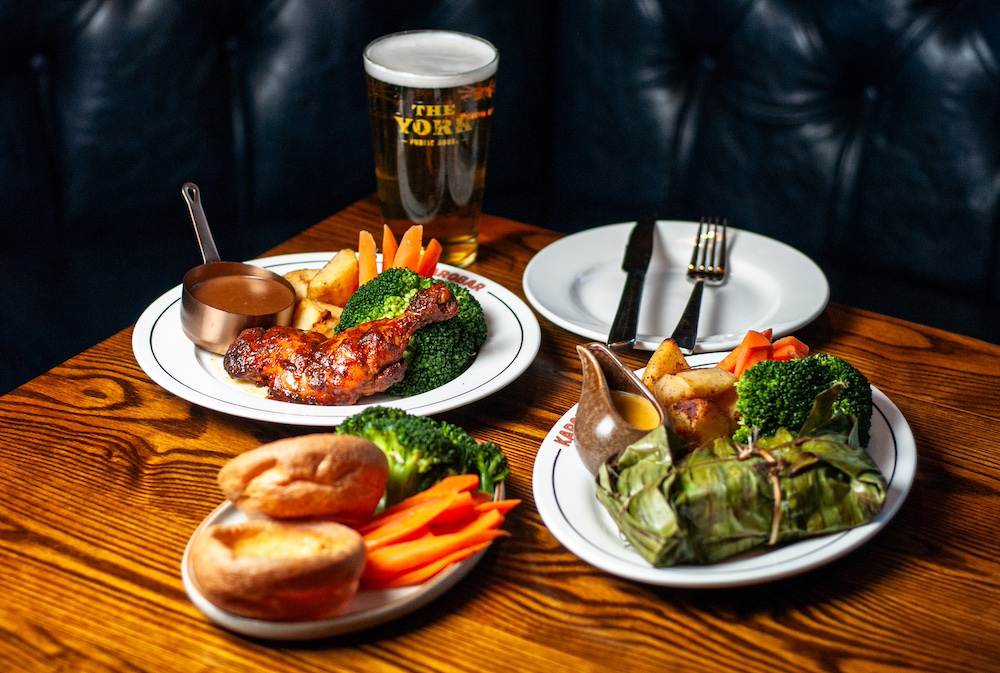
Photo Credit: @marcabarkerphotography
A longstanding fixture of the Broomhill pub scene and beloved by…