Sample Page
Hidden star found in unexpected dust zone
Written by
admin
in
7. Science
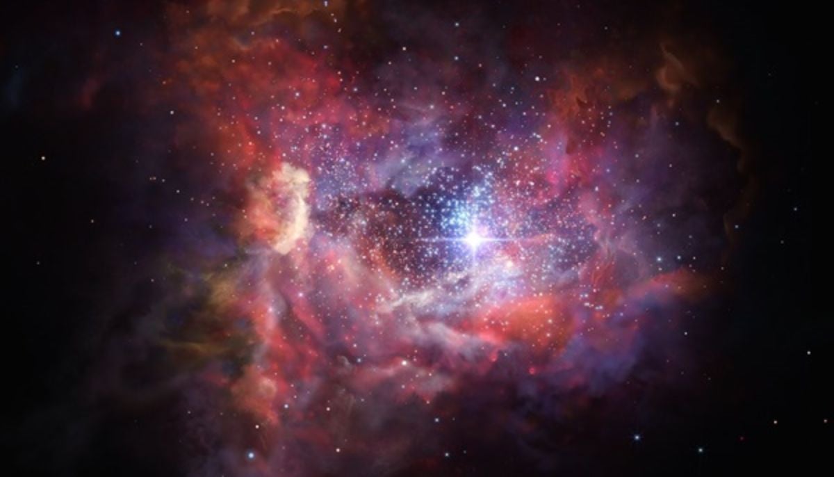
Rare comic discovery: Hidden star found in unexpected dust zone
Scientists in a record-breaking discovery have found a hidden star in a region…
Continue Reading
←
Katie Price’s ex-husband Kieran Hayler denies raping girl, 13
How to Watch the Final Meteor Shower of 2025
→
More posts
Savings and investment union: Council agrees position on revitalising the EU’s securitisation market – consilium.europa.eu
December 19, 2025
Insta360 X5 action camera dive bundle review
December 19, 2025
Applying 3D correlative structured illumination microscopy and X-ray tomography to characterise herpes simplex virus-1 morphogenesis
December 19, 2025
LDWF to Close Oyster Harvest in Portions of Drum Bay and Shell Point Reef in St. Bernard Parish
December 19, 2025