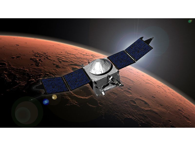
Comparison of EEG Burst Suppression and Hemodynamic Effects Between Re

Ying Cao,1,* Mi Gan,1,2,* Linling Wan,3,* Jun Lu,1,2 Ting Liu,1,2 Meiyan Liu,1,2 Di Wang,1 Sen Hong,1 Lin Zhou,1 Luying Deng,1 Zijun Wang,1 Jingjie Wang,1 Changyu Sun,1 Yang Liu,4 Yanqiu Liu,2,5 Meiwu Zhou1

1Department of Anesthesiology, The Second People’s Hospital of Guiyang, The Affiliated Jinyang Hospital of Guizhou Medical University, Guiyang, People’s Republic of China; 2School of Anesthesiology, Guizhou Medical University, Guiyang, People’s Republic of China; 3Chongqing Population and Family Planning Science and Technology Research Institute, Chongqing, People’s Republic of China; 4Department of Anesthesiology, The Affiliated Hospital of Guizhou Medical University, Guiyang, People’s Republic of China; 5Department of Anesthesiology, The Fourth People’s Hospital of Guiyang, Guiyang, People’s Republic of China

Correspondence: Yanqiu Liu, Department of Anesthesiology, The Fourth People’s Hospital of Guiyang, No. 91, Jiefang West Road, Nanming District, Guiyang, Guizhou, 550002, People’s Republic of China, Tel +86 13595161013, Email [email protected] Meiwu Zhou, Department of Anesthesiology, The Second People’s Hospital of Guiyang, The Affiliated Jinyang Hospital of Guizhou Medical University, No. 547, Jinyang South Road, Guanshanhu District, GuiYang, Guizhou, 550081, People’s Republic of China, Tel +86 15286065939, Email [email protected]

Background: Remimazolam tosilate, a novel ultra-short-acting benzodiazepine, demonstrates promising safety profiles in clinical settings. While both remimazolam tosilate and etomidate provide hemodynamic stability during anesthesia induction, limited research has directly compared their effects on electroencephalogram (EEG) burst suppression (periods of transient brain wave silence), a potential predictor of adverse neurological outcomes. This study aims to compare the incidence rate of EEG burst suppression (ESR) with remimazolam tosilate versus etomidate by reviewing the drug regimens used by different anesthesiologists in clinical practice.
Methods: This single-center retrospective study analyzed clinical anesthesia induction data from 161 patients from October 2023 to July 2024. Patients received either remimazolam tosilate (0.2 mg/kg, n=86, Group R) or etomidate (0.3 mg/kg, n=75, Group E) for general anesthesia induction. Primary outcomes included ESR and its duration during induction. Second outcomes comprised hemodynamic parameters: systolic blood pressure, diastolic blood pressure mean arterial pressure, and heart rate, measured at baseline (T0), 3 minutes post-induction (T1), immediately after intubation (T2), 5 minutes post-intubation (T3), 10 minutes post-intubation (T4), and adverse events occurrence.
Results: Baseline characteristics were comparable except ASA classification (higher ASA III proportion in Group R: 24.4% vs 2.7%, P< 0.001). No ESR occurred in Group R versus 29.34% in Group E (P< 0.01). Group R had a significantly lower incidence of intubation-related hypertension (10.5% vs 42.7%, P< 0.001) and maintained stable blood pressure and HR throughout induction, whereas Group E exhibited marked MAP and HR fluctuations. Other adverse events showed no significant inter-group differences.
Conclusion: Remimazolam tosilate demonstrated notable differences compared to etomidate during general anesthesia induction, including the absence of ESR and different hemodynamic response patterns. While these findings suggest potential advantages for certain patient populations, the retrospective design and ASA classification imbalance limit definitive conclusions, warranting prospective validation studies.

Keywords: EEG burst suppression, remimazolam tosilate, etomidate, hemodynamic stability, general anesthesia induction

Introduction

Enhanced recovery after surgery (ERAS) programs represent evidence-based multimodal care pathways designed to optimize perioperative outcomes and minimize physiological stress responses.1,2 Within these protocols, the choice of anesthetic agents, particularly ultra-short-acting drugs, is pivotal for facilitating faster recovery and better perioperative management.3,4

Electroencephalogram (EEG) burst suppression, first characterized by Derbyshire et al,5 manifests as a distinctive neural pattern alternating between high-voltage slow waves and periods of isoelectric suppression.6,7 This oscillating pattern demonstrates the brain’s transition between intense electrical activity and quiescent states.8,9 As a neurophysiological signature of severe cortical electrical inhibition, burst suppression can be triggered by various pathophysiological conditions, including hypothermia,10,11 hypoxia,12 hypoglycemia,13 and vascular brain injury.14,15 Furthermore, this pattern frequently emerges during deep anesthesia,16 serving as a reliable indicator of profound anesthetic states.17 Multiple studies have demonstrated robust associations between intraoperative burst suppression and adverse postoperative outcomes, particularly increased risk of perioperative neurocognitive disorders (PNDs), especially postoperative delirium (POD) and extended hospital stays.18–21 These associations likely involve disruption of normal cerebral homeostasis and potential neurotoxicity during periods of cortical suppression,22 which suggest that burst suppression patterns may serve as an important biomarker for identifying patients at increased risk for perioperative neurological complications.23

Etomidate, despite its reputation for hemodynamic stability, has raised concerns regarding its propensity to induce EEG burst suppression during induction and its suppressive effects on adrenal function.24 In contrast, remimazolam tosilate is a novel ultra-short-acting benzodiazepine with distinct pharmacokinetic and pharmacodynamic features.25 It exhibits favorable pharmacological properties including rapid onset and offset, predictable clearance through tissue esterases, and improved controllability.26,27 However, comparative data regarding the incidence rate of EEG burst suppression (ESR) between these agents during anesthesia induction remain limited.

Our team has long focused on brain function research during anesthesia, consistently implementing rigorous monitoring protocols including continuous EEG monitoring and automatic blood pressure recordings at 1-minute intervals during induction. Due to demonstrated hemodynamic advantages, more anesthesiologists have begun favoring remimazolam tosilate for induction in patients with higher ASA classifications. Through these meticulous practices in routine clinical care, we made an unexpected observation: EEG burst suppression patterns occurred predominantly when anesthesiologists employed their preferred etomidate-based protocols, while such patterns were notably absent with remimazolam tosilate regimens. This serendipitous finding prompted us to conduct a systematic retrospective analysis comparing ESR between these agents during anesthesia induction. Although remimazolam tosilate and etomidate differ in their pharmacological classifications—remimazolam being a benzodiazepine sedative and etomidate a potent hypnotic agent—our study focused on comparing their sedative effects during general anesthesia induction as routinely practiced in clinical settings. The choice of induction agent in each case was made by the attending anesthesiologist based on patient status and clinical preference. Sedation depth was quantitatively evaluated using the Patient State Index (PSI), providing an objective measure of anesthetic-induced cortical suppression. This approach allowed us to assess the real-world differences in sedative outcomes between the two drugs, despite their mechanistic distinctions.

Material and Methods

This single-center, retrospective study compared the ESR and its duration between remimazolam tosilate and etomidate during anesthesia induction, based on anesthetic regimens routinely selected by attending anesthesiologists. The study was approved by The Ethics Committee of the Second People’s Hospital of Guiyang (JYYY-2025-WZ-03), registered both in the National Medical Research Registration and Filing Information System (MR-52-25-014287) and Chinese Clinical Trial Registry (ChiCTR2500098018). The Ethics Committee waived the requirement for informed consent due to minimal patient risk and the impracticality of obtaining individual consent from patients lost to follow-up, with all procedures conducted in strict accordance with the Declaration of Helsinki and relevant ethical guidelines while ensuring patient anonymization and data security. We reviewed electronic records of 161 patients (aged 18–65 years, ASA I–III) who underwent elective surgery under general anesthesia between October 2023 and July 2024. Exclusion criteria included: 1. severe cardiovascular instability (eg, acute heart failure, severe arrhythmia, myocardial infarction); 2. uncontrolled hypertension; 3. anticipated difficult airway; 4. uncompleted or missing vital signs data; 5. records with documented protocol violations. To minimize potential selection bias, consecutive eligible cases during the study period were included. Data were extracted from the electronic medical record system by 4 independent investigators, with disagreements resolved through consultation with a senior anesthesiologist. The flowchart detailing the study design is illustrated in Figure 1.

Figure 1 Study flowchart depicting patient enrollment, inclusion, and exclusion criteria.

All patients observed standard fasting guidelines (6 hours for solids, 2 hours for clear fluids) preoperatively. Upon arrival in the operating room, routinely monitoring was initiated, including Electrocardiograph (ECG), peripheral oxygen saturation (SpO2), noninvasive blood pressure (NIBP) and continuous EEG monitoring using the Masimo SedLine® (PSA-4000, Masimo Corp., USA). This device recorded a four-channel EEG montage and generated the Patient State Index (PSI), a processed parameter reflecting sedation depth. During induction, PSI values were maintained between 20 and 50 to ensure an adequate level of unconsciousness. The use of this standardized EEG-based index enabled objective comparison of sedative effects between the two pharmacologically distinct agents. Following preoxygenation, general anesthesia was induced with either remimazolam tosilate (0.2 mg/kg, Group R) or etomidate (0.3 mg/kg, Group E) as the sole sedative hypnotic agent in their respective groups. The induction doses for both remimazolam tosilate and etomidate were calculated using adjusted body weight (ABW), determined by the formula: ABW = Ideal Body Weight (IBW) + 0.4 × (Actual Body Weight – IBW). The IBW was derived using the Devine formula: for males, IBW (kg) = 50 + 0.91 × (height in cm – 152.4); for females, IBW (kg) = 45.5 + 0.91 × (height in cm – 152.4). Subsequently, 0.5 µg/kg of sufentanil was administered, followed by 0.6 mg/kg of rocuronium after loss of eyelash reflex. Tracheal intubation was performed upon achieving complete muscle relaxation.

Burst suppression was defined as an EEG pattern characterized by alternating epochs of high-voltage activity and periods of near-isoelectric suppression. Suppression was detected via the Suppression Ratio (SR), indicating the percentage of time the EEG signal is suppressed within a 63-second analysis window. All burst suppression events flagged by SR (> 0%) were confirmed by direct visual inspection of the raw four-channel EEG waveforms. The onset was defined as the first instance of SR > 0% accompanied by visible suppression, and the end was marked when SR returned to 0% with normal EEG continuity. Cumulative burst suppression duration was calculated by summing all suppression periods within the induction window.

Outcome Assessment

The primary outcomes were ESR occurrence and duration during anesthesia induction. ESR was quantified by both occurrence rate and duration metrics, with duration measured from initial onset to resolution (defined as the time interval between the first and last burst suppression pattern observed on EEG). Cumulative duration was calculated by summing all periods of burst suppression during the induction phase. Secondary outcomes included: (1) Hemodynamic parameters and fluctuations, and (2) Adverse events. Systolic blood pressure (SBP), diastolic blood pressure (DBP), mean arterial pressure (MAP), and heart rate (HR) were recorded at predefined time points: 1 minute before induction as baseline (T0), 3 minutes after induction (T1), immediately after tracheal intubation (T2), 5 minutes post-intubation (T3), and 10 minutes post-intubation (T4). Cardiovascular comorbidity (CV), defined primarily as arterial hypertension, was specifically analyzed to determine its potential impact on hemodynamic responses during anesthesia induction. Hemodynamic fluctuations were measured as ΔMAP (difference between maximum or minimum MAP and baseline MAP) and ΔHR (difference between maximum or minimum HR and baseline HR). Adverse events were defined as follows: hypotension (MAP decrease >20% from baseline or <60 mmHg), hypertension (MAP increase >20% from baseline or >120 mmHg), severe bradycardia (HR <45 bpm), and tachycardia (HR >100 bpm). Hypotension was treated with 6 mg ephedrine or 1 mg methoxamine boluses as needed, with continuous norepinephrine infusion initiated when necessary. Severe bradycardia was treated with 0.25–0.5 mg atropine until HR normalized. Temporal correlation between ESR patterns and hemodynamic variables was analyzed to evaluate potential associations between neurophysiological changes and cardiovascular stability during induction.

Statistical Analysis

Data analysis was performed using SPSS version 22.0 (IBM Corp, Armonk, NY, USA). The Shapiro–Wilk test assessed normality of distribution. Continuous variables with normal distribution (age, height, weight, BMI, ΔMAP, ΔHR and the duration of ESR) were presented as mean ± standard deviation and compared using independent-sample t-tests. Changes in hemodynamic parameters (ΔMAP, ΔHR) from baseline within each group were analyzed using paired t-tests. Categorical data (gender, surgical procedure, adverse events) were expressed as n (%) and analyzed using Chi-square or Fisher’s exact tests as appropriate. For ordinal data (ASA classification and the duration of ESR between groups), the Mann–Whitney U-test was employed. Post-hoc power analysis was performed using G*Power 3.1.9.7 software to evaluate the statistical power for detecting differences in burst suppression rates between groups, based on the observed effect size and actual sample sizes with α=0.05. Statistical significance was set at P<0.05. To minimize potential selection bias in this retrospective study, we performed: 1. Detailed analysis of baseline characteristics; 2. Multivariate logistic regression to adjust for potential confounders (age, ASA status, and baseline hemodynamics); 3. Pre-specified subgroup analyses based on ASA classification and the presence of cardiovascular comorbidities. Given the study’s focus on single-point outcome comparisons during induction, Repeated Measures ANOVA was not applied.

Results

Patient Characteristics

Of the 161 patients reviewed, 86 received remimazolam tosilate (Group R) and 75 received etomidate (Group E). Baseline characteristics were generally well-balanced between groups. No significant differences were observed in age (45.5±12.9 vs 43.8±11.4 years, P=0.749), height (162±6.12 vs 162±8.46 cm, P=1.000), weight (63.9±8.89 vs 65.2±10.1 kg, P=0.778), BMI (24.20±2.99 vs 24.70±3.31 kg/m2, P=0.635). Gender distribution (male/female: 34/41 vs 36/50, P=0.651), or surgical procedure types (P=0.080). However, ASA classification differed significantly between groups, with Group R having a higher proportion of ASA III patients (24.4% vs 2.7%, P<0.001). To address potential selection bias in this retrospective study, multivariate logistic regression was performed to adjust for confounding factors (age, ASA status, and baseline hemodynamics), along with pre-specified subgroup analyses based on ASA classification and cardiovascular comorbidities (Table 1).

Table 1 Demographic and Baseline Characteristics of Patients

Subgroup analyses were conducted based on ASA classification, age, and CV comorbidity status. In the ASA subgroup analysis, BMI differed significantly among ASA III patients (26.4±3.3 kg/m² in Group R vs 24.1±3.5 kg/m² in Group E, P=0.045), whereas other baseline characteristics remained comparable. Age-stratified subgroup analysis showed broadly similar distributions of baseline characteristics between groups, except for the persistent imbalance in ASA classification (P<0.001) across both age categories. In the CV subgroups, significant differences were observed in age between groups (patients with CV comorbidities: 52.4±8.9 years in Group R vs 54.2±9.1 years in Group E; patients without CV comorbidities: 40.5±10.2 years in Group R vs 42.8±11.8 years in Group E; P=0.038) and ASA classification (P<0.001). Although multivariable regression was not employed, the consistency of ESR results across stratified subgroups mitigates the risk of baseline confounding. Notably, subgroup analysis specifically revealed significant differences between remimazolam and etomidate groups regarding age, BMI, and ASA classification among patients with CV comorbidities, predominantly arterial hypertension. This pattern indicates clinicians’ preferential selection of remimazolam tosilate for older patients with higher BMI and elevated ASA classifications, likely reflecting the drug’s favorable hemodynamic profile in this high-risk population. The persistent imbalance in ASA classification across subgroups, particularly among patients with arterial hypertension (higher prevalence of ASA III patients in Group R), necessitates cautious interpretation of the hemodynamic results and underscores the importance of appropriate statistical adjustments in our final analysis (Table 2).

Table 2 Subgroup Analysis Results

Primary Outcome

ESR demonstrated significant intergroup differences, with a complete absence in Group R (0.00%) versus 29.34% occurrence in Group E (P<0.01). Post-hoc power analysis confirmed that the study achieved >99% statistical power to detect the observed difference in burst suppression rates between groups, supporting the reliability of our primary findings. Within Group E, ASA III patients showed higher ESR rates than ASA I–II (50.00% vs 28.77%), though limited ASA III sample size (n=2 vs n=73) precluded definitive statistical inference. When burst suppression occurred, onset was typically observed at 45 seconds post-etomidate administration (IQR: 32–67 seconds), with a median duration of 63.5 seconds (IQR: 42.5–89.0 seconds, range: 40–96 seconds) (Table 3). All ESR patterns resolved before the T3 measurement point. Representative EEG waveforms from both groups are shown in Figure 2.

Table 3 ESR and Its Duration Between Two Groups

Figure 2 The collected EEG and BS patterns from both groups. (A) EEG patterns from patients receiving etomidate, with typical burst suppression highlighted in red boxes; (B) EEG patterns from patients receiving remimazolam tosilate, demonstrating continuous EEG activity without evidence of burst suppression.

Considering the observed baseline ASA imbalance, multivariate logistic regression analysis adjusting for age, ASA classification, and baseline hemodynamic parameters confirmed that the significantly lower ESR associated with remimazolam tosilate persisted (P<0.01). Subgroup analysis further revealed that within ASA III patients, burst suppression occurred only in the etomidate group (1 out of 2 patients), while none of the 21 ASA III patients in the remimazolam group exhibited burst suppression. Although subgroup numbers were limited, this finding qualitatively supports the robustness of the observed overall effect.

Secondary Outcomes

Hemodynamic Fluctuations

Analysis of hemodynamic changes from baseline demonstrated significant differences between the two groups. In Group E, MAP showed a significant decrease at T1 (−5.5±9.9 mmHg, P=0.016), followed by a marked increase at T2 (15.0±10.4 mmHg, P<0.001) and then a significant decrease at T3 (−8.1±15.2 mmHg, P=0.003). Similarly, HR in Group E demonstrated significant decrease at T1 (−5.0±8.5 bpm, P<0.001), followed by an increase at T2 (6.3±11.3 bpm, P=0.002) and a marked decrease at T3 (−10.9±14.1 bpm, P<0.001). In contrast, Group R maintained relatively stable MAP with no significant changes from baseline at any timepoint (T1: 2.1±9.5 mmHg, P=0.405; T2: 1.1±10.9 mmHg, P=0.114; T3: 1.4±18.7 mmHg, P=0.111; T4: −3.4±15.9 mmHg, P=0.641). HR in Group R showed minimal but significant changes at T1 (−0.4±9.0 bpm, P=0.019) and T2 (−0.1±10.9 bpm, P=0.001). Comparison between groups revealed no significant differences in baseline MAP (95.0±14.1 vs 93.6±14.0 mmHg, P=0.144) or HR (77.8±11.3 vs 75.9±11.5 bpm, P=0.376). However, significant differences emerged at T1, T2, and T3 for both MAP (T1: P<0.001; T2: P<0.001; T3: P=0.002) and HR (T1: P<0.001; T2: P<0.001; T3: P=0.001). By T4, both groups showed similar recovery patterns with no significant between-group differences in either MAP (−3.2±16.5 vs −3.4±15.9 mmHg, P=0.141) or HR (−3.3±13.8 vs −3.5±14.2 bpm, P=0.311) (Table 4). Figure 3.

Table 4 Hemodynamic Fluctuations Between Two Groups

Figure 3 Vital signs at different time points between two groups. (A) ∆MAP at different time points between two groups; (B) ∆HR at different time points between two groups.

Notes: Data are presented as mean ± SD. &P<0.05, P values were calculated using paired t-test comparing with baseline (T0); $P<0.05, P values were calculated using independent t-test between Group E and Group R.

Adverse Events

Analysis of adverse events demonstrated significant differences in hemodynamic stability between groups. Group R showed markedly lower incidence of hypertension compared to Group E (10.46% vs 42.67%, P<0.001), while other adverse events including hypotension (29.06% vs 41.33%, P=0.097), bradycardia (13.95% vs 22.67%, P=0.156), and tachycardia (15.11% vs 13.33%, P=0.745) showed no statistically significant differences. The pattern of adverse events demonstrates different hemodynamic response profiles between the agents, with remimazolam showing reduced hypertensive responses during induction, though both groups achieved similar hemodynamic recovery (Table 5).

Table 5 Adverse Events Between Two Groups

Subgroup analyses demonstrated that Group R maintained significantly lower hypertension rates across different patient populations, with notably reduced rates in ASA I–II patients (9.23% vs 42.47%, P<0.001) and consistent effects across both age groups (<60 years: 10.14% vs 41.38%, P<0.001; ≥60 years: 11.76% vs 47.06%, P=0.027). While the small sample size of ASA III patients in Group E (n=2) limited statistical comparison, Group R exhibited stable hemodynamics despite having more ASA III patients. Although older patients (≥60 years) showed slightly higher rates of hypotension in both groups, these differences were not statistically significant, suggesting remimazolam tosilate’s favorable hemodynamic profile persists across different risk stratifications (Table 6). The significant ASA classification imbalance across all subgroups represents a major study limitation that may confound interpretations, particularly given that anesthesiologist drug selection likely reflects patient risk assessment rather than randomized allocation.

Table 6 Adverse Events Analysis by Subgroups

Discussion

This retrospective analysis demonstrated three key findings regarding remimazolam tosilate compared to etomidate during anesthesia induction: (1) no occurrence of EEG burst suppression with remimazolam tosilate versus 29.34% incidence with etomidate; (2) modest hemodynamic differences between groups, with both agents demonstrating reasonable cardiovascular stability; and (3) significantly lower incidence of hypertensive events in the remimazolam tosilate group (10.46% vs 42.67%). Although this study did not employ a formal multivariable regression model, we addressed confounding through structured subgroup analyses across ASA classification, age, and cardiovascular comorbidities. Despite a higher proportion of ASA III patients in the remimazolam group, ESR incidence remained consistently lower in all strata. These findings, supported by post-hoc power analysis and large effect size, strengthen the internal validity of our conclusions.

EEG monitoring has become increasingly important in anesthetic practice, with guidelines emphasizing its role in optimizing anesthetic depth and preventing adverse outcomes.17,28,29 Recent studies have revealed that continuous monitoring of alpha oscillatory activity and other EEG parameters enables more precise individualization of anesthetic administration,30 which is especially valuable given the variability of anesthetic responses among patients. Our findings of zero ESR with remimazolam tosilate compared to 29.34% with etomidate demonstrate clinically relevant differences in neurophysiological effects during induction. Incorporating advanced EEG monitoring into routine practice has significantly enhanced our ability to deliver personalized anesthetic care, potentially lowering the risk of adverse neurological outcomes through more precise drug titration. Burst suppression has been associated with adverse perioperative neurological outcomes,31,32 particularly postoperative delirium which increases ICU stays and hospital costs.33,34 The observed differences likely reflect the distinct pharmacological mechanisms underlying these agents.

The pharmacological basis for our findings can be attributed to different GABAa (γ-aminobutyric acid type A) receptor (GABAaR) binding sites and mechanisms between etomidate and remimazolam tosilate. Etomidate binds between alpha and beta subunits in the transmembrane domain, demonstrating enhanced potency at beta3 subunit-containing receptors and exhibiting distinct subunit selectivity that drives diverse clinical effects.35 Literature suggests that high-dose GABAa agonists such as etomidate can directly induce burst suppression through profound reduction of cerebral metabolic rate and direct cortical effects, independent of hemodynamic changes.36 Moreover, its selective action on alpha5 subunit-containing receptors forming “tonic” GABAaRs specifically mediates amnestic effects, highlighting the subtype-dependent nature of its pharmacological profile.37 Etomidate’s potent GABAaR-mediated cortical inhibition triggers profound neurophysiological changes, resulting in MAP fluctuations (ΔT1: −5.5±9.9 mmHg, P=0.016; ΔT2: 15.0±10.4 mmHg, P<0.001; ΔT3: −8.1±15.2 mmHg, P=0.003) coinciding with ESR onset at 45 seconds (IQR: 32–67 seconds), with a median duration of 63.5 seconds (IQR: 42.5–89.0 seconds, range: 40–96 seconds). This temporal relationship aligns with findings that deeper anesthesia may increase POD risk.38–40

In contrast, remimazolam tosilate binds at the interface between alpha and gamma subunits and exhibits a ceiling effect on EEG depression characteristic of benzodiazepines, with burst suppression rarely observed when used alone.41 This inherent pharmacological characteristic makes remimazolam tosilate less likely to produce burst suppression compared to potent hypnotics like etomidate, independent of hemodynamic effects, thus explaining the absence of burst suppression and stable MAP throughout induction. These findings suggest that the differences in burst suppression rates are primarily attributable to pharmacodynamic properties rather than hemodynamic stability alone.

A clinically significant finding was the four-fold difference in hypertensive events between groups (42.67% vs 10.46%, P<0.001). This disparity may be attributed to etomidate’s relatively short duration of action, potentially resulting in insufficient anesthetic depth by the time of laryngoscopy and inadequate suppression of sympathetic stress response to airway manipulation. Conversely, remimazolam tosilate appears to provide more sustained anesthetic effect during induction, maintaining adequate depth throughout intubation and attenuating sympathetic response.42,43 Importantly, although we initially speculated that remimazolam tosilate’s more stable hemodynamic profile might explain its lower BS incidence, further analysis suggests this is unlikely to be the sole factor. Both groups demonstrated similar rates of hypotensive events (29.06% vs 41.33%, P=0.097), with MAP increasing following intubation in both groups, though notably greater with etomidate (approximately +15 mmHg vs +1 mmHg at T2). Thus, we emphasize that pharmacodynamic differences in EEG suppression potential are a more plausible mechanistic explanation. Despite statistical significance, the magnitude of these differences (approximately 5–15 mmHg for MAP) was modest and likely clinically insignificant for healthy adults.44 Nevertheless, this observation carries practical clinical implications, particularly given etomidate’s reputation for hemodynamic stability. Our findings suggest that while etomidate may preserve baseline cardiovascular parameters, remimazolam tosilate demonstrated different patterns in controlling intubation-related hypertensive responses, which may be relevant for perioperative cardiovascular management in susceptible patients. Although our results demonstrated a complete absence of EEG burst suppression in the remimazolam group, it is important to note that our study only captured a brief observation window during anesthesia induction. The burst suppression episodes observed in the etomidate group were short in duration—typically lasting seconds to a few minutes—rather than prolonged. Existing literature suggests that sustained or repeated burst suppression, particularly during cardiopulmonary bypass or deep anesthesia maintenance, is more clearly associated with adverse postoperative neurocognitive outcomes. By contrast, transient burst suppression occurring at induction may carry less clinical significance. Nevertheless, even brief burst suppression episodes could be undesirable in high-risk populations such as the elderly or patients with cerebrovascular disease. Therefore, the clinical advantage of remimazolam tosilate may be better understood not as a result of superior hemodynamic stability—which was modestly different between groups—but as a consequence of its pharmacodynamic ceiling effect on EEG suppression. Avoiding even momentary burst suppression during induction could help reduce neurological risk in susceptible individuals. Notably, Group R showed consistently lower ESR across ASA classifications (ASA I–II: 0% vs 28.77%, P<0.01), aligning with evidence that patients with fewer comorbidities are more susceptible to anesthetic depth-related cognitive effects,45 particularly given POD’s association with increased one-year mortality (12% vs 6%).46 Consequently, while etomidate can serve as a general anesthetic, benzodiazepines like remimazolam tosilate are primarily employed as sedatives and anxiolytics.47–49

As a retrospective analysis, this study has inherent limitations. First, potential underreporting or incomplete documentation of ESR events during anesthesia induction may affect the accuracy of the findings. Patients often exhibit involuntary movements during general anesthesia induction, complicating the acquisition of clear, noise-free EEG signals, which contributed to the relatively limited number of cases included in this study. Second, although our study population had a relatively normal BMI distribution (mean approximately 24 kg/m², with no patients exceeding 35 kg/m²), thus minimizing dosing discrepancies, we acknowledge an important methodological consideration regarding dosage calculations for anesthetic induction agents. In our study, remimazolam tosilate and etomidate doses were calculated using ABW, accounting for the difference between actual and ideal body weights. However, it is essential to emphasize the recommended practice of dosing based on ideal or lean body weight for markedly obese patients, as overdosing based on actual weight in these populations could increase the risk of excessive sedation, hemodynamic instability, and EEG burst suppression. Previous pharmacokinetic studies indicate that anesthetic doses based on ideal or adjusted body weight effectively reduce drug accumulation and adverse events in obese patients.50 Third, the lack of postoperative follow-up precluded evaluation of long-term neurological outcomes. Lastly, despite careful statistical adjustment for potential confounders including ASA classification, age, and baseline hemodynamics, the significant imbalance in ASA classification—specifically the higher prevalence of ASA III patients in the group R-remains an important methodological consideration. This baseline imbalance likely reflects anesthesiologists’ clinical preference for remimazolam tosilate in older, hypertensive, and higher-risk patients due to its favorable hemodynamic profile. Although our adjusted analyses and subgroup assessments confirmed the robustness of the primary findings, the limited sample size within certain subgroups, particularly ASA III patients receiving etomidate, necessitates cautious interpretation of the results. We speculate that the lower ESR associated with remimazolam tosilate may be related to its more stable hemodynamic profile during anesthesia induction, potentially minimizing cerebral perfusion fluctuations and reducing the risk of adverse neurological outcomes. However, future prospective randomized controlled trials with balanced ASA classifications, rigorous monitoring protocols, standardized dosage strategies across diverse populations, and long-term neurological follow-up are essential to validate these preliminary observations and definitively establish the clinical advantages of remimazolam in specific high-risk populations.

Conclusion

Our findings suggest that remimazolam tosilate offers advantages over etomidate in terms of a reduced incidence of EEG burst suppression and more stable hemodynamics during induction, in a retrospective analysis. However, given the study’s limitations, further prospective research is warranted to confirm these observations.

Abbreviations

EEG, electroencephalogram; BS, burst suppression; ESR, the incidence rate of EEG burst suppression; SBP, systolic blood pressure; DBP, diastolic blood pressure; MAP, mean arterial pressure; HR, heart rate; ERAS, Enhanced recovery after surgery; PNDs, perioperative neurocognitive disorders; POD, postoperative delirium; ECG, Electrocardiograph; SpO2, peripheral oxygen saturation; NIBP, noninvasive blood pressure; PSI, Patient State Index; Group E, 0.3 mg/kg etomidate group; Group R, 0.2 mg/kg remimazolam tosilate group; BMI, body mass index; ASA, American Society of Anaesthesiologists; n, number; Gender (M/F), Gender Male/Female; bpm, beat per minute; CV, cardiovascular comorbidities; GABAa, γ-aminobutyric acid type A.

Data Sharing Statement

The datasets generated during and/or analyzed during the current study are not publicly available due to the privacy policy but are available from the corresponding authors on reasonable requests.

Author Contributions

All authors made a significant contribution to the work reported, whether that is in the conception, study design, execution, acquisition of data, analysis and interpretation, or in all these areas; took part in drafting, revising or critically reviewing the article; gave final approval of the version to be published; have agreed on the journal to which the article has been submitted; and agree to be accountable for all aspects of the work.

Funding

This work was supported by the Fund of Guiyang Municipal Health Commission Science and Technology Program, High-Level Innovative Young Health Professionals Training Project, (YC, [2020] ZhuWeijianKejiHetong, Contract No. 006), the Fund of Chongqing Municipal Health Commission Medical Research Project, (LLW, 2023WSJK030), the Science and Technology Fund Project of Guizhou Health Commission in 2025 (YC, gzwkj2025-083), the Regional Science Fund Project from the National Natural Science Foundation of China (YL, Grant No. 82460244), the Seed Foundation of Guiyang Second People’s Hospital Doctoral Research Project (YC, Contact No. ZhuErYiKeYan-BS[2024]06).

Disclosure

The authors report no conflicts of interest in this work.

References

1. Wang SK, Wang QJ, Wang P, et al. The impact of frailty on clinical outcomes of older patients undergoing enhanced recovery after lumbar fusion surgery: a prospective cohort study. Int J Surg. 2024;110(8):4785–4795. doi:10.1097/JS9.0000000000001594

2. Kehlet H. Multimodal approach to control postoperative pathophysiology and rehabilitation. Br J Anaesth. 1997;78(5):606–617. doi:10.1093/bja/78.5.606

3. Kim KM. Remimazolam: pharmacological characteristics and clinical applications in anesthesiology. Anesth Pain Med (Seoul). 2022;17(1):1–11. doi:10.17085/apm.21115

4. Zargar-Shoshtari K, Hill AG. Optimization of perioperative care for colonic surgery: a review of the evidence. ANZ J Surg. 2008;78(1–2):13–23. doi:10.1111/j.1445-2197.2007.04350.x

5. Belsey R. Resection and reconstruction of the intrathoracic trachea. Br J Surg. 1950;38(150):200–205. doi:10.1002/bjs.18003815008

6. Pawar N, Barreto Chang OL. Burst Suppression During General Anesthesia and Postoperative Outcomes: mini Review. Front Syst Neurosci. 2021;15:767489. doi:10.3389/fnsys.2021.767489

7. Amzica F. What does burst suppression really mean? Epilepsy Behav. 2015;49:234–237. doi:10.1016/j.yebeh.2015.06.012

8. Fleischmann A, Pilge S, Kiel T, Kratzer S, Schneider G, Kreuzer M. Substance-Specific Differences in Human Electroencephalographic Burst Suppression Patterns. Front Hum Neurosci. 2018;12:368. doi:10.3389/fnhum.2018.00368

9. Cartailler J, Parutto P, Touchard C, Vallée F, Holcman D. Alpha rhythm collapse predicts iso-electric suppressions during anesthesia. Commun Biol. 2019;2:327. doi:10.1038/s42003-019-0575-3

10. Saczynski JS, Marcantonio ER, Quach L, et al. Cognitive trajectories after postoperative delirium. N Engl J Med. 2012;367(1):30–39. doi:10.1056/NEJMoa1112923

11. Seder DB, Fraser GL, Robbins T, Libby L, Riker RR. The bispectral index and suppression ratio are very early predictors of neurological outcome during therapeutic hypothermia after cardiac arrest. Intensive Care Medicine. 2010;36(2):281–288. doi:10.1007/s00134-009-1691-1

12. Liu N, Chazot T, Mutter C, Fischler M. Elevated burst suppression ratio: the possible role of hypoxemia. Anesth Analg. 2006;103(6):1609–1610. doi:10.1213/01.ane.0000246268.58067.99

13. Declerck A, Liu N, Chazot T, Fischler M. BIS values during resuscitation: the role of the suppression ratio (case report). J Clin Monit Comput. 2009;23(5):307–309. doi:10.1007/s10877-009-9197-5

14. Johansen JW. Esmolol promotes electroencephalographic burst suppression during propofol/alfentanil anesthesia. Anesth Analg. 2001;93(6):1526–1531. doi:10.1097/00000539-200112000-00039

15. Umegaki N, Hirota K, Kitayama M, Yatsu Y, Ishihara H, Mtasuki A. A marked decrease in bispectral index with elevation of suppression ratio by cervical haematoma reducing cerebral perfusion pressure. J Clin Neurosci. 2003;10(6):694–696. doi:10.1016/j.jocn.2002.11.001

16. Hayashida M, Sekiyama H, Orii R, et al. Effects of deep hypothermic circulatory arrest with retrograde cerebral perfusion on electroencephalographic bispectral index and suppression ratio. J Cardiothorac Vasc Anesth. 2007;21(1):61–67. doi:10.1053/j.jvca.2006.02.029

17. Shanker A, Abel JH, Schamberg G, Brown EN. Etiology of Burst Suppression EEG Patterns. Front Psychol. 2021;12:673529. doi:10.3389/fpsyg.2021.673529

18. Fang PP, Shang ZX, Xu J, et al. Contribution of intraoperative electroencephalogram suppression to frailty-associated postoperative delirium: mediation analysis of a prospective surgical cohort. Br J Anaesth. 2023;130(2):e263–e271. doi:10.1016/j.bja.2022.11.002

19. Fritz BA, Kalarickal PL, Maybrier HR, et al. Intraoperative Electroencephalogram Suppression Predicts Postoperative Delirium. Anesth Analg. 2016;122(1):234–242. doi:10.1213/ane.0000000000000989

20. Tymianski M, Tator CH. Normal and abnormal calcium homeostasis in neurons: a basis for the pathophysiology of traumatic and ischemic central nervous system injury. Neurosurgery. 1996;38(6):1176–1195. doi:10.1097/00006123-199606000-00028

21. Hesse S, Kreuzer M, Hight D, et al. Association of electroencephalogram trajectories during emergence from anaesthesia with delirium in the postanaesthesia care unit: an early sign of postoperative complications. Br J Anaesth. 2019;122(5):622–634. doi:10.1016/j.bja.2018.09.016

22. Lewis LD, Ching S, Weiner VS, et al. Local cortical dynamics of burst suppression in the anaesthetized brain. Brain. 2013;136(Pt 9):2727–2737. doi:10.1093/brain/awt174

23. Yang T, Velagapudi R, Terrando N. Neuroinflammation after surgery: from mechanisms to therapeutic targets. Nat Immunol. 2020;21(11):1319–1326. doi:10.1038/s41590-020-00812-1

24. Butovas S, Rudolph U, Jurd R, Schwarz C, Antkowiak B. Activity patterns in the prefrontal cortex and hippocampus during and after awakening from etomidate anesthesia. Anesthesiology. 2010;113(1):48–57. doi:10.1097/ALN.0b013e3181dc1db7

25. J DU, Licheri M, Barucco G, et al. Remimazolam for anesthesia and sedation in cardiac surgery and for cardiac patients undergoing non-cardiac surgery: a systematic-narrative hybrid review. Minerva Anestesiol. 2024;90(7–8):682–693. doi:10.23736/S0375-9393.24.17943-6

26. Monaco F, Bottussi A, Barucco G, D’Andria UJ. Remimazolam in Cardiac Anesthesia: not So “Ultra-short Acting” After All. J Cardiothorac Vasc Anesth. 2025;39(1):327–328. doi:10.1053/j.jvca.2024.10.008

27. Zhang H, Li H, Zhao S, Bao F. Remimazolam in General Anesthesia: a Comprehensive Review of Its Applications and Clinical Efficacy. Drug Des Devel Ther. 2024;18:3487–3498. doi:10.2147/dddt.S474854

28. Aldecoa C, Bettelli G, Bilotta F, et al. European Society of Anaesthesiology evidence-based and consensus-based guideline on postoperative delirium. Eur J Anaesthesiol. 2017;34(4):192–214. doi:10.1097/eja.0000000000000594

29. Brown EN, Lydic R, Schiff ND. General anesthesia, sleep, and coma. N Engl J Med. 2010;363(27):2638–2650. doi:10.1056/NEJMra0808281

30. Obert DP, Hight D, Sleigh J, et al. The First Derivative of the Electroencephalogram Facilitates Tracking of Electroencephalographic Alpha Band Activity During General Anesthesia. Anesth Analg. 2022;134(5):1062–1071. doi:10.1213/ane.0000000000005783

31. Muhlhofer WG, Zak R, Kamal T, et al. Burst-suppression ratio underestimates absolute duration of electroencephalogram suppression compared with visual analysis of intraoperative electroencephalogram. Br J Anaesth. 2017;118(5):755–761. doi:10.1093/bja/aex054

32. Reese M, Christensen S, Anolick H, et al. EEG pre-burst suppression: characterization and inverse association with preoperative cognitive function in older adults. Front Aging Neurosci. 2023;15:1229081. doi:10.3389/fnagi.2023.1229081

33. Hshieh TT, Saczynski J, Gou RY, et al. Trajectory of Functional Recovery After Postoperative Delirium in Elective Surgery. Ann Surg. 2017;265(4):647–653. doi:10.1097/sla.0000000000001952

34. Vasunilashorn SM, Fong TG, Albuquerque A, et al. Delirium Severity Post-Surgery and its Relationship with Long-Term Cognitive Decline in a Cohort of Patients without Dementia. J Alzheimers Dis. 2018;61(1):347–358. doi:10.3233/jad-170288

35. Trudell JR, Bertaccini E, Maciver MB. Teaching an old GABA receptor new tricks. Anesth Analg. 2012;115(2):270–273. doi:10.1213/ANE.0b013e31824a0b3c

36. Newman MF, Murkin JM, Roach G, et al. Cerebral physiologic effects of burst suppression doses of propofol during nonpulsatile cardiopulmonary bypass. CNS Subgroup of McSPI. Anesth Analg. 1995;81(3):452–457. doi:10.1097/00000539-199509000-00004

37. Cheng VY, Martin LJ, Elliott EM, et al. Alpha5GABAA receptors mediate the amnestic but not sedative-hypnotic effects of the general anesthetic etomidate. J Neurosci. 2006;26(14):3713–3720. doi:10.1523/jneurosci.5024-05.2006

38. Chan MT, Cheng BC, Lee TM, Gin T. BIS-guided anesthesia decreases postoperative delirium and cognitive decline. J Neurosurg Anesthesiol. 2013;25(1):33–42. doi:10.1097/ANA.0b013e3182712fba

39. Radtke FM, Franck M, Lendner J, Krüger S, Wernecke KD, Spies CD. Monitoring depth of anaesthesia in a randomized trial decreases the rate of postoperative delirium but not postoperative cognitive dysfunction. Br J Anaesth. 2013;110(Suppl 1):i98–105. doi:10.1093/bja/aet055

40. Whitlock EL, Torres BA, Lin N, et al. Postoperative delirium in a substudy of cardiothoracic surgical patients in the BAG-RECALL clinical trial. Anesth Analg. 2014;118(4):809–817. doi:10.1213/ane.0000000000000028

41. Bae MI, Bae J, Song Y, Kim M, Han DW. Comparative Analysis of the Performance of Electroencephalogram Parameters for Monitoring the Depth of Sedation During Remimazolam Target-Controlled Infusion. Anesth Analg. 2024;138(6):1295–1303. doi:10.1213/ANE.0000000000006718

42. Huang X, Cao H, Zhang C, et al. The difference in mean arterial pressure induced by remimazolam compared to etomidate in the presence of fentanyl at tracheal intubation: a randomized controlled trial. Front Pharmacol. 2023;14:1143784. doi:10.3389/fphar.2023.1143784

43. Zong J, Yuan P, Zhang R, Wu S, Liu M, Qu L. Effect of remimazolam tosylate on the response to endotracheal intubation under general anesthesia in patients undergoing catheter placement for peritoneal dialysis. Am J Transl Res. 2025;17(2):974–982. doi:10.62347/XWSD5681

44. Hu B, Zhang M, Wu Z, et al. Comparison of Remimazolam Tosilate and Etomidate on Hemodynamics in Cardiac Surgery: a Randomised Controlled Trial. Drug Des Devel Ther. 2023;17:381–388. doi:10.2147/DDDT.S401969

45. Sieber F, Neufeld KJ, Gottschalk A, et al. Depth of sedation as an interventional target to reduce postoperative delirium: mortality and functional outcomes of the Strategy to Reduce the Incidence of Postoperative Delirium in Elderly Patients randomised clinical trial. Br J Anaesth. 2019;122(4):480–489. doi:10.1016/j.bja.2018.12.021

46. Oh ES, Fong TG, Hshieh TT, Inouye SK. Delirium in Older Persons: advances in Diagnosis and Treatment. JAMA. 2017;318(12):1161–1174. doi:10.1001/jama.2017.12067

47. Ge RL, Pejo E, Haburcak M, Husain SS, Forman SA, Raines DE. Pharmacological studies of methoxycarbonyl etomidate’s carboxylic acid metabolite. Anesth Analg. 2012;115(2):305–308. doi:10.1213/ANE.0b013e318239c6ca

48. Wiltshire HR, Kilpatrick GJ, Tilbrook GS, Borkett KM. A placebo- and midazolam-controlled Phase I single ascending-dose study evaluating the safety, pharmacokinetics, and pharmacodynamics of remimazolam (CNS 7056): part II. Population pharmacokinetic and pharmacodynamic modeling and simulation. Anesth Analg. 2012;115(2):284–296. doi:10.1213/ANE.0b013e318241f68a

49. Antonik LJ, Goldwater DR, Kilpatrick GJ, Tilbrook GS, Borkett KM. A placebo- and midazolam-controlled phase I single ascending-dose study evaluating the safety, pharmacokinetics, and pharmacodynamics of remimazolam (CNS 7056): part I. Safety, efficacy, and basic pharmacokinetics. Anesth Analg. 2012;115(2):274–283. doi:10.1213/ANE.0b013e31823f0c28

50. Ingrande J, Lemmens HJ. Dose adjustment of anaesthetics in the morbidly obese. Br J Anaesth. 2010;105(Suppl 1):i16–23. doi:10.1093/bja/aeq312

Continue Reading