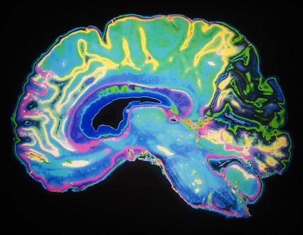
Most people recognize Alzheimer’s from its devastating symptoms such as memory loss, while new drugs target pathological aspects of disease manifestations, such as plaques of amyloid proteins. Now a sweeping new study in the Sept. 4 edition of Cell by MIT researchers shows the importance of understanding the disease as a battle over how well brain cells control the expression of their genes. The study paints a high-resolution picture of a desperate struggle to maintain healthy gene expression and gene regulation where the consequences of failure or success are nothing less than the loss or preservation of cell function and cognition.
The study presents a first-of-its-kind, multimodal atlas of combined gene expression and gene regulation spanning 3.5 million cells from six brain regions, obtained by profiling 384 post-mortem brain samples across 111 donors. The researchers profiled both the “transcriptome,” showing which genes are expressed into RNA, and the “epigenome,” the set of chromosomal modifications that establish which DNA regions are accessible and thus utilized between different cell types.
The resulting atlas revealed many insights, described in a paper that appears in the September 4th issue of Cell, and shows that the progression of Alzheimer’s is characterized by two major epigenomic trends. The first is that vulnerable cells in key brain regions suffer a breakdown of the rigorous nuclear “compartments” they normally maintain to ensure some parts of the genome are open for expression but others remain locked away. The second major finding is that susceptible cells experience a loss of “epigenomic information,” meaning they lose their grip on the unique pattern of gene regulation and expression that gives them their specific identity and enables their healthy function.
Accompanying the evidence of compromised compartmentalization and the erosion of epigenomic information are many specific findings pinpointing molecular circuitry that breaks down by cell type, by region, and gene network. They found, for instance, that when epigenomic conditions deteriorate, that opens the door to expression of many genes associated with disease, whereas if cells manage to keep their epigenomic house in order, they can keep disease-associated genes in check. Moreover, the researchers clearly saw that when the epigenomic breakdowns were occurring people lost cognitive ability but where epigenomic stability remained, so did cognition.
To understand the circuitry, the logic responsible for gene expression changes in Alzheimer’s disease, we needed to understand the regulation and upstream control of all the changes that are happening, and that’s where the epigenome comes in. This is the first large-scale single-cell multi-region gene-regulatory atlas of AD, systematically dissecting the dynamics of epigenomic and transcriptomic programs across disease progression and resilience.”
Manolis Kellis, senior author, professor in the Computer Science and Artificial Intelligence Lab and head of MIT’s Computational Biology Group
By providing that detailed examination of the epigenomic mechanisms of Alzheimer’s progression, the study provides a blueprint for devising new Alzheimer’s treatments that can target factors underlying the broad erosion of epigenomic control or the specific manifestations that affect key cell types such as neurons and supporting glial cells.
“The key to developing new and more effective treatments for Alzheimer’s disease depends on deepening our understanding of the mechanisms that contribute to the breakdowns of cellular and network function in the brain,” said Picower Professor and co-corresponding author Li-Huei Tsai, director of The Picower Institute for Learning and Memory and a founding member of MIT’s Aging Brain Initiative, along with Kellis. “This new data advances our understanding of how epigenomic factors drive disease.”
Kellis Lab members Zunpeng Liu and Shanshan Zhang are the study’s co-lead authors.
Compromised compartments and eroded information
Among the post-mortem brain samples in the study, 57 came from donors to the Religious Orders Study or the Rush Memory and Aging Project (collectively known as “ROSMAP”) who did not have AD pathology or symptoms, while 33 came from donors with early-stage pathology and 21 came from donors at a late stage. The samples therefore provided rich information about the symptoms and pathology each donor was experiencing before death.
In the new study, Liu and Zhang combined analyses of single cell RNA sequencing of the samples, which measures which genes are being expressed in each cell, and ATACseq, which measures whether chromosomal regions are accessible for gene expression. Considered together, these transcriptomic and epigenomic measures enabled the researchers to understand the molecular details of how gene expression is regulated across seven broad classes of brain cells (e.g. neurons or other glial cell types) and 67 subtypes of cells types (e.g. 17 kinds of excitatory neurons or 6 kinds of inhibitory ones).
The researchers annotated more than 1 million gene-regulatory control regions that different cells employ to establish their specific identities and functionality using epigenomic marking. Then, by comparing the cells from Alzheimer’s brains to the ones without, and accounting for stage of pathology and cognitive symptoms, they could produce rigorous associations between the erosion of these epigenomic markings and ultimately loss of function.
For instance, they saw that among people who advanced to late-stage AD, normally repressive compartments opened up for more expression and compartments that were normally more open during health became more repressed. Worryingly, when the normally repressive compartments of brain cells opened up, they became more afflicted with disease.
“For Alzheimer’s patients, repressive compartments opened up, and gene expression levels increased, which was associated with decreased cognitive function,” explained first author Zunpeng Liu.
But when cells managed to keep their compartments in order such that they expressed the genes they were supposed to, people remained cognitively intact.
Meanwhile, based on the cells’ expression of their regulatory elements, the researchers created an epigenomic information score for each cell. Generally, information declined as pathology progressed but that was particularly notable among cells in the two brain regions affected earliest in Alzheimer’s: the entorhinal cortex and the hippocampus. The analyses also highlighted specific cell types that were especially vulnerable including microglia that play immune and other roles, oligodendrocytes that produce myelin insulation for neurons, and particular kinds of excitatory neurons.
Risk genes and ‘chromatin guardians’
Detailed analyses in the paper highlighted how epigenomic regulation tracked with disease-related problems, Liu noted. The e4 variant of the APOE gene, for instance, is widely understood to be the single biggest genetic risk factor for Alzheimer’s. In APOE4 brains, microglia initially responded to the emerging disease pathology with an increase in their epigenomic information, suggesting that they were stepping up to their unique responsibility to fight off disease. But as the disease progressed the cells exhibited a sharp drop off in information, a sign of deterioration and degeneration. This turnabout was strongest in people who had two copies of APOE4, rather than just one. The findings, Kellis said, suggest that APOE4 might destabilize the genome of microglia, causing them to burn out.
Another example is the fate of neurons expressing the gene RELN and its protein Reelin. Prior studies, including by Kellis and Tsai, have shown that RELN- expressing neurons in the entorhinal cortex and hippocampus are especially vulnerable in Alzheimer’s, but promote resilience if they survive. The new study sheds new light on their fate by demonstrating that they exhibit early and severe epigenomic information loss as disease advances, but that in people who remained cognitively resilient the neurons maintained epigenomic information.
In yet another example, the researchers tracked what they colloquially call “chromatin guardians” because their expression sustains and regulates cells’ epigenomic programs. For instance, cells with greater epigenomic erosion and advanced AD progression displayed increased chromatin accessibility in areas that were supposed to be locked down by Polycomb repression genes or other gene expression silencers. While resilient cells expressed genes promoting neural connectivity, epigenomically eroded cells expressed genes linked to inflammation and oxidative stress.
“The message is clear: Alzheimer’s is not only about plaques and tangles, but about the erosion of nuclear order itself,” Kellis said. “Cognitive decline emerges when chromatin guardians lose ground to the forces of erosion, switching from resilience to vulnerability at the most fundamental level of genome regulation.
“And when our brain cells lose their epigenomic memory marks and epigenomic information at the lowest level deep inside our neurons and microglia, it seems that Alheimer’s patients also lose their memory and cognition at the highest level.”
Other authors of the paper are Benjamin T. James, Kyriaki Galani, Riley J. Mangan, Stuart Benjamin Fass, Chuqian Liang, Manoj M. Wagle, Carles A. Boix, Yosuke Tanigawa, Sukwon Yun, Yena Sung, Xushen Xiong, Na Sun, Lei Hou, Martin Wohlwend, Mufan Qiu, Xikun Han, Lei Xiong, Efthalia Preka, Lei Huang, William F. Li, Li-Lun Ho, Amy Grayson, Julio Mantero, Alexey Kozlenkov, Hansruedi Mathys, Tianlong Chen, Stella Dracheva, and David A. Bennett.
Funding for the research came from The National Institutes of Health, The National Science Foundation, the Cure Alzheimer’s Fund, the Freedom Together Foundation, the Robert A. and Renee E. Belfer Family Foundation, Eduardo Eurnekian, and Joseph P. DiSabato.
Source:
Journal reference:
Liu, Z., et al. (2025). Single-cell multiregion epigenomic rewiring in Alzheimer’s disease progression and cognitive resilience. Cell. doi.org/10.1016/j.cell.2025.06.031
