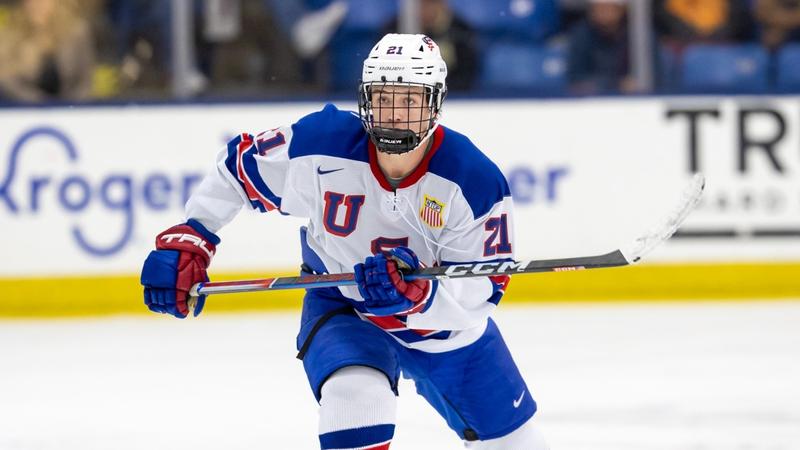
Introduction
Acute myocarditis (AM) is characterized by inflammation of the myocardium and exhibits a wide spectrum of clinical manifestations and prognoses, primarily triggered by viral infections or autoimmune diseases.1 Fulminant myocarditis (FM), as a severe form of myocarditis, presents with severe left ventricular (LV) systolic dysfunction and cardiogenic shock, necessitating the use of inotropic agents or temporary mechanical circulatory support (tMCS).2,3 The acute phase mortality rate of FM can reach as high as 45%-56%.4–6 Nonetheless, the “Chinese Protocol”, which combines tMCS with immunomodulatory therapy (IT), has significantly reduced the in-hospital mortality rate of patients with FM to 3.7%-8.1%.7–9 Although tMCS combined with IT was widely used in the early stages, studies have demonstrated that during long-term follow-up, 24.2–29% of FM patients have a left ventricular ejection fraction (LVEF) < 55%, the heart transplant rates range from 25.5% to 47.7%, and some patients even experience cardiac death, particularly in patients with systolic dysfunction at admission.10–13 Therefore, how to prevent the progression of FM to chronic persistent cardiac dysfunction remains a significant clinical challenge that urgently needs to be addressed.
Although there is currently no established long-term strategy for the management of cardiac function in patients with FM, both the European Society of Cardiology guidelines and the “Chinese Protocol” advocate the use of standard treatments for heart failure with reduced ejection fraction (HFrEF) regimens for managing these patients.9,14 Angiotensin-converting enzyme (ACE) inhibitors are the fundamental therapeutic agents for treating HFrEF and dilated cardiomyopathy.15,16 A small observational study involving 35 patients with AM revealed that the continuous utilization of ACE inhibitors during long-term follow-up can prevent the deterioration of LVEF.17 Another study by Tara et al demonstrated that the utilization of ACE inhibitors or angiotensin II receptor antagonists (ARBs) during hospitalization can reduce 90-day mortality and heart transplant rates in FM patients.18 However, currently there is limited understanding of the natural course of chronic persistent cardiac dysfunction secondary to FM. It remains unclear whether patients with FM should continue to receive treatment with ACE inhibitors after undergoing tMCS combined with IT. Additionally, the efficacy of ACE inhibitors in preventing chronic persistent cardiac dysfunction in FM patients with an LVEF ≤ 40% has not been verified. Although ACE inhibitors are widely employed in FM patients, the absence of definitive evidence limits our understanding of their precise role in the management of FM. Therefore, this study seeks to assess if ACE inhibitors can prevent the progression of chronic persistent cardiac dysfunction in FM patients under tMCS combined with IT, with the aim of offering preliminary insights for optimizing treatment plans for this high-risk population.
Methods
Study Design and Population
This is a multicenter, retrospective, observational study. From April 2016 to December 2023, a retrospective screening was carried out on 281 patients suspected of having AM who were admitted to Tongji Hospital of Tongji Medical College, Huazhong University of Science and Technology in Wuhan, China as well as relevant patients from Shanxi Bethune Hospital in Taiyuan, China, and patients from Minda Hospital of Hubei Minzu University in Enshi, China (Figure 1). The diagnosis of AM was based on one of the following criteria: (1) A diagnosis of myocarditis made by endomyocardial biopsy (EMB); (2) Elevated levels of cardiac troponin and both “Lake Louise” cardiac magnetic resonance (CMR) criteria being met.19 In this study, among 124 patients diagnosed with FM, 82 (66.13%) underwent EMB before discharge, and 85 (68.55%) underwent CMR before discharge. Chronic persistent cardiac dysfunction was defined as the persistence of cardiac symptoms for more than one month and an LVEF < 55% during follow-up.20,21
Figure 1 Flowchart of the study design and patient inclusion.
|
The diagnosis of FM must satisfy the following inclusion criteria:9,22,23 (1) Meeting the diagnostic criteria for AM; (2) Acute clinical manifestations characterized by the onset of cardiac symptoms within 30 days before admission; (3) Presenting with prodromal symptoms of the virus and rapidly progressing to severe heart failure (HF) within the subsequent two weeks; (4) The presence of significant hemodynamic instability that necessitates the use of inotropic drugs and even mechanical circulatory support;24,25 (5) The first echocardiogram indicating an LVEF ≤ 40%; (6) Performing coronary angiography on patients aged ≥ 25 years to exclude acute myocardial infarction (AMI).
The exclusion criteria were as follows: (1) Patients diagnosed with non-fulminant myocarditis (NFM); (2) The appearance of cardiac symptoms more than 30 days before admission; (3) The initial echocardiogram showing an LVEF > 40%; (4) Cases of death during hospitalization; (5) Patients diagnosed with ischemic heart disease in the past or present, or discharged with other diagnoses; (6) Long-term use of ACE inhibitors; (7) Patients with incomplete clinical data or those who decline to participate in follow-up.
This study received approval from the Institutional Review Board of the Ethics Committee of Tongji Hospital (TJ-C20160202) and was conducted in accordance with the principles outlined in the Declaration of Helsinki. Due to the retrospective nature of this study and the use of de-identified patient data, the requirement for informed consent was waived by the Institutional Review Board.
Patient Management
All selected patients received the “Chinese Protocol”,9 which includes: (1) The tMCS using an intra-aortic balloon pump (IABP) with or without extracorporeal membrane oxygenation (ECMO); (2) IT use of adequate doses of glucocorticoids and/or immunoglobulins; (3) The use of antiviral drugs alone or in combination (including ganciclovir, penciclovir, oseltamivir). IABP is typically used as the initial option for tMCS. If the implementation of the IABP fails to correct persistent hemodynamic disorders, ECMO is then used in combination.
Echocardiographic Protocol and Measurements
Standard transthoracic echocardiography was performed on patients in a resting state using a single echocardiography system (Vivid E9; GE Vingmed; Horten, Norway). This examination was performed by two experienced, board-certified echocardiography cardiologists who were blinded to patient data and analyzed the images independently. All echocardiographic assessments were completed during the patients’ admission and follow-up periods.
The end-diastolic interventricular septum thickness (IVSd), left ventricular posterior wall thickness (LVPWd), and left ventricular end-diastolic dimension (LVEDD) were measured from the parasternal long-axis view. The LVEF was calculated using the modified biplane Simpson’s method.26 When observing the four-chamber view, the ratio of the early (E-wave) to the late (A-wave) diastolic filling of the LV, denoted as E/A, was assessed with pulsed Doppler. The velocity of the septal mitral annulus (e’) was evaluated using tissue Doppler, and subsequently, the E/e’ was calculated.27 In the parasternal long-axis view, the anteroposterior diameter of the left atrium (LA) was measured.28
Primary Outcomes and Follow-Up
Based on previous studies in FM patients, LVEF < 55% was considered incomplete recovery.11,12,23 The primary outcome measure of this study was defined as an LVEF < 55% at the last follow-up. The follow-up time (in months) was defined as the duration from the date of diagnosis to the last follow-up date. The data collection was carried out by a professionally trained physician through reviewing medical records or conducting telephone interviews with patients or their relatives. The last follow-up date was December 31, 2023.
Statistical Analysis
Categorical variables are expressed as numbers (percentages), and normally distributed continuous data are reported as the mean ± standard deviation. Non-normally distributed continuous data are presented as the median with interquartile range (IQR). The Kolmogorov–Smirnov test was used to evaluate the normality of the data distribution. Based on the normality of the variables, Student’s t-test or the Mann–Whitney U-test was used to analyze the inter-group differences of continuous variables. Categorical variables were compared using the chi-square test or Fisher’s exact test. This study conducted prespecified subgroup analyses to explore heterogeneity of treatment effects among specific groups. To avoid multicollinearity, covariates were evaluated using the variance inflation factor (VIF) and tolerance. Variables with a VIF exceeding 5 were excluded. Univariate logistic regression analysis was used to determine the clinical variables associated with an LVEF < 55% at the last follow-up in the entire group and the ACE inhibitor-using group. Multivariable logistic regression was adjusted for age and sex, and included variables with P < 0.05 from the univariate logistic regression, using backward stepwise selection to identify independent risk factors. Subsequently, this study incorporated confounding factors into the models: Model 1 was not adjusted for any confounding variables. Model 2 was adjusted for age and sex. Model 3 was adjusted for age, sex, smoking status, drinking status, time from onset to admission (TFOTA), length of stay (LOS), systolic blood pressure (SBP), coronary artery disease (CAD), hypertension, IABP, ECMO, β-blockers, and ACE inhibitors (only adjusted in the entire group). Restricted cubic spline (RCS) regression was used to evaluate the nonlinear relationships between the baseline LVEDD and an LVEF < 55% at the last follow-up and to visualize the potential dose-response relationships. All analyses were conducted using RStudio (version 4.2.0) and GraphPad Prism (version 9.0). The statistical tests used a two-tailed test, and a P value < 0.05 was considered statistically significant.
Results
Demographic and Clinical Characteristics of the Entire Group and Subgroups with or Without Angiotensin-Converting Enzyme Inhibitors
Among 281 patients diagnosed with AM, 157 (55.87%) were excluded for the following reasons: 105 had NFM, 21 had incomplete data that affected the analysis results, 15 had an LVEF > 40% at admission, 7 had ischemic heart disease, 6 had a history of long-term use of ACE inhibitors, and 3 died during hospitalization. Finally, the study included 124 patients (44.13%) with FM and an LVEF ≤ 40%. Among these patients, 90 cases (72.58%) took ACE inhibitors, while 34 cases (27.42%) did not take them (Figure 1).
The median age of the study population was 38 years (IQR: 28.5–53). Females accounted for 52.42% of the population. The median SBP was 98 (88.5, 108) mmHg, the median TFOTA was 3 days (IQR: 2–5), and the median LOS was 12 days (IQR: 10–17). Electrocardiographic findings revealed that 12.9% of patients had third-degree atrioventricular block, and 5.65% of patients experienced ventricular flutter/ventricular fibrillation. The median level of cardiac troponin I (cTnI) was 20698.8 (IQR: 5632.43–44,975.2) pg/mL, and the median level of N-terminal pro-B-type natriuretic peptide (NT-proBNP) was 6291 (IQR: 3064.5–15,618.03) pg/mL. Echocardiographic results showed that the median LVEF was 27% (IQR: 21–31%), and the median LVEDD was 4.84 ± 0.6 cm. There were no significant differences in other clinical characteristics between the subgroups (Table 1).
![]() |
Table 1 Demographic and Clinical Characteristics of the Entire Group and Subgroups with or Without Angiotensin-Converting Enzyme Inhibitors
|
Regarding the management during hospitalization, all patients followed the “Chinese Protocol”. Among them, 18.55% of patients received cardiopulmonary resuscitation (CPR) defibrillation treatment due to cardiac arrest. Additionally, 37.9% and 88.71% of patients received ECMO and/or IABP support treatment, respectively. Temporary pacemakers were utilized in 30.65% of patients. Moreover, 95.97% and 98.39% of patients received immunoglobulin and glucocorticoids, respectively. Vasoactive drugs were administered to 55.65% of patients, and as many as 72.58% of patients used ACE inhibitors. There were no significant differences in other clinical characteristics between the subgroups. The specific treatment strategies are detailed in Table 2.
![]() |
Table 2 Inpatient Management of the Entire Group and Subgroups with or Without Angiotensin-Converting Enzyme Inhibitors
|
Change in Left Ventricular Ejection Fraction from the Baseline to the Last Follow-Up in Patients with or Without Angiotensin-Converting Enzyme Inhibitors, Along with Subgroup Analysis
Over a median follow-up period of 12 (6, 18) months, the median LVEF of 124 patients with FM increased from 27% (IQR: 21–31%) at admission to 58% (IQR: 50–63%) at the last follow-up (Figure 2A and B). However, among the 124 patients, 46 (37.1%) had cardiac dysfunction with an LVEF < 55% at the last follow-up. Interestingly, compared with the ACE inhibitors group (27.78%), the non-ACE inhibitors group had a higher percentage of patients with persistent cardiac dysfunction (61.76%) (P < 0.001; Table 1 and Figure 2C). Moreover, the median LVEF of patients in the non-ACE inhibitors group exhibited a continuous downward trend over the 24-month follow-up period, with values of 24.3 ± 12.3% at 1 month, 25.6 ± 20.1% at 6 months, 25.1 ± 16.3% at 12 months, and 15.1 ± 14.7% at 24 months. In contrast, the median LVEF of the ACE inhibitors group continued to improve, starting from 29.5 ± 12.8% at 1 month, reaching 32.2 ± 10.5% at 6 months, 33.8 ± 10.6% at 12 months, and ending at 32.5 ± 12.6% at 24 months (Figure 2D, Table 3). However, we observed that 49 patients (39.52%) had an LVEDD ≥ 5cm at admission (Supplementary Figure 1). Among these 49 patients, 29 (59.18%) had an LVEF < 55% at the last follow-up, 15 (51.72%) were receiving ACE inhibitors treatment, and 14 (48.28%) were not. Additionally, 3 patients (2.42%) underwent permanent pacemaker implantation, and 1 patient (0.81%) received a heart transplant during the follow-up period, and none of them received ACE inhibitors treatment.
![]() |
Table 3 The Change Values in LVEF from Baseline to 24 Months of Follow-Up
|
![]() |
Figure 2 Changes in LVEF over a 24-month follow-up period among patients with FM and LVEF ≤ 40%. (A) Patients with FM and an LVEF ≤ 40% had a significant improvement in LV systolic function from the baseline to the last follow-up based on ACE inhibitors and (B) non-ACE inhibitors; (C) The proportion of patients using or not using ACE inhibitors with an LVEF < 55% at the last follow-up; (D) Changes in LVEF from the baseline to the 24-month mark.
|
Subgroup analyses were predefined based on demographic and clinical characteristics of interest. The results indicated that the use of ACE inhibitors had a sustained beneficial effect on the primary outcome compared with those without ACE inhibitors. The P-value for interaction > 0.05 indicates no significant interaction across groups (Figure 3).
![]() |
Figure 3 Primary outcomes of prespecified subgroups. The primary outcome was defined as an LVEF < 55% at the last follow-up.
|
Primary Outcome Impact Factors in the Entire and Angiotensin-Converting Enzyme Inhibitors Group
The demographic and clinical characteristics of the ACE inhibitors group, along with its subgroups of LVEF ≥ 55% and LVEF < 55%, are presented in Supplementary Table 1. To further investigate the potential factors influencing LVEF < 55% at the last follow-up, a logistic regression analysis was performed on the entire group and the ACE inhibitors group. All covariates included in the analysis were measured at admission. The univariate logistic regression model indicated that age, SBP, LVEDD, LA, lactate dehydrogenase (LDH), and ACE inhibitors use were significantly associated with LVEF < 55%. However, in the entire group, multivariate logistic regression adjusted for age, sex, SBP, LVEDD, LA, LDH, and ACE inhibitor use, identified only ACE inhibitor use (HR = 0.19, 95% CI: 0.04–0.96, P = 0.045) and LVEDD (HR = 9.18, 95% CI: 2.73–30.83, P < 0.001) as significantly associated with LVEF < 55% (Table 4). Similarly, in the ACE inhibitors group, multivariate logistic regression analysis also identified the LVEDD (HR = 9.61, 95% CI: 2.42–38.19, P < 0.001) as a risk factor for LVEF < 55% (Supplementary Table 2).
![]() |
Table 4 Univariate and Multivariate Logistic Regression of Influencing Factors for LVEF < 55% at the Last Follow-Up in the Entire Group
|
Restricted Cubic Spline Curve Fitting
Restricted cubic spline (RCS) analysis was employed to examine the relationship between the LVEDD and LVEF < 55% in the entire group and the subgroup of patients using ACE inhibitors. RCS analysis indicated that the nonlinear relationship between the two variables was not statistically significant (P > 0.05). In the unadjusted and partially adjusted weighted logistic regression models, the risk of LVEF < 55% at the last follow-up increased approximately linearly with increasing levels of the LVEDD, even after adjusting for the use of ACE inhibitors (Figure 4).
![]() |
Figure 4 The RCS curve fit between LVEDD and the LVEF < 55%. Blue lines represent the estimated OR of LVEDD and the primary outcome. Blue-shaded areas represent the 95% CI. (A) Model 1 was unadjusted in the entire group; (B) Model 2 was adjusted for age and sex in the entire group; (C) Model 3 was adjusted for age, sex, smoking status, drinking status, TFOTA, LOS, SBP, CAD, hypertension, IABP, ECMO, β-blockers, and ACE inhibitors in the entire group; (D) Model 1 was unadjusted in the ACE inhibitors group; (E) Model 2 was adjusted for age and sex in the ACE inhibitors group; (F) Model 3 was adjusted for age, sex, smoking status, drinking status, TFOTA, LOS, SBP, CAD, hypertension, IABP, ECMO, and β-blockers in the ACE inhibitors group.
|
Discussion
In this multicenter retrospective study, we emphasized the importance of long-term cardiac function management for patients with FM who received tMCS combined with IT and had an LVEF ≤ 40%, even for those whose cardiac function had recovered to LVEF ≥ 55% at the time of discharge. In this study, more than 3 out of 4 patients were treated with ACE inhibitors, and there were no significant differences in baseline blood pressure, renal function, and serum potassium levels among these patients. Long-term follow-up results revealed that the use of ACE inhibitors may contribute to improving LV function in patients with FM and preventing chronic persistent cardiac dysfunction. However, during long-term follow-up, an increase in LVEDD may diminish the efficacy of ACE inhibitors.
Patients with FM are characterized by sudden onset, rapid progression, and deterioration of the clinical course, which can lead to severe hemodynamic compromise, cardiogenic shock, and even death.29 In the early phase of acute sustained injury, there might be a substantial initial decline in the LVEF. However, if appropriate therapeutic measures are promptly implemented, the systolic function of the heart can rapidly recover. Ammirati et al demonstrated that median LVEF increased from 21% at baseline to 54% at follow-up in patients who survived the acute phase of FM.11 Following the “Chinese Protocol”, Zhou et al reported an increase in median LVEF from 40% to 57%,8 while in this study, LVEF increased from 27% to 58%. Notably, despite aggressive treatment with tMCS and IT during the acute phase, several studies have reported varying degrees of LV impairment in patients with FM. For instance, Ammirati et al indicated that despite improvement in LVEF, 29% of patients had LVEF < 55% at a median follow-up of 22 months.11 A multicenter study involving 216 patients with FM demonstrated that 16% had LVEF ≤ 50% at 1-year follow-up, indicating persistent cardiac dysfunction in some cases.30 Among patients who received tMCS combined with IT, Jiang et al observed that 22% had LVEF < 55% at 2-year follow-up, with no improvement compared to discharge.23 Another study reported 24.2% of the patients with FM had persistent LVEF < 55% at a median follow-up of 12 months.12 In the present study, the incidence was higher, reaching 37.1%. All the aforementioned studies indicate that patients with FM may experience impaired cardiac systolic function during long-term follow-up, suggesting that the current interventions may be insufficient to prevent the progression to chronicity. Previous studies have demonstrated that FM patients complicated by LV systolic dysfunction have significantly elevated heart transplant levels (25.5–47.7%) and cardiac mortality rates (28–35.5%).10,11 This renders it an urgent imperative to prevent patients with FM from developing chronic persistent cardiac dysfunction.
According to reports, immunosuppressive therapy has a positive impact on chronic inflammatory cardiomyopathy.31,32 The preliminary results of the Clinical Assessment of New Treatment Regimen for Adult FM trial (NCT03268642) conducted by our center demonstrated that, compared with routine treatment, the “Chinese Protocol” (without cytotoxic drugs) significantly reduced the in-hospital mortality rate of FM patients [3.7% (3/81) compared to 46.6% (41/88)].7 Therefore, the “Chinese Protocol” recommends its application only in FM patients who continue to exhibit HF symptoms after standard anti-HF treatment and require EMB to determine the underlying myocarditis pathology and assess the need for immunosuppressive therapy.9 The standard treatment protocol for HF in AM has been recognized by the guidelines of the European Society of Cardiology and the Chinese Society of Cardiology.9,14 ACE inhibitors are the fundamental therapeutic agents for HFrEF. They are widely used to treat other cardiovascular diseases, including dilated cardiomyopathy and hypertension. Moreover, they contribute to favorable LV remodeling and are effective in reducing LV enlargement.33–36 Although there is a lack of observational studies evaluating the therapeutic efficacy of ACE inhibitors on patients with FM and their role in preventing the development of chronic persistent cardiac dysfunction, recent survey data indicate that a considerable number of physicians prescribe ACE inhibitors for FM patients, with utilization rates ranging from 59% to 74% in other study groups and reaching 73% in this study.11,13 Basic research has revealed that losartan or captopril significantly reduced inflammation, necrosis, and fibrosis in mice with autoimmune myocarditis.37 Another study demonstrated that ACE inhibitors and/or ARBs not only reduce complications associated with myocarditis but also downregulate the potential autoimmune component of the disease.38 A multicenter study found that the administration of ACE inhibitors or ARBs during hospitalization in FM patients was associated with a reduced 90-day mortality rate and heart transplant rate.18 A small observational study followed up 35 patients with AM who were taking ACE inhibitors and reported that continuous use of ACE inhibitors reduced the deterioration of the LVEF during long-term follow-up.17 Consistent with previous findings, this study suggests that ACE inhibitor use may provide potential benefits in improving LV function and preventing chronic persistent cardiac dysfunction in FM patients. In contrast, patients who did not receive ACE inhibitors exhibited a persistent decline in cardiac systolic function during long-term follow-up. These benefits may be attributed to the reduction in chronic myocardial inflammatory and the improvement of ventricular remodeling.38,39 Further investigation is warranted to elucidate the mechanism by which ACE inhibitors exert their effects on FM patients.
Several studies have demonstrated that an increase in LVEDD is associated with persistent LV dysfunction.40,41 Ammirati et al reported that 17.3% of patients with complicated AM underwent progressive LV dilation during the follow-up period, and 14.5% of the patients had an LVEF < 50%.2 Jiang et al’s research showcased that 12.1% of FM patients encountered LV dilation after discharge, and 24.2% of the patients had a sustained LVEF < 55%.12 A single-center retrospective study revealed that during a 2-year follow-up period, 28% of patients with FM had an LVEDD ≥ 5 cm, and 22% of the patients had an LVEF < 55%.23 Based on this study, we discovered that 49 patients (39.52%) had an LVEDD ≥ 5cm at admission, and 29 patients (59.18%) had an LVEF < 55% at the last follow-up. 15 patients (51.72%) had been on ACE inhibitors treatment, whereas 14 patients (48.28%) had not. Although the utilization of ACE inhibitors may partially reverse the increase in LVEDD among patients, reducing the number from 30 to 14 (Supplementary Figure 1), we observed that patients with a larger LVEDD at admission exhibited weaker responses to ACE inhibitors during long-term follow-up, and there was no significant difference in the diagnosis and treatment time among these patients (Table 1). Furthermore, during follow-up, we observed that eight patients with an LVEDD ≥5 cm at admission who were receiving ACE inhibitor therapy initiated spironolactone treatment. Of these, four patients exhibited LVEF <55% at the last follow-up. The limited cohort size precludes definitive conclusions regarding the therapeutic potential of aldosterone antagonists in FM. Ammirati et al’s study demonstrated that there were disparities in the baseline LVEDD among different pathological subtypes of FM. Specifically, the median LVEDD of patients with giant cell myocarditis was 52 mm, that of patients with eosinophilic myocarditis was 50 mm, and that of patients with lymphocytic myocarditis was 49 mm. In comparison with other subtypes, patients with giant cell FM exhibit higher mortality and heart transplantation rates, which may potentially explain the results of this study.10 Moreover, the underlying or hereditary predisposition to cardiomyopathy is also a possible explanation that warrants future investigation. This research underscores the significance of ACE inhibitors in patients with FM, as they contribute to the restoration of cardiac structure and function. However, some patients did not respond to ACE inhibitor therapy. For these individuals, following current guidelines, substitution with angiotensin receptor-neprilysin inhibitor (ARNI), adding other standardized HF medicines, such as aldosterone antagonists, β-blockers, and sodium-glucose cotransporter 2 (SGLT2) inhibitors, may be considered.9,14,42 In addition, immunosuppressants can be added appropriately according to the pathological type of myocarditis.9 Given the limited clinical evidence, the effects of these therapies in FM patients warrant further investigation.
Limitations
This study exhibits several limitations. Firstly, it was a retrospective study characterized by a relatively small sample size and inherent bias. The majority of patients were referred to the cardiology departments of three centers during the severe stages of the disease. This circumstance led to the inclusion of patients with FM whose initial echocardiogram indicated an LVEF ≤ 40%, thereby causing selection bias. Secondly, this study was conducted in China. The background of the patient population and clinical practices may vary from those in other countries, which could potentially restrict the generalizability of the research findings in other regions or populations. Thirdly, based on the utilization of ACE inhibitors, 124 patients with FM were further divided into subgroups. This division led to a decline in the number of patients in each subgroup, diminished statistical power, and further constrained the study’s capacity to assess the benefits of ACE inhibitors use. Furthermore, owing to the absence of data regarding the dosage of ACE inhibitors and post-medication blood pressure changes, a more comprehensive analysis cannot be performed. Fourthly, the initial analysis of this study solely concentrated on patients who survived and were discharged, without including in-hospital deaths. Fifthly, only 66.13% of patients underwent EMB and were not tested for specific virus types. This situation made it challenging to ascertain the influence of different histological subtypes and/or viral subtypes on cardiac function during the follow-up period. Finally, the study design only centered on patients with FM and did not conduct a comparative analysis between FM cases and NFM cases. This situation needs to be considered in combination with the background of the study population.
Conclusion
ACE inhibitors may hold the potential to improve LV function and impede chronic persistent cardiac dysfunction in patients with FM under tMCS combined with IT. However, an increase in the LVEDD may potentially undermine the benefits during long-term follow-up. These findings offer evidence in support of background ACE inhibitor therapy for FM patients and highlight the significance of preventing the development of chronic persistent cardiac dysfunction.
Abbreviations
tMCS: Temporary mechanical circulatory supports; IT: Immunoregulatory therapy; FM: Fulminant myocarditis; ACE: Angiotensin-converting enzyme; LVEF: Left ventricular ejection fraction; LVEDD: Left ventricular end-diastolic dimensions; AM: Acute myocarditis; HFrEF: Heart failure with reduced ejection fraction; EMB: Endomyocardial biopsy; CMR: cardiac magnetic resonance; AMI: Acute myocardial infarction; NFM: non-fulminant myocarditis; IABP: Intra-aortic balloon pump; ECMO: Extracorporeal membrane oxygenation; IVSd: End-diastolic interventricular septum thickness; LVPWd: Left ventricular posterior wall thickness; IQR: Interquartile range; VIF: Variance inflation factor; TFOTA: Time from onset to admission; LOS: Length of stay; SBP: Systolic blood pressure; CAD: Coronary artery disease; RCS: Restricted cubic spline; cTnI: Troponin I; NT-proBNP: N-terminal pro-B-type natriuretic peptide; CPR: Cardiopulmonary resuscitation; LDH: Lactate dehydrogenase; ARNI: Angiotensin receptor-neprilysin inhibitor; SGLT2: Sodium-glucose cotransporter 2.
Data Sharing Statement
The datasets generated during and/or analyzed during the current study are available from the corresponding author, Jiangang Jiang, upon reasonable request.
Ethics Approval and Informed Consent
This study received approval from the Institutional Review Board of the Ethics Committee of Tongji Hospital (TJ-C20160202) and was conducted in accordance with the principles outlined in the Declaration of Helsinki. Due to the retrospective nature of this study and the use of de-identified patient data, the requirement for informed consent was waived by the Institutional Review Board.
Author Contributions
All authors made a significant contribution to the work reported, whether that is in the conception, study design, execution, acquisition of data, analysis and interpretation, or in all these areas; took part in drafting, revising or critically reviewing the article; gave final approval of the version to be published; have agreed on the journal to which the article has been submitted; and agree to be accountable for all aspects of the work.
Funding
Open access publication fees were funded by the Tongji Hospital, Huazhong University of Science and Technology, with no other sources of funding.
Disclosure
All authors declare that they have no conflicts of interest in this work.
References
1. Caforio AL, Pankuweit S, Arbustini E, et al. Current state of knowledge on aetiology, diagnosis, management, and therapy of myocarditis: a position statement of the European society of cardiology working group on myocardial and pericardial diseases. Eur Heart J. 2013;34(33):2636–48,2648a–2648d. doi:10.1093/eurheartj/eht210
2. Ammirati E, Cipriani M, Moro C, et al. Clinical presentation and outcome in a contemporary cohort of patients with acute myocarditis: multicenter lombardy registry. Circulation. 2018;138(11):1088–1099. doi:10.1161/circulationaha.118.035319
3. Pahuja M, Adegbala O, Mishra T, et al. Trends in the incidence of in-hospital mortality, cardiogenic shock, and utilization of mechanical circulatory support devices in myocarditis (analysis of national inpatient sample data, 2005–2014). J Card Fail. 2019;25(6):457–467. doi:10.1016/j.cardfail.2019.04.012
4. Lee CH, Tsai WC, Hsu CH, Liu PY, Lin LJ, Chen JH. Predictive factors of a fulminant course in acute myocarditis. Int J Cardiol. 2006;109(1):142–145. doi:10.1016/j.ijcard.2005.04.014
5. Sawamura A, Okumura T, Ito M, et al. Prognostic value of electrocardiography in patients with fulminant myocarditis supported by percutaneous venoarterial extracorporeal membrane oxygenationx- analysis from the CHANGE PUMP study. Circ J. 2018;82(8):2089–2095. doi:10.1253/circj.CJ-18-0136
6. Hung Y, Lin WH, Lin CS, et al. The prognostic role of QTc interval in acute myocarditis. Acta Cardiol Sin. 2016;32(2):223–230. doi:10.6515/acs20150226a
7. Li S, Xu S, Li C, et al. A life support-based comprehensive treatment regimen dramatically lowers the in-hospital mortality of patients with fulminant myocarditis: a multiple center study. Sci China Life Sci. 2019;62(3):369–380. doi:10.1007/s11427-018-9501-9
8. Zhou N, Zhao Y, Jiang J, et al. Impact of mechanical circulatory support and immunomodulation therapy on outcome of patients with fulminant myocarditis: chinese registry of fulminant myocarditis. Signal Transduct Target Ther. 2021;6(1):350. doi:10.1038/s41392-021-00700-6
9. Jiang J, Shu H, Wang DW, et al. Chinese Society of Cardiology guidelines on the diagnosis and treatment of adult fulminant myocarditis. Sci China Life Sci. 2024;67(5):913–939. doi:10.1007/s11427-023-2421-0
10. Ammirati E, Veronese G, Brambatti M, et al. Fulminant versus acute nonfulminant myocarditis in patients with left ventricular systolic dysfunction. J Am Coll Cardiol. 2019;74(3):299–311. doi:10.1016/j.jacc.2019.04.063
11. Ammirati E, Cipriani M, Lilliu M, et al. Survival and left ventricular function changes in fulminant versus nonfulminant acute myocarditis. Circulation. 2017;136(6):529–545. doi:10.1161/circulationaha.117.026386
12. Jiang JG, Liu C, Cui GL, et al. Long term prognosis of fulminant myocarditis and predictors related to impaired cardiac function post discharge. Zhonghua Xin Xue Guan Bing Za Zhi. 2022;50(3):263–269. doi:10.3760/cma.j.cn112148-20211206-01056
13. Kanaoka K, Onoue K, Terasaki S, et al. Features and outcomes of histologically proven myocarditis with fulminant presentation. Circulation. 2022;146(19):1425–1433. doi:10.1161/circulationaha.121.058869
14. McDonagh TA, Metra M, Adamo M, et al. 2021 ESC guidelines for the diagnosis and treatment of acute and chronic heart failure. Eur Heart J. 2021;42(36):3599–3726. doi:10.1093/eurheartj/ehab368
15. McDonagh TA, Metra M, Adamo M, et al. 2023 focused update of the 2021 ESC guidelines for the diagnosis and treatment of acute and chronic heart failure. Eur Heart J. 2023;44(37):3627–3639. doi:10.1093/eurheartj/ehad195
16. Heidenreich PA, Bozkurt B, Aguilar D, et al. 2022 AHA/ACC/HFSA guideline for the management of heart failure: a report of the American college of cardiology/american heart association joint committee on clinical practice guidelines. Circulation. 2022;145(18):e895–e1032. doi:10.1161/cir.0000000000001063
17. Anguita-Sánchez M, Castillo-Domínguez JC, Mesa-Rubio D, Ruiz-Ortiz M, López-Granados A, Suárez de Lezo J. [Should Angiotensin-converting enzyme inhibitors be continued over the long term in patients whose left ventricular ejection fraction normalizes after an episode of acute myocarditis?]. Se deben mantener los inhibidores de la enzima de conversión de la angiotensina a largo plazo en pacientes que normalizan la fracción de eyección tras un episodio de miocarditis aguda? Rev Esp Cardiol. 2006;59(11):1199–1201.
18. Tara S, Yamamoto T, Kanaoka K, et al. Effects of cardioprotective drugs on 90-day mortality or heart transplantation in patients with fulminant myocarditis. Circ Rep. 2024;6(8):322–332. doi:10.1253/circrep.CR-24-0059
19. Ferreira VM, Schulz-Menger J, Holmvang G, et al. Cardiovascular magnetic resonance in nonischemic myocardial inflammation: expert recommendations. J Am Coll Cardiol. 2018;72(24):3158–3176. doi:10.1016/j.jacc.2018.09.072
20. Ammirati E, Frigerio M, Adler ED, et al. Management of acute myocarditis and chronic inflammatory cardiomyopathy: an expert consensus document. Circ Heart Fail. 2020;13(11):e007405. doi:10.1161/circheartfailure.120.007405
21. Maisch B, Ruppert V, Pankuweit S. Management of fulminant myocarditis: a diagnosis in search of its etiology but with therapeutic options. Curr Heart Fail Rep. 2014;11(2):166–177. doi:10.1007/s11897-014-0196-6
22. Wang J, He M, Li H, et al. Soluble ST2 is a sensitive and specific biomarker for fulminant myocarditis. J Am Heart Assoc. 2022;11(7):e024417. doi:10.1161/jaha.121.024417
23. Jiang L, Zhang K, Zhang C, et al. Left ventricular function changes and echocardiographic predictors in adult survivors of fulminant myocarditis treated with the Chinese protocol. Sci Rep. 2023;13(1):6274. doi:10.1038/s41598-023-33285-x
24. Hang W, Chen C, Seubert JM, Wang DW. Fulminant myocarditis: a comprehensive review from etiology to treatments and outcomes. Signal Transduct Target Ther. 2020;5(1):287. doi:10.1038/s41392-020-00360-y
25. Sharma AN, Stultz JR, Bellamkonda N, Amsterdam EA. Fulminant myocarditis: epidemiology, pathogenesis, diagnosis, and management. Am J Cardiol. 2019;124(12):1954–1960. doi:10.1016/j.amjcard.2019.09.017
26. Gottdiener JS, Bednarz J, Devereux R, et al. American society of echocardiography recommendations for use of echocardiography in clinical trials. J Am Soc Echocardiogr. 2004;17(10):1086–1119. doi:10.1016/j.echo.2004.07.013
27. Nagueh SF, Smiseth OA, Appleton CP, et al. Recommendations for the evaluation of left ventricular diastolic function by echocardiography: an update from the American society of echocardiography and the European association of cardiovascular imaging. Eur Heart J Cardiovasc Imaging. 2016;17(12):1321–1360. doi:10.1093/ehjci/jew082
28. Leung DY, Boyd A, Ng AA, Chi C, Thomas L. Echocardiographic evaluation of left atrial size and function: current understanding, pathophysiologic correlates, and prognostic implications. Am Heart J. 2008;156(6):1056–1064. doi:10.1016/j.ahj.2008.07.021
29. Wang D, Li S, Jiang J, et al. Chinese society of cardiology expert consensus statement on the diagnosis and treatment of adult fulminant myocarditis. Sci China Life Sci. 2019;62(2):187–202. doi:10.1007/s11427-018-9385-3
30. Kondo T, Okumura T, Shibata N, et al. Differences in prognosis and cardiac function according to required percutaneous mechanical circulatory support and histological findings in patients with fulminant myocarditis: insights from the CHANGE PUMP 2 study. J Am Heart Assoc. 2022;11(4):e023719. doi:10.1161/jaha.121.023719
31. Wojnicz R, Nowalany-Kozielska E, Wojciechowska C, et al. Randomized, placebo-controlled study for immunosuppressive treatment of inflammatory dilated cardiomyopathy: two-year follow-up results. Circulation. 2001;104(1):39–45. doi:10.1161/01.cir.104.1.39
32. Merken J, Hazebroek M, Van Paassen P, et al. Immunosuppressive therapy improves both short- and long-term prognosis in patients with virus-negative nonfulminant inflammatory cardiomyopathy. Circ Heart Fail. 2018;11(2):e004228. doi:10.1161/circheartfailure.117.004228
33. Baker WL, Coleman CI, Kluger J, et al. Systematic review: comparative effectiveness of angiotensin-converting enzyme inhibitors or angiotensin II-receptor blockers for ischemic heart disease. Ann Intern Med. 2009;151(12):861–871. doi:10.7326/0003-4819-151-12-200912150-00162
34. Arsenault M, Zendaoui A, Roussel E, et al. Angiotensin II-converting enzyme inhibition improves survival, ventricular remodeling, and myocardial energetics in experimental aortic regurgitation. Circ Heart Fail. 2013;6(5):1021–1028. doi:10.1161/circheartfailure.112.000045
35. Patel J, Rassekh N, Fonarow GC, et al. Guideline-directed medical therapy for the treatment of heart failure with reduced ejection fraction. Drugs. 2023;83(9):747–759. doi:10.1007/s40265-023-01887-4
36. Ahad A, Al-Mohizea AM, Al-Jenoobi FI, Aqil M. Transdermal delivery of angiotensin II receptor blockers (ARBs), angiotensin-converting enzyme inhibitors (ACEIs) and others for management of hypertension. Drug Deliv. 2016;23(2):579–590. doi:10.3109/10717544.2014.942444
37. Bahk TJ, Daniels MD, Leon JS, Wang K, Engman DM. Comparison of angiotensin converting enzyme inhibition and angiotensin II receptor blockade for the prevention of experimental autoimmune myocarditis. Int J Cardiol. 2008;125(1):85–93. doi:10.1016/j.ijcard.2007.04.062
38. Godsel LM, Leon JS, Engman DM. Angiotensin converting enzyme inhibitors and angiotensin II receptor antagonists in experimental myocarditis. Curr Pharm Des. 2003;9(9):723–735. doi:10.2174/1381612033455440
39. Juan W, Nakazawa M, Watanabe K, et al. Quinapril inhibits progression of heart failure and fibrosis in rats with dilated cardiomyopathy after myocarditis. Mol Cell Biochem. 2003;251(1–2):77–82. doi:10.1023/A:1025433900034
40. Merlo M, Ammirati E, Gentile P, et al. Persistent left ventricular dysfunction after acute lymphocytic myocarditis: frequency and predictors. PLoS One. 2019;14(3):e0214616. doi:10.1371/journal.pone.0214616
41. Kanaoka K, Onoue K, Terasaki S, et al. Changes in cardiac function following fulminant myocarditis. Circ Heart Fail. 2024;17(4):e010840. doi:10.1161/circheartfailure.123.010840
42. Kitai T, Kohsaka S, Kato T, et al. JCS/JHFS 2025 guideline on diagnosis and treatment of heart failure. J Card Fail. 2025. doi:10.1016/j.cardfail.2025.02.014