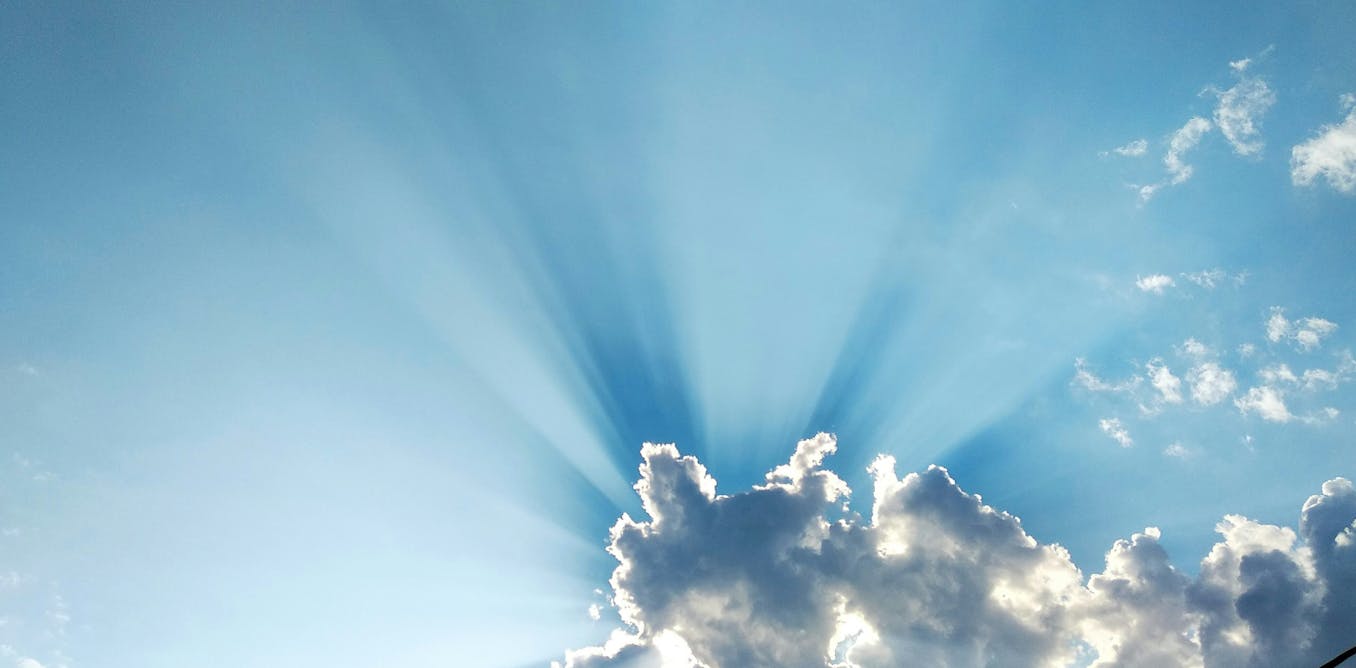
Recurrent Subcutaneous Abscess caused by Co-infection with Prototheca

Introduction

Prototheca is a type of chlorophyll-free algae that cannot undergo photosynthesis.1 They are widely detected in the natural environment and can occasionally cause human infections.2 Although clinically managed similarly to fungi and often classified among opportunistic fungal pathogens in medical contexts, Prototheca is taxonomically non-photosynthetic, achlorophyllous algae. In 1964, it was first reported that caused human infection.3 In the past 10 years, the incidence rate of human protothecosis has gradually increased with the development of molecular diagnostic technology. After reviewing domestic and foreign literature, it was found that 305 cases have been reported in the world. Protothecosis has been reported to be distributed on all continents around the world, except for Antarctica. In China and Japan, most patients were located in coastal areas.2 So far, 18 species of Prototheca have been identified,4 and Prototheca wickerhamii (P. wickerhamii) is the main pathogen in human infections. The main manifestations of infections caused by Prototheca are skin and subcutaneous tissue infections, synovitis and fibrous tissue inflammation, and disseminated infections.5 Due to its rarity, unclear pathogenesis, and lack of standard treatment guidelines, this infection is prone to neglect and misdiagnosis in clinical practice.6,7

Mycobacterium haemophilum (M. haemophilum) is a slow-growing nontuberculous mycobacteria, which can cause different types of infections ranging from localized to disseminated diseases in humans. In children and immunocompromised individuals, it presents as localized lymphadenitis, while in immunocompromised patients, it is more common to present as cutaneous, synovial, and disseminated infections.8 It is difficult to grow in routine tissue cultures, which require ferric iron or haemin to the culture media and lower temperature incubation in vitro at 30–32.8,9 So, conventional microbial culture methods always do not provide positive results, and molecular diagnostic techniques are crucial for rapid diagnosis. Metagenomic next-generation sequencing (mNGS) has been increasingly used for diagnosis of infectious diseases due to its advantages of unbiased, rapid and high sensitivity for detecting microbial gene sequences in samples. However, it also has some known limitations which includes databases lack accuracy or completeness, interpretation challenges, difficulty with low biomass or contaminants, bioinformatics pipeline biases. In this study, we reported a case of co-infection with P. wickerhamii and M. haemophilum which was misidentified M. haemophilum as M. leprae and overlooking P. wickerhamii by mNGS.

Case Presentation

A 39-year-old man from southern China presented with recurrent swelling and tenderness on his fingers bilaterally for 1 year at our hospital. Clinical symptoms mainly include mild pain in the abscess area, no joint pain, no fever and other symptoms. He reported a history of finger injury several times while using firewood and handling live chickens and ducks. He did not go to the hospital for proper treatment and used some ointments by himself (specific details were unknown).The wound repeatedly became red and swollen, and pus gradually appeared. He had no other underlying diseases. Physical examination vital signs were unremarkable and dark red abscesses on the dorsum of several fingers on both hands, with pus in an egg-sized abscess on the left middle finger (Figure 1A). Laboratory tests revealed a normal white blood cell count, and both the (1-3)-β-D-glucan assay (BDG) and galactomannan tests were negative. HIV antibodies were undetectable, and CD4+ and CD8+ T-lymphocyte counts were within normal ranges. X ray showed no damage to the bones and joints of the hands.

Figure 1 Clinical presentation. (A) Clinical image of the patient showed dark red abscesses on the dorsum of several fingers on both hands, with pus in one abscess on the left middle finger. (B) The figure showed the redness and swelling on the patient’s hands have subsided obviously and mild scars were remained after 6 months of treatment.

The pus smears from the fingers found acid-fast bacilli (AFB) by Ziehl-Neelsen staining (Figure 2A). Tissue biopsy from abscesses was performed simultaneously for histopathologic examination and microbiological culture as well as metagenomic next-generation sequencing (mNGS, Illumina platform) detection. The first time of mNGS detection showed only Mycobacterium leprae (M. leprae). The result of histopathology showed granulomatous inflammation, and periodic acid-Schiff staining was positive for morula bodies (Figure 2B) but negative for AFB. At the same time, irregular granulomatous around the vessels and nerves, tuberculous granuloma and foam cell granuloma were not found in the histopathology. Six days later, white, yeast-like colonies grew under SDA medium with abscess tissue and pus samples (Figure 2C). Endospores in typical sporangia were seen under the microscope, which were confirmed as P. wickerhamii by mass spectrography (MS), internal-transcribed-spacer (ITS) and cytB gene sequencing (Figure 2D). In combination with clinical manifestations of the patient, which did not have lion-like face, peripheral nerve enlargement or sensory impairment, we considered that the patient should not be diagnosed as leprosy. So, we requested a re-analysis of the raw sequence data detected by mNGS, further analyzed the detected sequences and blasted with the database on NCBI. We subsequently identified that the sequence was 100% identical to M. haemophilum but only 98% closely matched M. leprae. As the gene sequences of M. haemophilum and M. leprae were very similar, it was easy to cause misdiagnosis. Meanwhile, the presence of another pathogen was also discovered by mNGS, it was consistent with the results of tissue microbial culture which were both confirmed as P. wickerhamii. Finally, we revised the result of mNGS to be both M. haemophilum and P. wickerhami in samples from the abscess tissue and pus. A chronic subcutaneous abscess caused by the co-infection of P. wickerhamii and M. haemophilum was diagnosed. The patient received amikacin (0.8g daily), clarithromycin (500mg twice daily), and moxifloxacin (400mg daily) for M. haemophilum therapy, and itraconazole (200mg twice daily) for P. wickerhami. No surgical intervention was performed during the treatment process. Symptoms improved and the patient was discharged after 2 weeks. Regular maintain treatment with clarithromycin, moxifloxacin, and itraconazole after discharge. After 6 months of treatment, the redness and swelling on his hands have subsided obviously and mild scars were remained (Figure 1B). Currently, we are still following up on the patient.

Figure 2 Laboratory test results of the patient. (A) Ziehl-Neelsen staining revealed positive acid-fast bacilli in the pus (100×Oil). (B) Histopathologic Examination showed granulomatous inflammation, and periodic acid-Schiff staining was positive for morula bodies (PAS×400). (C) Fungal culture: SDA, 6 days, white yeast-like colonies with smooth or wrinkled surface. (D) Microscopic examination of fungal culture showing many endospores in typical sporangia, morula-like, strawberry-like or wheel-like (Lactophenol-cotton blue stain ×1000).

Discussion

As M. haemophilum and P. wickerhamii are both rare pathogens that can infect both immunocompromised and immunocompetent hosts.10,11 The infection usually occurs by traumatic inoculation from the natural environment reservoirs includes slime fux of trees, grass, wastewater, dairy farms, soil or insect bites with the pathogen.12 Both of the pathogens could cause atypical clinical manifestations, which led to the misdiagnosis of the patient. To our knowledge, the co-infection of P. wickerhami and M. haemophilum has not been reported in the literature. We speculate that the possible reason for the co-infection in this case is that the patient was stabbed on the skin several times by live poultry and firewood from different sources.

The advantages of mNGS include a wide detection range and high sensitivity, especially for those pathogens, which were difficult to cultivate or rare. However, mNGS was also exposed to some drawbacks. The diagnosis was complicated by mNGS misidentifying M. haemophilum as M. leprae (98% sequence similarity) and overlooking P. wickerhamii in our case. The problems have firstly appeared in the diagnosis of Protothecosis. Although mNGS can detect all the gene sequence information of pathogens from the specimens of patients, it was overlooked during the initial bioinformatic analysis of pathogens due to the rarity of P. wickerhami. We speculated that possible reasons may include missing sequences of rare pathogens in the database or the sequence was filtered out due to its extreme rarity. The missed diagnosis of P. wickerhami also indirectly indicates that the diagnostic efficiency of mNGS partially depends on whether its database platform is complete.

In addition, this case was misdiagnosed as M. leprae in the results of mNGS for first time. In our case, the pus smear was AFB positive which is possible for both M. leprae and M. haemophilum. The tissue histopathology was AFB negative, which may be related to sampling difference, bacilli low load, staining technique variation. Due to the inconsistency between mNGS detection results and clinical manifestations, we have blasted with NCBI and consulted literature. We found that the leprosy bacilli are phylogenetically closely related to M. haemophilum and their gene sequences are very similar.13 It is easy to cause confusion even with molecular detection techniques such as mNGS. The case underscores the critical need to correlate mNGS results with clinical features and use complementary diagnostic methods to avoid errors. The combination of traditional and molecular methods can improve diagnostic accuracy.

Conclusion

This case highlights the following points. The limitations of mNGS can lead to misdiagnosis between genetically similar pathogens or oversight of rare organisms. In addition, combining traditional methods (microbial culture, histopathology) with molecular techniques (mNGS, ITS, 16S rRNA) is essential for accurate diagnosis of complex infections. Finally, clinical correlation is critical because of mNGS results must be cross-verified with patient symptoms and epidemiological history.

Data Sharing Statement

The datasets generated analysed during the current study are not publicly available to protect patient privacy but are available from the corresponding author on reasonable request.

Ethics Approval and Consent to Participate

Written informed consent has been provided by the patient for the case details and images to be published. Details of the case can be published without ethical committee approval.

Consent for Publication

The patient’s written consent has been obtained regarding publishing his data and photographs.

Acknowledgments

We thank the patient for granting permission to publish this information. We would like to acknowledge the Next-Generation Sequencing platform for whole-genome sequencing and analysis.

Author Contributions

All authors made a significant contribution to the work reported, whether that is in the conception, study design, execution, acquisition of data, analysis and interpretation, or in all these areas; took part in drafting, revising or critically reviewing the article; gave final approval of the version to be published; have agreed on the journal to which the article has been submitted; and agree to be accountable for all aspects of the work.

Funding

This study was supported by Supported by the National Key Research and Development Program of China (2022YFC2504800), the Natural Science Foundation of Guangxi Province of China (AD23026325), Science and Technology plan project of Fangchenggang City (AB23006038), and The scientific research project of Guangxi Health Commission (No. Z20201166). (Li Meng and Cao Cunwei).

Disclosure

The authors declare that they have no conflicts of interest in this work.

References

1. Dziurzynski M, Decewicz P, Iskra M, et al. Prototheca-ID: a web-based application for molecular identification of Prototheca species. Database. 2021;2021. doi:10.1093/database/baab073

2. Xu C, Pan C, Xu C, et al. Successful treatment of cutaneous protothecosis with fluconazole: a case report and epidemiology study of Prototheca infection in China. J Cosmet Dermatol. 2024;23:2190–2198. doi:10.1111/jocd.16272

3. Davies RR, Spencer H, Wakelin PO. A case of human protothecosis. Trans R Soc Trop Med Hyg. 1964;58:448–451. doi:10.1016/0035-9203(64)90094-X

4. Proskurnicka A, Iskra M, Wronka S, et al. Genotyping and drug susceptibility profiling of Prototheca sp. strains isolated from cases of protothecosis in dogs. J Vet Intern Med. 2025;39(1):e17173. doi:10.1111/jvim.17173

5. Mcmullan B, Muthiah K, Stark D, et al. Prototheca wickerhamii mimicking yeast: a cautionary tale. J Clin Microbiol. 2011;49(8):3078–3081. doi:10.1128/JCM.00487-11

6. Yagnik K, Bosse R, Reppucci J, et al. Case report: Olecranon bursitis due to Prototheca wickerhamii in an immunocompromised patient. Am J Trop Med Hyg. 2019;100(3):703–705. doi:10.4269/ajtmh.18-0818

7. Sykora T, Horakova J, Buzzasyova D, et al. Protothecal peritonitis in child after bone marrow transplantation: case report and literature review of paediatric cases. New Microbes New Infect. 2014;2(6):156–160. doi:10.1002/nmi2.61

8. El ZS, Abu SO, Wengenack NL, et al. Clinical manifestations, treatment and outcomes of patients infected with Mycobacterium haemophilum with a focus on immune reconstitution inflammatory syndrome: a retrospective multi-site study. Infect Dis. 2023;55(7):467–479. doi:10.1080/23744235.2023.2208210

9. Yuan R. Swollen thumb and bead-like nodules on the forearm. Lancet Infect Dis. 2021;21(6):887. doi:10.1016/S1473-3099(21)00047-5

10. Pisitpayat P, Sirikul T, Hongmanee P, et al. Mycobacterium haemophilum scleritis: two case reports and review of literature. BMC Ophthalmol. 2020;20(1):378. doi:10.1186/s12886-020-01649-w

11. Kano R. Emergence of fungal-like organisms: Prototheca. Mycopathologia. 2020;185(5):747–754. doi:10.1007/s11046-019-00365-4

12. Trespalacios SA, Arroyo-Salgado B, Rodriguez-Blanco J, et al. Systemic protothecosis in an immunocompetent patient. Trop Dis Travel Med Vaccines. 2022;8(1):23. doi:10.1186/s40794-022-00180-8

13. Silva FJ, Santos-Garcia D, Zheng X, et al. Construction and analysis of the complete genome sequence of leprosy agent Mycobacterium lepromatosis. Microbiol Spectr. 2022;10(3):e169221. doi:10.1128/spectrum.01692-21

Continue Reading