1 January 2026
Take action early and seek good advice when it comes to transferring the family farm, writes Ruth Fennell, Collaborative Farming Specialist, who discusses how the O’Connor family from Roscommon are availing of reliefs that will allow the family farm to pass between generations with minimal or even no tax burden whatsoever.
Transferring the family farm is often approached with nervousness and hesitation, largely due to fears of triggering substantial tax liabilities. As a result, some families choose to postpone the process. The bad news is that putting something on the long finger rarely helps. The good news is that with early planning and the right financial and legal guidance, there are several valuable tax reliefs available for the transfer of agricultural assets. These reliefs can allow many family farms to pass between generations with minimal or even no tax burden whatsoever.
The case of the O’Connors shows what can be achieved. The family have been farming at Cloonfad, Willsbrook, Co. Roscommon for generations. Currently, the farm is owned by Gerard O’Connor. It was transferred to him as a lifetime gift from his father John when Gerard was in his early 20s. The farm is all in grass and has been run by Gerard as a successful suckler beef farm for decades. Gerard has three children. His son Shane had always shown a keen interest in the farm and was identified early on as the likely successor.
Shane has been actively involved in the running of the farm, even during his many years in education. He initially completed a diploma in degree in planning in Edinburgh. He is currently completing a Master’s degree in Spatial Planning in Dublin. Despite his education, Shane maintained his strong interest in farming and went on to complete the Green Cert at the local Teagasc office in Roscommon in 2012. At the time, Ireland was in a deep recession, and Shane began making plans to develop the farm. By generating more output and income he hoped ultimately to be able to return to farming, at least part-time.
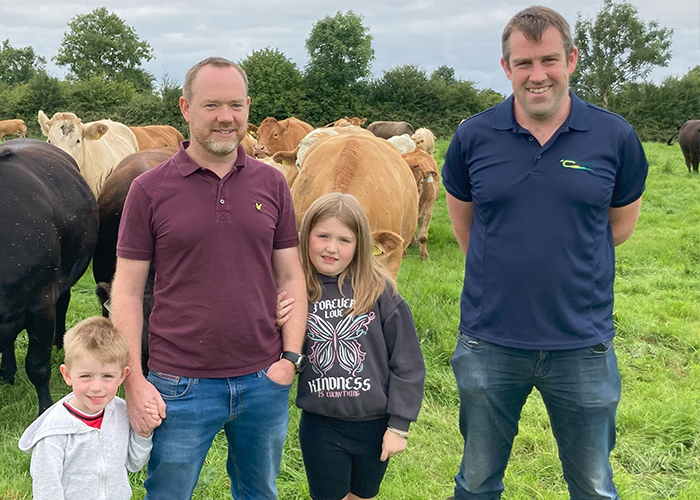
Aidan, Shane and Evelyn O’Connor with Colm Murray, Teagasc advisor
Plans on hold
Initial discussions began regarding the farm transfer from Gerard to Shane, but as the economy improved and construction work became more available, the plans were put on hold. In 2015, with the launch of a new CAP funding round, Shane was added to Gerard’s herd number, forming what is commonly known as a joint herd number or joint venture. This was the minimum requirement for Shane to qualify for the Young Farmers’ Scheme that was available at the time.
While the land transfer was paused due to Shane’s career uncertainties, he remained actively involved in farm operations alongside Gerard. “Alternatively, they could have considered a Registered Farm Partnership and potentially, a Succession Farm Partnership at this stage that would have allowed Shane to qualify for the Young Farmers’ Scheme and it would have also provided additional TAMS funding and significant tax reliefs to the partners,” says Ruth Fennell.
At the outset of the joint venture, suckler cow numbers were in the high 20s and progeny were sold as weanlings. The system evolved over time; with Gerard now less involved in day-to-day tasks, Shane has simplified operations due to his full-time, off-farm job. “We reduced cow numbers and decided to keep all progeny up to the forward store stage,” he says. “The system is working well and allows us to maintain a closed herd.”
Plans are now well advanced to transfer the farm from Gerard to Shane. It is expected that Shane will transfer the herd number into his sole name and that the 2026 BISS application will be submitted under his name rather than through the current joint venture. When assessing the tax implications of this lifetime transfer, several issues must be addressed. As it is a gift rather than an inheritance, both Gerard and Shane must consider taxes and potential reliefs.
From Gerard’s perspective – as the disposer of the asset – the relevant tax is Capital Gains Tax (CGT). This is calculated based on the asset’s value when Gerard received it from his father and its value now. Any increase is subject to CGT at 33%. However, Retirement Relief may apply. As Gerard has owned and farmed the land for over 10 years, is under 70 at the time of transfer and is transferring it to his son, he can claim this relief. Under Retirement Relief, up to €10 million in agricultural assets can be transferred tax-free. This should ensure Gerard avoids CGT on the transfer. If Gerard was over 70 when transferring, the Retirement Relief would be capped at €3 million when disposing to a son/ daughter. For Shane – the beneficiary – two taxes are relevant. The First is Stamp Duty, currently 7.5% of the value of non-residential land. Had this been an inheritance, Stamp Duty would not apply.
Thresholds
Since Shane is over 35, he doesn’t qualify for Young Trained Farmer Relief, which would have reduced the rate of Stamp Duty to 0%. To avail of the Young Trained Farmer 0% relief, the applicant must be under 35 and also hold a recognised agricultural qualification and actively farm the land themselves. However, Shane does qualify for Consanguinity Relief, as he is receiving the farm from a blood relative and will actively farm it. This reduces the Stamp Duty rate to 1%, a significant saving.
The second relevant tax for Shane is Capital Acquisitions Tax (CAT). This applies to the value of the gifted asset. Under a Category A relationship (parent to child), Shane can receive up to €400,000 tax-free. Anything above that is liable for CAT at a rate of 33%. Agricultural Relief may apply, and this reduces the value of the agricultural asset by 90% for CAT. To avail of this, two conditions must be met:
- At least 80% of Shane’s total assets on the date of the gift must be agricultural assets.
- He must hold a relevant qualification and/or spend at least half of his time farming, or alternatively lease the land to someone who does for a minimum of six years.
Where the conditions for Retirement Relief or Agricultural Relief cannot be met, there are alternatives such as Entrepreneurial Relief and Business Asset Relief. These possibilities should be discussed with a financial advisor. In addition to these tax considerations Shane will need to submit an ER1 form to the local District Veterinary Office to transfer the herd number to his sole name as it is currently in joint names with his father. Shane must also complete an ‘Auth’ form to grant his advisor access to the DAFM online system and complete a transfer of entitlements from the joint venture to the new sole herd number.
Careful planning is essential to ensure a successful and tax-efficient transfer of farm assets. Professional legal and financial advice should be sought, and the Succession Planning Advice Grant (SPAG) can help finance this support. “If you get good advice you can negotiate the transition without too much hassle or cost,” says Shane who now looks forward to continuing the O’Connor family farming tradition for many years to come.
This article first appeared in the November/ December 2025 edition of Today’s Farm
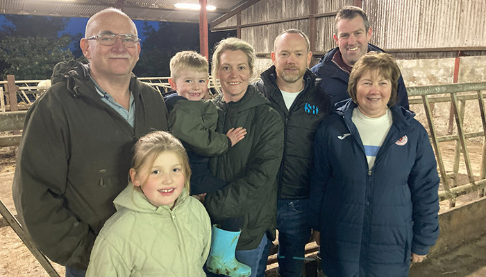
Featured image: Gerard, Evelyn, Aidan, Alisha, Shane, and Memie O’Connor. Included in the photo is their Teagasc advisor, Colm Murray.