Stay informed with free updates
Simply sign up to the EU energy myFT Digest — delivered directly to your inbox.
The boss of one of Europe’s largest grid operators has warned that too many speculative and unprepared projects are holding up grid connections for critical energy projects and causing years-long queues.
Bernard Gustin, chief executive of Elia Group, which operates the Belgian and parts of the German grid, said that operators of network infrastructure should be able to allocate connections to projects that are ready, rather than those that applied first.
‘‘I think in Belgium we have 10 times more projects [than] needed until 2030,” he said, referring to battery storage projects. “If you change from first come, first served to first ready, first served, then you will focus on the ones who are really serious because they have everything [ready].”
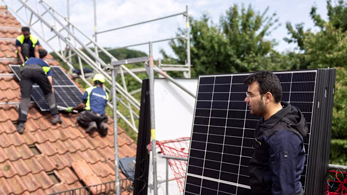
Grid connections have become a huge issue for European countries. Many are trying to manage a rapid increase in demand for grid access as more industrial plants and households install wind and solar power that can go into the grid, as well as an increasing number of applications from data centres to use energy from it.
In some countries, such as the Netherlands, queues to be connected to the grid stretch more than seven years. In Slovakia, about 50 per cent of capacity reserved for connection remains unused, according to commission figures. In Germany, there are twice as many requests to add battery storage to the grid as is planned in the country’s grid development plan, an Elia Group report found.
The rollout of renewables in the EU has outpaced the infrastructure needed to support it, as countries race to meet renewable energy targets set by Brussels and move away from imported fossil fuels. The European Commission has estimated that €1.2tn needs to be spent on the EU’s grids by 2040 in order to support the transition.
Gustin said that grid operators are competing for funding to rapidly build out networks and upgrade infrastructure to balance the volatility of wind and solar energy.
After years of stagnant investment levels, “we all have huge capex plans, so big that you need to be able to finance them, which is a challenge”, he said.
Costs from grid congestion — where cheap electricity cannot flow to where demand is so people have to pay for higher cost sources — are rising. Acer, the EU energy regulator, has said that they reached €5.2bn in 2022 and could rise to €26bn by 2030.
In a document published in December, Brussels set out recommendations to prioritise connections to the grid. The commission also said that it would take a more top-down approach to energy infrastructure planning in order to accelerate the build-out and ensure costs were shared between EU countries.
“In Europe it’s a huge problem and we lose billions every year in lost value because of curtailment and bottlenecks,” EU energy commissioner Dan Jørgensen told the Financial Times.
In a report on energy storage, Elia Group found that the first 100GW of installed batteries in Europe would reduce the curtailment of renewable power by 13 per cent, meaning that 13 per cent more power would be available.

Elia plans to spend €31.6bn on grid upgrades until 2028, split roughly one-third to Belgium and two-thirds to its German arm. To deal with connection demands from batteries, data centres and renewable energy installations could cost an additional €10bn, Gustin estimated.
“These are not small amounts . . . you have a lot of people saying we don’t want tariffs to go up on energy, electricity is not competitive. And so we have a first challenge [which] is how do we make sure, given the amounts we need to raise, that we have a competitive return on equity?”
Gustin, who was formerly chief executive of Brussels Airlines, oversaw a €2.2bn capital raise earlier this year, bringing in investors such as BlackRock and the Canadian pension fund CPP.
Often the length of time it takes to grant permits for infrastructure projects is seen as a risk factor by investors, he said, with permitting times in Belgium running up to eight years.
“By [that] time inflation and the price have increased and some investors are telling you these were not the conditions we had at the start, we cannot continue,” he said.
The EU’s recent legislation aims to speed up permitting times by setting time limits for permit deliberations and proposing that energy projects should be seen as having an overriding interest.