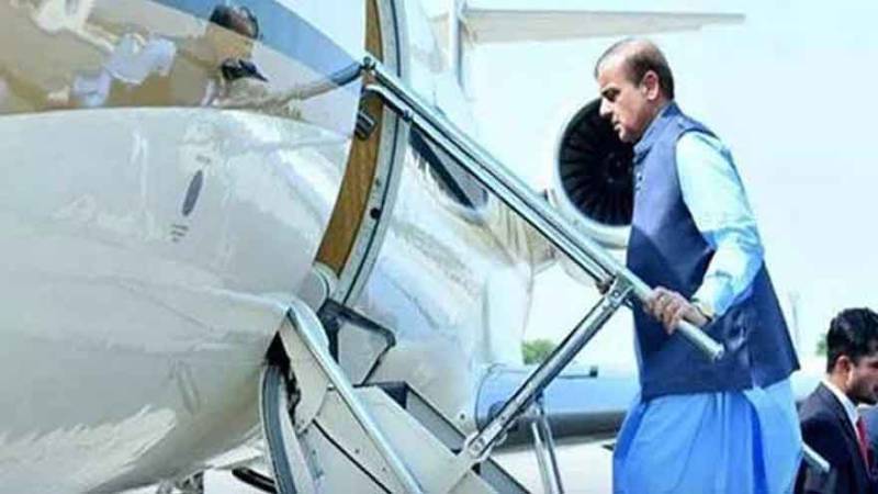
Introduction
Fungal keratitis (FK), also called keratomycosis, is an invasive fungal disease with significant global morbidity, accounting for approximately 50% of infectious keratitis cases.1–4 The prevalence of FK has increased in recent years.5 Delayed treatment of FK exacerbates vision loss and blindness.5,6 Ocular trauma, particularly that caused by vegetative material, is believed to be a specific risk factor for FK.3,7 Contact lens wear, immunocompromised states and being from a rural or indigenous population are other risk factors.3,7 Etiological studies revealed that over 150 fungal genera or species account for FK, whereas the dominant pathogens vary by geographical distribution and climate.1,8 Among these FK causal agents, yeast Candida and filamentous fungi Fusarium and Aspergillus are relatively common globally, while Curvularia, Alternaria, Acremonium, Penicillium, Bipolaris, Mucor, Scedosporium, Cladosporium and other filamentous fungi are uncommon fungal genera or species for FK.6–8 FK pathogenesis involves fungal adhesion, invasion, and immune evasion.3 Different fungal species can cause different FK clinical manifestations owing to various levels of virulence, hyphal growth patterns in the cornea, and host immune status.6,7 Conducting etiological studies on corneal infections is thus necessary. Here, we describe two clinical cases of FK that were infected with two atypical pathogens belonging to dematiaceous fungi. Notably, the pathogen species Achaetomium luteum has never been previously reported in humans, and the pathogen Exserohilum rostratum is also rare.9,10 The diagnosis, etiology, and treatment of these two rare pathogens could improve our knowledge of mycotic keratitis.
Case Report
In case one, a previously healthy 51-year-old married urban male presented with a two-day history of a painful red left eye with foreign body sensation and blurred vision. The patient had a three-day history of outdoor trauma to the left eye caused by a twig from a tree in a city park. The patient had lived locally for a long time and had no family history of similar eye diseases. The medical examination results upon admission were normal, and the patient had no underlying diseases. His left eye had a visual acuity of 2/25 and presented with diffuse corneal edema, central circular-like grayish-white corneal infiltrates, and corneal pseudopodia on ophthalmic examination (Figure 1A). There were 2+ anterior chamber cells with a normal depth and no pus accumulation in the anterior chamber. Corneal scraping was promptly performed on admission and sent for microbiological examination.
Figure 1 For case one, the clinical photograph on admission (A) one SDA plate inoculated with corneal scraping and cultured at 35°C for 48 hours (B) mycelia picked from one SDA plate onto a glass slide and dyed by the lactate phenol cotton blue staining solution (C) and ITS sequence of pathogen Achaetomium luteum sequenced (D).
|
For microscopic examination of corneal scraping, Gram staining for bacterial detection was negative, whereas microscopic examination by fungal fluorescence staining detected filamentous fungal hyphae, which proved the diagnosis of fungal keratitis rather than other forms of microbial keratitis. Corneal scraping was inoculated on Sabouraud Dextrose Agar (SDA) plates and cultured separately at 25°C and 35°C. After 48 hours, yellowish-white fluffy mycelia (filamentous fungus) grew on both SDA plates (Figure 1B). To identify the species, mycelia were picked from one SDA plate onto a glass slide and dyed with the lactated phenol blue staining solution. Microscopic examination revealed non-septate hyphae; however, no typical conidial heads or spores were detected (Figure 1C). We then applied matrix-assisted laser desorption ionization time-of-flight mass spectrometry (MALDI-TOF MS) (Bruker, Germany) with Bruker Filamentous Fungi Library v2.0 to identify its taxonomic species but failed to do so. The failure to identify the filamentous fungus using MALDI-TOF MS also indicated that it was not a typical clinical strain. Finally, targeted DNA sequencing (RuiBio BioTech, China) of the internal transcribed spacer (ITS) sequence was performed, and the fungal strain was identified as Achaetomium luteum with a sequence identity of 99.41% (Figure 1D).
Before admission to our hospital, the patient was treated with levofloxacin eye drops (dosage unknown), fluconazole eye drops (dosage unknown), and oral itraconazole capsule (dosage unknown) for 2 days, but the improvement of the condition was not obvious. The patient was then transferred to our hospital. After admission, the patient was diagnosed with FK by microscopic examination of corneal scraping. For antimicrobial therapy, the patient was empirically treated with fluconazole eye drops q15min, oral itraconazole capsule 200 mg/d, and tobramycin eye drops 6h/d combined with hourly topical voriconazole 2% initially. The condition improved 48 h after admission; that is, the grayish-white corneal infiltrates were smaller and thinner than before. After about four days, the species name of the fungal pathogen A. luteum was reported, which is a novel pathogen for FK and belongs to dematiaceous fungi. A corresponding change was made by replacing fluconazole with natamycin eye drops 2h/d from then on. On the seventh day after admission, the improvement in corneal infiltrate lesions was obvious, and the patient was discharged with medication.
In case two, a 51-year-old male farmer presented with a two-day history of right eye photophobia, tearing, foreign body sensation, and visual loss. The patient had been injured in the right eye by a blade of grass one week prior. The patient had lived locally for a long time and had no family history of similar eye diseases. Medical examination upon admission was normal, except for a 17-year history of hypertension and gout. On admission, visual acuity was hand motion at 20 centimeters in the right eye. The right eye presented with monocular visual loss, mixed congestion, central grayish-white corneal infiltrates, and pus accumulation of approximately 2 mm in the anterior chamber (Figure 2A). Corneal scraping was performed, and the specimen was sent for microbiological examination.
![]() |
Figure 2 For case two, the clinical photograph on admission (A) one SDA plate inoculated with corneal scraping and cultured at 35°C for 48 hours (B) mycelia picked from one SDA plate onto a glass slide and dyed by the lactate phenol cotton blue staining solution (C) and ITS sequence of pathogen Exserohilum rostratum sequenced (D).
|
The laboratory diagnostic process in case two was similar to that in case one. For corneal scraping, Gram staining for bacterial detection was negative, whereas microscopic examination by fungal fluorescence staining and fungal cultivation was positive. The diagnosis of FK was made by the positive microscopic examination. Grayish-white fluffy mycelia (filamentous fungus) were grown on both SDA plates and were cultivated for 48 hours (Figure 2B). Dyed mycelia presented septate hyphae and no typical conidial heads or spores (Figure 2C), making it difficult to identify the fungal species. MALDI-TOF MS (Bruker, Germany) with Bruker Filamentous Fungi Library v2.0 was used, but the taxonomic species was not identified. ITS sequence sequencing (RuiBio BioTech, China) classified the pathogen as Exserohilum rostratum, with a sequence identity of 100.00% (Figure 2D).
Two days before admission to our ward, the patient was treated with tobramycin eye drops (dosage unknown) and levofloxacin gel (dosage unknown) at the outpatient department of another hospital, but the condition did not improve. After admission to our ward, the microscopic examination of corneal scraping supported the diagnosis of FK, and antifungal therapy was developed in a timely manner. The therapy included fluconazole eye drops 2h/d, oral itraconazole capsules 200 mg/d, levofloxacin eye drops 6h/d and tobramycin eye drops once daily. Forty-eight hours after admission, the corneal infiltrate lesions had healed and no pus had accumulated in the anterior chamber. After the species name of the fungal pathogen E. rostratum was reported, the clinical treatment continued as previously. The patient recovered well and was discharged on the sixth day of hospitalization.
Discussion
Invasive fungal disease (IFD) is emerging as a significant disease, given the increasing number of people with severely immunocompromised conditions, such as transplant recipients, HIV and cancer patients, premature neonates and the elderly.5,6 Fungal infection of the human cornea (fungal keratitis) is a unique type of IFD that affects both the immunocompromised and immunocompetent.7 Ocular trauma, particularly agricultural trauma, is a risk factor for FK.3,7 Herein, we report two clinical cases of immunocompetent patients from urban and rural areas. Two patients presented with ocular trauma caused by a twig or blade of grass before admission, which reminded us that protecting ourselves from vegetative trauma is important for avoiding corneal infection.
Culture-proven cases of FK have shown that pathogenic filamentous fungi mainly involve Fusarium spp. (37.6%), Aspergillus spp. (30.4%), and dematiaceous fungi (15.7%).6,7 Dematiaceous fungi are generally distributed worldwide and are often found in natural environments such as soil and vegetation.11 Melanin in the cell walls of dematiaceous fungi affords them unique pathogenic mechanisms, in addition to proteases or toxins degrading the corneal stroma.7,11 Here, we report two atypical dematiaceous fungi for FK, Achaetomium luteum and Exserohilum rostratum, both of which are dematiaceous. The species A. luteum has never been reported to cause human infections, whereas E. rostratum is a rare pathogen. A. luteum, belonging to the family Chaetomiaceae of the class Sordariomycetes of phylum Ascomycota, was first isolated from soils.12 A literature review has shown that A. luteum is not a typical pathogen.12 Notably, FK caused by A. luteum presented with acute infection within 24 h, and the patient nearly lost his monocular vision (visual acuity 2/25; left eye), indicating that the invasiveness and virulence of A. luteum are relatively high and deserve attention. Another pathogenic species, E. rostratum, belongs to the family Pleosporaceae of the class Dothideomycetes of the phylum Ascomycota. E. rostratum is a rare but medically important fungal pathogen that is a major source of new infectious dermatoses, endocarditis, rhinosinusitis, keratitis, and plant diseases.13,14 In contrast to the rapidly progressing infections caused by A. luteum, the pathogen E. rostratum caused relatively slow infectious progress within 5 days. However, the visual acuity of the right eye was lost, and the anterior chamber accumulated approximately 2 mm of pus, which indicated that the infectious progress of E. rostratum was also severe. Regarding different FK clinical manifestations, studies have shown that various virulence and hyphal growth patterns of different fungal pathogens, as well as host immune status, may play roles in infectious progress.6,7
To date, to distinguish FK from other forms of microbial keratitis like viral keratitis or bacterial keratitis is still a challenge.4,8 Meanwhile, novel causal agents of FK are emerging, like novel pathogens A. luteum here, Lasiodiplodia theobromae,15 Myrothecium verrucaria,16 Scedosporium apiospermum17 and Blastoschizomyces capitatus.18 It is another challenge to distinguish divergent fungal pathogens and their clinical grounds.4 Diagnosis made by microscope and culture could face difficulties. Some molecular techniques like MALDI-TOF MS, PCR amplification, ITS sequencing and next-generation sequencing (NGS) gave a better choice.4,5 Early diagnosis, especially species identification by molecular techniques, could improve therapeutic strategies effectively.4,8 Experience accumulations on the pathogenesis of corneal infections, including the diversity of pathogenic species and therapeutic strategies, are important but still have a long way to go.
Here, both patients received timely treatment and recovered well. For case one, caused by A. luteum, earlier antifungal therapy with the combined use of topical fluconazole and oral itraconazole delayed progression of the condition at another institution. However, the combined use of topical voriconazole, natamycin, and oral itraconazole in our ward was effective. For case two, caused by E. rostratum, antifungal therapy using topical fluconazole combined with oral itraconazole was effective, and improvement of the condition was obvious within 48 h. Topical natamycin or voriconazole have traditionally been used as first-line therapy for FK, but their drug toxicity and patient tolerance should be assessed.19 In some cases, voriconazole monotherapy leads to treatment failure, and combination therapy is recommended in some severe cases.19 Furthermore, antibacterial drugs, including tobramycin in case one and levofloxacin combined with tobramycin in case two, were used for preventive therapy against post-traumatic bacterial infection. As damages to the eye caused by the fungal pathogen had happened, combination therapy with antifungal and antibacterial drugs is recommended in FK infections. From these two cases, we conclude that rapid diagnosis of pathogenic species is the cornerstone of targeted anti-infective therapy.
Ultimately, we report two rare fungal species causing opportunistic FK infections in Shandong Province, China. Among them, A. luteum (family Chaetomiaceae) is a novel pathogen for FK, while E. rostratum is a rare, but medically important fungal species with a broad clinical spectrum. Some experiences on the diagnosis and the treatment of rare fungal pathogens were discussed. This study revealed the medical importance of opportunistic fungal pathogens for FK infections. Furthermore, more knowledge of the etiology and treatment therapies for FK is required, and standardized treatment protocols are in need.
Ethics and Consent Statements
This study was conducted according to the Declaration of Helsinki and approved by the Institutional Review Board (IRB) at the Affiliated Yantai Yuhuangding Hospital of Qingdao University. The ethics permit number is 2025-618. The authors certify that they have obtained all appropriate patient consent forms from both patients. All patient data were handled with strict confidentiality and care to ensure privacy.
Consent to Publish
Written informed consent was provided by the patients to have the case details and any accompanying images published. The study participants gave consent to publish.
Acknowledgments
This study was funded by Shandong Medical and Health Science and Technology Development Plan Project (202411000676).
Disclosure
The authors declare that they have no conflicts of interest.
References
1. Mythili A, Shobana CS, Krizsan K, et al. Molecular identification, phylogeny and antifungal susceptibilities of dematiaceous fungi isolated from human keratomycosis. J Infect Public Health. 2023;16(1):25–33. doi:10.1016/j.jiph.2022.11.018
2. Fang W, Wu J, Cheng M, et al. Diagnosis of invasive fungal infections: challenges and recent developments. J Biomed Sci. 2023;30(1):42. doi:10.1186/s12929-023-00926-2
3. Whitcher JP, Srinivasan M, Upadhyay MP. Corneal blindness: a global perspective. Bull World Health Organ. 2001;79(3):214–221.
4. Hoffman JJ, Burton MJ, Leck A. Mycotic Keratitis-A global threat from the filamentous fungi. J Fungi. 2021;7(4). doi:10.3390/jof7040273
5. Giannella M, Lanternier F, Delliere S, et al. Invasive fungal disease in the immunocompromised host: changing epidemiology, new antifungal therapies, and management challenges. Clin Microbiol Infect. 2025;31(1):29–36. doi:10.1016/j.cmi.2024.08.006
6. Vallabhaneni S, Mody RK, Walker T, Chiller T. The global burden of fungal diseases. Infect Dis Clin North Am. 2016;30(1):1–11. doi:10.1016/j.idc.2015.10.004
7. Garg P, Gopinathan U, Choudhary K, Rao GN. Keratomycosis: clinical and microbiologic experience with dematiaceous fungi. Ophthalmology. 2000;107(3):574–580. doi:10.1016/s0161-6420(99)00079-2
8. Gautam M, Lal B, Patel S, et al. An emerging global threat of mycotic keratitis caused by uncommon fungal species: a systematic review and Meta-Analysis. Transl Vis Sci Technol. 2025;14(4):4. doi:10.1167/tvst.14.4.4
9. Chaidaroon W, Phaocharoen N, Srisomboon T, Vanittanakom N. Exserohilum rostratum keratitis in a patient with human immunodeficiency virus. Case Rep Ophthalmol. 2019;10(1):127–133. doi:10.1159/000499688
10. Jayasudha R, Sharma S, Kalra P, Mishra DK. Exserohilum keratitis: clinical profile of nine patients and comparison of morphology versus ITS-Based DNA sequencing for species identification of the fungal isolates. Indian J Med Microbiol. 2018;36(4):564–568. doi:10.4103/ijmm.IJMM_19_51
11. Revankar SG. Dematiaceous fungi. Mycoses. 2007;50(2):91–101. doi:10.1111/j.1439-0507.2006.01331.x
12. Wang XW, Han PJ, Bai FY, et al. Taxonomy, phylogeny and identification of Chaetomiaceae with emphasis on thermophilic species. Stud Mycol. 2022;101:121–243. doi:10.3114/sim.2022.101.03
13. Sharma K, Goss EM, Dickstein ER, et al. Exserohilum rostratum: characterization of a cross-kingdom pathogen of plants and humans. PLoS One. 2014;9(10):e108691. doi:10.1371/journal.pone.0108691
14. Tang Z, Lou J, Mo J, et al. First worldwide report of exserohilum rostratum causing leaf spot on strawberry (Fragaria x ananassa Duch.). Plant Dis. 2023. doi:10.1094/PDIS-07-22-1724-PDN
15. Lazrak A, Desbois N, Bonnafous M, Ouamrane K, Merle H. Favorable outcome of lasiodiplodia theobromae keratomycosis: a clinical case and systematic review. BMC Ophthalmol. 2024;24(1):332. doi:10.1186/s12886-024-03599-z
16. Liu H, Lei X, Chen L, Hu S, Li G, Deng Z. Keratomycosis caused by a rare pathogen, myrothecium verrucaria. Mycopathologia. 2021;186(6):893–895. doi:10.1007/s11046-021-00587-5
17. Lenk J, Raiskup F, Pillunat LE, Rossler S. Scedosporium apiospermum-a rare pathogen of keratomycosis. Ophthalmologe. 2020;117(12):1225–1228. doi:10.1007/s00347-020-01073-x
18. Sahu SK, Dora J, Hota G. Keratomycosis caused by blastoschizomyces capitatus. Indian J Pathol Microbiol. 2016;59(1):117–118. doi:10.4103/0377-4929.178232
19. Arora R, Gupta D, Goyal J, Kaur R. Voriconazole versus natamycin as primary treatment in fungal corneal ulcers. Clin Exp Ophthalmol. 2011;39(5):434–440. doi:10.1111/j.1442-9071.2010.02473.x