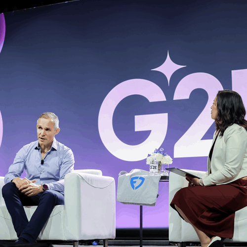
Inside the C-Suite: Gaming’s Future in Focus
At G2E 2025, Peter Jackson, CEO of Flutter Entertainment, joined journalist Hope King for an engaging, in-depth fireside chat, delving into Flutter’s global growth, shifting consumer behaviors, the future of sports betting, and why responsible gaming is more than just a talking point for the industry’s biggest player.
Hope: Since becoming CEO in 2018, you’ve led at least eight major deals. Why is scale so important to Flutter’s strategy?
Peter: We’re focused on being #1 in the U.S. and having “gold medal” positions globally. Like other digital industries, we believe scale is very important, but diversification matters too. What’s key for us is being nimble. We operate on a federal basis, so leaders have full control of their businesses but can draw on Flutter’s global capabilities when needed.
Hope: Flutter is the #1 gaming operator in the world and you’re expanding fast, in places like Italy, Brazil, and Southern Europe & Africa. How much more of the world is left for Flutter to conquer?
Peter: We’re delighted with our position in the U.S. as the #1 sports betting and gaming business. But it’s important to remember that the international market is even bigger. By 2030, we estimate the global addressable market will be nearly $300 billion. That’s why we’re very focused on maintaining our leading positions in markets like the UK, Italy, and Australia. Our international experience actually strengthens our U.S. business – through the Flutter Edge we share technology, expertise, and best practices across regions.
Hope: From your global vantage point, what are you learning in more mature markets like Italy or Australia? And how does that inform your approach in emerging ones?
Peter: We’re always learning from new markets, especially when we acquire businesses. Often, we find product ideas or technologies we can bring elsewhere. Responsible gaming is one area where we’re determined to lead. For example, our Real-Time Check-In tool (RTCI) in the U.S. is based on a solution we developed in Australia. It’s about creating a race to the top when it comes to consumer protection.

Hope: You’re now listed on the New York Stock Exchange. How has that helped?
Peter: We’ve definitely seen a step up in liquidity since the U.S. listing, which is great for our investors. I’m meeting many of them over the course of today and tomorrow here at G2E. It’s a very efficient way of getting to see a lot of shareholders.
Hope: Let’s talk about FanDuel. You’ve raised your outlook and that’s despite what we’re hearing about consumers feeling less rosy about the economy. Where does sports betting or online gaming fall in the spectrum of consumer spending?
Peter: Our experience from operating around the world is that sports betting and online gaming is actually very defensive. People might trade down and watch games at home instead of going to bars, but they still want to get their parlay on and have that excitement of seeing whether they can guess what’s going to happen in the game.
Ultimately, we believe that our core business is very defensive. We’ve got good tailwinds from a growth perspective and we’re still in the early days of growth in the industry.
Hope: One big future-facing move is your partnership with CME Group that you announced this summer. Can you explain the origin and intent behind the deal, especially given that sports events aren’t currently included? Why did you pick CME Group? Why did they pick you?
Peter: We are reasonably familiar with this space. We operate the Betfair Exchange – our peer-to-peer platform – in many markets around the world. It has a lot of similar characteristics to the way in which we’re seeing prediction markets unfold here in the States. So we were obviously very thoughtful in terms of where the regulations are going, and when I met with Terry and the team, it was a very like-minded conversation.
We’re very excited about the opportunities that we’ve got in front of us, for a range of products that we can bring to the market here in the US, leveraging our expertise and the capabilities of CME as well.
Hope: CME has described the partnership as a ‘distribution play’ and an ‘expansion of their retail trading strategy’ -what do you make of this?
Peter: CME Group has been very clear about what they’re trying to achieve with this partnership. Whether it’s trading financial products or leisure or entertainment experiences, some of which might be closer to home for us, we see strong possibilities to bring our capabilities here to the U.S. market, and that’s something which we will make available for consumers soon. For CME, they do want to get closer to the retail consumers, and we’ve got a terrific brand and lots of customers in the market. So, I think it’s a very exciting opportunity for us both.
Hope: There’s been a flurry of activity in this space with ICE most recently investing $2bn into Polymarket. What’s your reaction?
Peter: There’s a lot of news flow in this space and I’m pretty sanguine about it. When I look at the sort of outcomes that are possible, I think we’re very well placed irrespective of which path we end up following. That’s thanks to our capabilities, partnerships, and brand reach here in the U.S. I’m excited about it, and I can see why others are wanting to get into the market.
Hope: When you saw that Kalshi introduced a builder combo feature which is very much like same game parlay, did you view that as a loophole to sports betting?
Peter: Building Same Game Parlay products isn’t easy. Competitors are spending hundreds of millions trying to catch up with us, and we’re powering ahead, we’re not standing still. There’s complex pricing involved – positive and negative correlations, etc. I think it’s very difficult to deploy that through a prediction market. We’ll get some clarity on the regulators standpoint, but the combo feature looks suspiciously like sports betting to me.
Hope: With the +20 years’ experience you have with the Betfair Exchange, how do you think that will help your strategy going forward with more peer-to-peer products?
Peter: The Betfair Exchange definitely appeals to a subset of consumers, but it’s worth recognizing that there are some challenges with it. You can’t offer generosity through the Betfair Exchange – it’s very difficult to technically do that on these platforms because you’re sat in the middle and you’re not the principal, and it’s the principal that needs to effectively offer generosity. Consumers love generosity. They’re in it for the entertainment and the excitement, so it’s worth recognizing that.
It’s also very difficult to find liquidity in all the different permutations and combinations that you see in the product. If I look at the work we’re doing with Your Way here in the U.S. with FanDuel, we’re getting to the point where we have effectively limitless assortments of products available for consumers to choose from.
And we believe it’s very difficult to come anywhere close to that.
Hope: How are you viewing the convergence of this new category overlap? What does this say about the way people view money?
Peter: There’s a clue in how we describe ourselves, right? We call ourselves Flutter Entertainment, and ultimately that’s what we bring to life for our consumers around the world.
But that’s our lens on it, that’s how we’re thinking about it and how we go to market. Whether that’s with Paddy Power, which is iconic for the irreverent humor it brings to its advertising. The same is true with Sportsbet in Australia. We have different brand positionings but ultimately it’s about bringing entertainment to life and I think that’s what we strive to do.
Hope: Do you see this type of product bringing a fundamentally different kind of consumer into a category that you haven’t seen yet?
Peter: For those of us who delve into Reddit and other similar platforms, you can see what our consumers are saying and thinking. People are trying to figure out how they can get an edge and how they can get ahead and that’s part of the game for them. And whether you apply that to sports or some of the other leisure style products or the financial markets, you can see that there’s a very strong overlap between these things.
Hope: Talking more about how you see growth evolving. A big part of this is from media and media partnerships. With your expanded deal with Amazon Prime Video and native broadcast integration, is this a more cost-effective customer acquisition strategy?
Peter: We’re very excited about the partnership with Amazon. As you say, the integration of the odds, whether that’s on a personalized basis so we can actually show the bets that you’ve got live on the stream that you’re watching, or the ability to overlay in real time what the betting markets are saying about what’s going to happen with the player that you’re seeing on the screen, it’s very compelling in terms of adding that excitement to the action. So we’re pleased.
Hope: Taxes have become a hot topic. You’re passing through some costs in places like Illinois but there’s also the upcoming change with the limiting of losses happening in January. Do you expect that to have a big impact on Flutter and on the industry?
Peter: If I look at our businesses around the world, it’s our scale that helps us in these situations. We have a bigger business that we can defray our costs across, and it allows us to mitigate the potential impact of tax changes.
We were very disappointed with the Illinois tax. I think it disproportionately impacts the very low spending, recreational consumer base and I think it’s wrong. Nor do I think it’s a particularly smart way of raising revenues. It’s true in lots of other industries you’ll follow, but as a scale player, you can weather this stuff better. We’ll see how other markets evolve, but I think we’re well placed.
Hope: At G2E last year you referenced ‘societal license’ as a risk to your business or something that you worry about with the growth of Flutter. With new surveys suggesting the ‘quiet middle’ might be shrinking, how are you thinking about innovation and operating at a time when there is much more attention on this area of entertainment and gaming innovation?
Peter: It’s really important that we showcase all of the good work that we’re doing across our business to ensure we provide a safe and secure environment for our consumers so they can have fun. This includes the $100m+ that we’re spending on responsible gaming tools, like Real-Time Check-In we have here in the U.S.
Show me a social media business that’s stopping their consumers using their products because they’re worried they’re using it too much. It doesn’t happen. But that’s what we’re doing and we’re proud of it.
You can watch Peter’s full interview here.