Surgeons and doctors often rely on artificial models to practice delicate procedures. Yet most training tissues feel stiff, simple, and far from realistic.
That gap between training models and real organs has limited how well medical professionals can prepare before entering the operating room. Now, researchers at the University of Minnesota Twin Cities have developed a new 3D printing technique that creates lifelike human tissue structures.
Their work could reshape surgical training by offering models that look, feel, and respond more like real human tissue.
Previous 3D-printed tissues lacked the complexity of natural organs. The Minnesota team found a way to control the shape and size of microscopic patterns inside the printed material.
Those patterns directly influence the strength and stretchiness of the tissues, giving them realistic mechanical properties.
They also built a mathematical formula to predict how the tissues behave under stress.
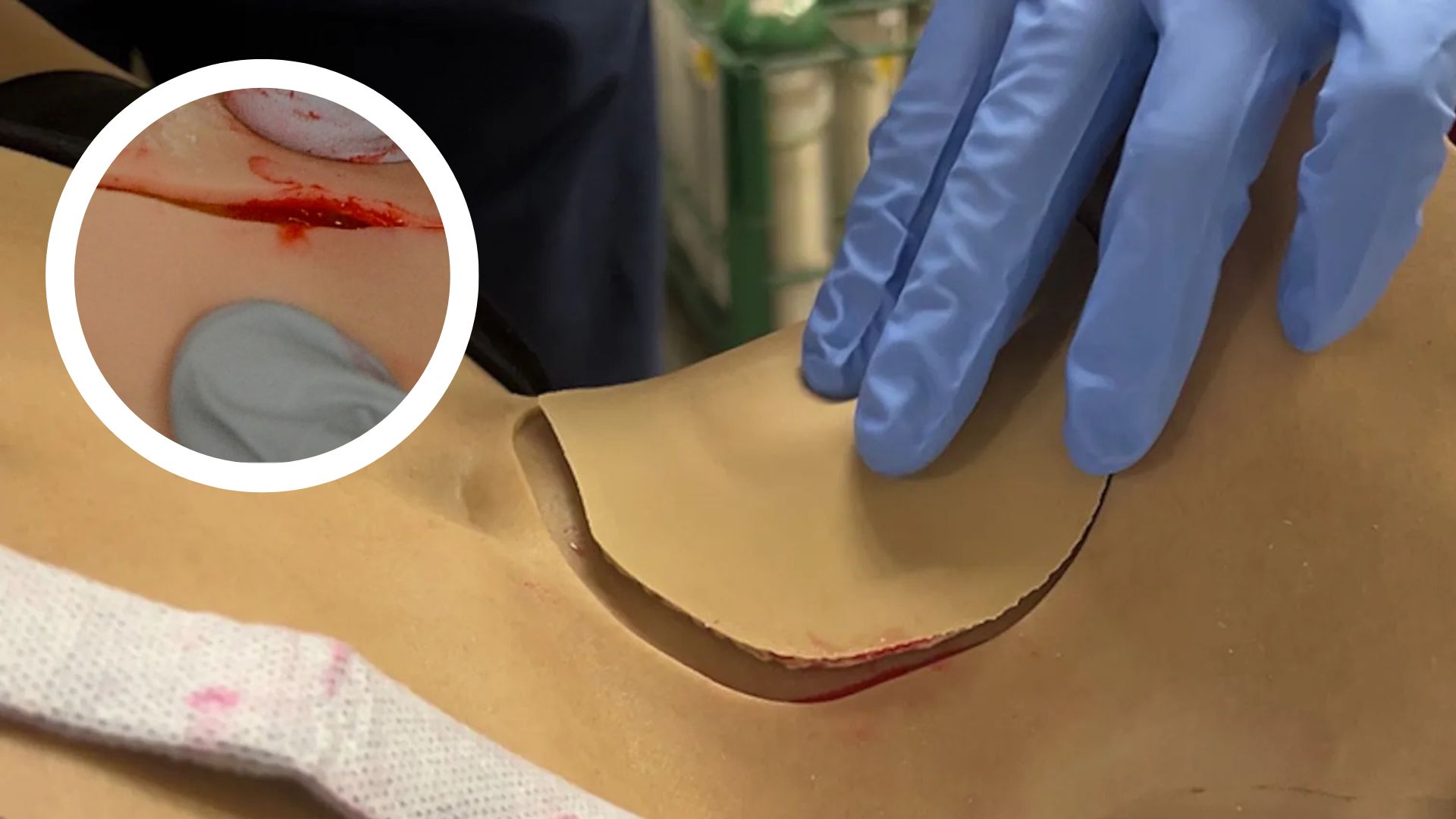
The team added blood-like liquids to make the models even more convincing during the printing process. Microcapsules seal the fluid so it wouldn’t evaporate or interfere with printing.
Adarsh Somayaji, the paper’s first author and a Ph.D. graduate from the Department of Mechanical Engineering, said the new method “opens the door to creating more realistic training models for surgery, which could ultimately improve medical outcomes.”
He explained that scaling up the process will take time, but stressed that the technique holds strong potential for “low-volume, high-complexity training scenarios.”
Surgeons rate models higher
The study also tested the models with surgeons. The new tissues scored higher on tactile feedback and cutting response than conventional replicas.
For doctors in training, that difference could mean more accurate practice and smoother transitions to working with real patients.
Researchers believe that by closing the gap between practice models and living tissue, the new method could improve safety and effectiveness in surgery.
Next steps in research
The team plans to expand their work to mimic different organ shapes and functions.
They are also exploring ways to develop bionic organs and add materials that respond to advanced surgical tools such as electrocautery, which uses heat to remove small growths.
Alongside Somayaji, the study involved Matthew Lawler from Biomedical Engineering, Zachary Fuenning, and Michael McAlpine from Mechanical Engineering.
The research also drew on collaborations with the CREST Lab and Wang Lab at the University of Washington.
Funding came from the U.S. Department of Defense, supported by the University of Minnesota’s MnDRIVE Initiative on Robotics, Sensors, and Advanced Manufacturing (RSAM) and the Minnesota Nano Center.
The researchers see their method as a key step toward surgical models that truly replicate the human body.
If successful, their approach could raise the standard of medical training and, ultimately, patient care.
The study is published in the journal Science Advances.