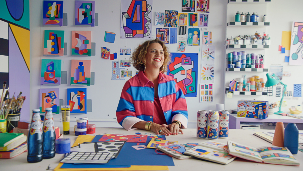
Ae. albopictus GT strain showed similar microbial diversity but different composition to GUA strain
Following size filtering, quality control, and chimera removal, a total of 1,575,628 high-quality sequences were obtained from 30 samples from two Ae. albopictus strains. Rarefaction analysis showed that the number of sequences increased sharply before reaching a plateau (Fig. 1a), indicating comprehensive coverage of bacterial diversity in these samples. Good’s coverage was 99.9% in all the sequencing samples (Fig. 1b), indicating sufficient sequencing depth. To evaluate alterations in microbial diversity between the GUA and GT strains, microbial alpha diversity was shown using Shannon and Chao diversity indices (Fig. 1c, d), with no significant differences observed (P = 0.2924 and P = 0.9153, respectively). In addition, 3653 OTUs were identified, including 45 phyla, 1048 genera, and 1953 species, which were annotated for subsequent analyses.
Microbial diversity and composition in GT and GUA strains of Aedes albopictus. a Rarefaction analysis of bacterial communities. b The good’s coverage of bacterial communities. c The boxplots of Shannon index; d The Chao index. The Shannon index and the Chao index were constructed to evaluate microbial diversity. e Venn diagram analysis of microbial communities at the OTU level. f CIRCOS plot showing the distribution of phylum between the GT and GUA strain mosquitoes. g Community bar chart at the genus level. h PCoA score plot of the bacteria in the GUA and GT strains on OTU level. i PLS-DA score plot of the bacteria in the GUA and GT strains on OTU level
Venn diagram analysis based on the OTU abundance was performed to identify the common and unique OTUs between the GUA and GT strains (Fig. 1e). A total of 1672 OTUs shared by both strains, referred to as core microorganisms, were predominantly composed of Proteobacteria (64.27%), Firmicutes (16.09%), Actinobacteriota (11.22%) and Bacteroidota (4.96%) (Fig. 1f), accounting for over 96.00% of the total microbial in the two strains. The unique OTU number of the GUA strain was lower than that of the GT strain (Fig. 1e). The relative abundance for the top ten genera varied in the two strains. For example, Asaia was the most dominant genus in both strains (GUA: 47.33%, GT: 32.69%), though no significant difference observed (P = 0.1466) (Fig. 1g, Suppl Fig. 1a). Similarity, no significant difference was observed on Rhodococcus (P = 0.1150) (Suppl Fig. 1b), unclassified_f__Acetobacteraceae (P = 0.1710) (Suppl Fig. 1c), Elizabethkingia (P = 0.1250) (Suppl Fig. 1d), Ralstonia (P = 0.1150) (Suppl Fig. 1e), Burkholderia-Caballeronia-Paraburkholderia (P = 0.0740) (Suppl Fig. 1f) and Pseudomonas (P = 0.4800) (Suppl Fig. 1 g) in the two strains (Fig. 1g). However, significant difference was observed on Pseudonocardia (P = 0.0101) (Suppl Fig. 1 h), Enterococcus (P = 0.0062) (Suppl Fig. 1i) and Wolbachia (P = 0.0000) (Suppl Fig. 1j). Wolbachia was only observed in the GUA strain, while Enterococcus showed a higher relative abundance in GT strain compared to GUA strain (22.85 vs 7.75%) (Fig. 1g).
To further explore the microbial compositions between GUA and GT strains, we used PCoA (based on Weighted-Unifrac distances) at the OTU level. This analysis considered species relationships, composition abundance and evolutionary relationships, revealing clear segregation between the microbiota of the two strains (Fig. 1h, P = 0.0010). Additionally, Partial Least Squares Discriminant Analysis (PLS-DA) and the PERMANOVA analysis (Table 1) were conducted, showing distinct separation between the two strains (Fig. 1i). The two results from the beta-diversity analysis suggested that GUA and GT strains maintained different bacterial composition, and the differences might be due to the variation in the relative abundance of the major genera that were observed. (Fig. 1g). Our results suggested that the Ae. albopictus GT strain showed similar microbial diversity but different composition to GUA strain. Additionally, we conducted KEGG pathway prediction analysis on samples from the two strains and found no significant differences in their major metabolic pathways (Suppl Fig. 2).
GT females showed similar microbial diversity and composition to GUA females
To understand the microbial diversity and composition among the samples, the 30 samples were divided into six groups: GUA_2M, GUA_5M, GUA_5F, GT_2M, GT_5M and GT_5F. Regarding to female mosquitoes, no significant difference was observed on the microbial diversity between the GT and GUA strains based on Shannon (P = 0.5969) and Chao (P = 0.4521) indices (Fig. 2a, b). Additionally, similar bacterial composition was also observed in females of these two strains based on the PCoA analysis (Fig. 2c, P = 0.3460). The PERMANOVA analysis also indicated no significant differences between the two groups (Table 1).

Microbial diversity and composition in GT and GUA female Aedes albopictus at age of 5-day old. a The boxplots of Shannon index. b The Chao index. The Shannon index and the Chao index were constructed to evaluate microbial diversity. c PCoA score plot of the bacteria on OTU level. d The LEfSe diagram of bacteria in GT and GUA strains. Red indicates significant enrichment in GUA, blue indicates significant enrichment in GT, and both indicate microbial taxa that have a significant impact on inter-group differences. Light yellow nodes indicate no significant difference or no significant impact on inter-group differences for microbial taxa. e Indicator bacteria with LDA scores of 4 or greater in bacterial communities in GT and GUA females. LDA score on the x-axis represents score changes in GT females compared to GUA females at the bottom left of the figure, showing the varying impacts of identified indicator species on the differences between GT and GUA females
Linear discriminant analysis effect size (LEfSe) method was further employed to identify the species/genus-specific difference in females. The GT females were characterized by a preponderance of Ralstonia (Burkholderiales), Paracoccus (Rhodobacterales), and Pelomonas (Sphingobacteriales) (Fig. 2d) while Wolbachia, Anaplasmataceae, Pseudonocardia (Rickettsiales), and Massilia (Pseudonocardiales) were more consistently present in the GUA females (Fig. 2e). Specifically, sequence analysis showed that the top five genera in the GUA females were Asaia (41.72%), Enterococcus (20.78%), Wolbachia (8.27%), Rhodococcus (2.44%) and Ralstonia (1.42%) (Suppl Fig. 3a) while the four predominant genera in the GT females were Asaia (55.08%), Enterococcus (19.22%), Rhodococcus (6.76%) and Ralstonia (3.63%) (Suppl Fig. 3b). Among of these genera bacteria, the presence of Wolbachia was the most differences between GT and GUA females.
GT males showed similar microbial diversity but different composition to GUA males
In terms of male mosquitoes, no significant difference was observed on the microbial diversity based on Shannon (Fig. 3a, P = 0.1197) and Chao indices (Fig. 3b, P = 0.8302) between the GT and GUA strains. However, significant difference was observed on the composition based on the PLS-DA (Fig. 3c) and the PERMANOVA analysis (Table 1).

Microbial diversity and composition in GT and GUA male Aedes albopictus at age of 2-day and 5-day old. a The boxplots of Shannon index. b The Chao index. The Shannon index and the Chao index were constructed to evaluate microbiome diversity. c PLS-DA score plot of the bacteria in the GUA and GT strains on OTU level. d The LEfSe diagram of bacteria in GT and GUA strains. Red indicates significant enrichment in GUA, blue indicates significant enrichment in GT, and both indicate microbial taxa that have a significant impact on inter-group differences. Light yellow nodes indicate no significant difference or no significant impact on inter-group differences for microbial taxa. e Indicator bacteria with LDA scores of 4 or greater in bacterial communities in GT and GUA males. LDA score on the x-axis represents score changes in GT males compared to GUA males at the bottom left of the figure, showing the varying impacts of identified indicator species on the differences between GT and GUA males. f Kruskal-Wallis H test bar plot on genus level of GT and GUA males at 2 and 5 days old. g PCoA score plot of the bacteria in the four male groups on OTU level. h Kruskal-Wallis H test of Wolbachia, Asaia, Enterococcus and Elizabethkingia in GT and GUA males at 2 and 5 days old
LEfSe was employed to identify the species/genus-specific difference in males. The GT males were characterized by a preponderance of Bacilli, Enterococcus (Firmicutes), Enterococcaceae and Lactobacillales (Fig. 3d, e), while Alphaproteobacteria, Asaia (Proteobacteria), Acetobacteraceae (Acetobacterales) and Wolbachia (Anaplasmataceae) were more consistently present in the GUA males (Fig. 3d, e). Specifically, sequence analysis showed that the abundance of the top four genera in GUA males were Asaia, Wolbachia, Rhodococcus and Raistonia, with Wolbachia being the second most abundant and Enterococcus nearly absent (Suppl Fig. 3c, d). In GT males, Asaia, Enterococcus, Rhodococcus, Elizabethkingia and Ralstonia were prominent, whereas Enterococcus being the second most abundant genus (Suppl Fig. 3e, f). Among of these genera bacteria, the presence of Wolbachia (P = 0.0010) or Enterococcus (P = 0.0063) showed significant statistical differences between GT and GUA males (Fig. 3f).
Age significantly affected microbial composition in male mosquitoes
This study further revealed age-related differences in bacterial composition among the four male groups. The PCoA results have shown significant differences among the four groups (P = 0.0010) (Fig. 3g). In the GUA strain, the proportion of Asaia decreased slightly with age, while the proportion of Wolbachia had a substantial increase (Suppl Fig. 3c, d). Meanwhile, in the GT strain, the proportion of Asaia and Enterococcus had a considerable increase with age (Suppl Fig. 3e, f). In 2-day male group, the distribution of Asaia was highest in the GUA strain, whereas the bacterial composition in the GT strain remained relatively well-distributed among the top five genera (Suppl Fig. 3c, e). Additionally, the proportion of Asaia was consistently higher in the GUA strain compared to the GT strain (Suppl Fig. 3c–f). Additionally, significant difference in bacteria like Wolbachia and Enterococcus were observed between the 2-day male groups and 5-day male groups (P < 0.05) (Fig. 3f). Compared to the 5-day male group, the 2-day male group had a significantly higher prevalence of bacterial genera, such as Sphingomonas (P = 0.0203), Streptomyces (P = 0.0097), Nocardioides (P = 0.0288), and Qipengyuania (P = 0.0388) (Fig. 3f).
We found significant differences in the abundance of Wolbachia (2-day male groups: P = 0.0060; 5-day male groups: P = 0.0249) and Asaia (P = 0.0151) bacteria between GT and GUA strains, with Wolbachia being eliminated and its abundance increasing with age in the GUA males, while the abundance of Asaia decreased in GT males (Fig. 3h). However, Enterococcus (2-day male groups: P = 0.1095; 5-day male groups: P = 0.06123) and Elizabethkingia (2-day male groups: P = 0.3136; 5-day male groups: P = 0.3738) showed no significant differences between GT and GUA males (Fig. 3h), but Enterococcus was identified as a biomarker in the LEfSe results (Fig. 3d, e).
The relative abundance of Asaia, Wolbachia, Enterocossus and Elizabethinga
qPCR was further used to measure the relative density of 4 major bacterial genera. For Asaia, higher density was observed in GUA_2M group as compared to GT_2M group (Two-tailed Mann-Whitney U-test, P = 0.0079), while there was no difference in both 5-day male groups (GUA_5M vs GT_5M, P = 0.0556) and 5-day female groups (GUA_5F vs GT_5F, P = 0.0556) (Fig. 4a). GT males showed higher abundance of Enterococcus than GUA males, either at age of 2 days (P = 0.0079) or 5 days (P = 0.0476). However, this difference was not observed in 5-day female groups (P = 0.2381) (Fig. 4b). No significant difference was observed on Elizabethkinga between GUA and GT mosquitoes (P = 0.1508) (Fig. 4c). All GUA mosquito groups (GUA_2M, GUA_5M and GUA_5F) were positive with Wolbachia while the bacterium was not amplified in GT mosquitoes, confirming that Wolbachia was completely eliminated in the GT strain.

Relative abundance of Asaia, Wolbachia, Enterococcus, and Elizabethkingia in GUA and GT strains of Aedes albopictus mosquitoes. a Relative abundance of four genera in the 2-day-old male group. b Relative abundance of four genera in the 5-day-old male group. c Relative abundance of four genera in the 5-day-old female group
Wolbachia wAlbB can serve as a biomarker to distinguish the sterile from wild male Ae. albopictus
Three vegetation-rich sites in downtown Guangzhou, China, were selected for human-baited experiments to collect adult male Ae. albopictus mosquitoes (Fig. 5a). These sites included shrubbery near a parking lot, shrubbery near a basketball court, and shrubbery near a monument. The experiments were carried out monthly on the 15th day of each month, from July 2023 to November 2023, at all three sites. At each site, 30 male mosquitoes were collected, and 12 were selected for DNA extraction and PCR identification experiments. Male mosquitoes infected with both Wolbachia strains (wAlbA & wAlbB) were exclusively found, but in different proportions, in both the lab-GUA and the wild strains (Fig. 5b, c). The lab-GUA strain were 100% (12/12) double-infections with wAlbA and wAlbB. In terms of the wild strain, the wAlbB exhibited a mean of 96.7% (± 4.6%) (58/60) infection proportion and 61.7% (± 32.1%) (37/60) for wAlbA, though the difference was not statistically significant (Wilcoxon matched-pairs signed rank test, P = 0.1250) due to high variation observed in wAlbA. Conversely, both the lab-GT (0/12) and the mass-reared GT (0/60) strains were negative with Wolbachia in both wAlbA and wAlbB. All four tested strains were positive for Enterococcus (Fig. 5d). No significant difference was observed on the infection proportion of Enterococcus between the mass-reared and the wild strains (Wilcoxon matched-pairs signed rank test, P = 0.0625).

Proportion of Wolbachia and Enterococcus in male Aedes albopictus. a Spatial distribution of the monitoring method in Yuexiu District, Guangzhou City. Yellow points represent the positions to perform human-baited collection. N represents the North. b Proportion of wAlbA infection. c Proportion of wAlbB infection. d Proportion of Enterococcus infection. Within a, b, c, from left to right showed the laboratory strain GUA (lab-GUA), the laboratory strain GT (lab-GT), the wild strain collected from July to November 2024, and the mass-reared GT strain collected from July to November 2024
A total number of 90 irradiated GT males were recaptured with obvious fluorescent powder observed in these males, corresponding to a 4.5% (90/2000) recaptured rate within 4 days (Fig. 6a, b). The number of daily captured male mosquitoes with or without marking was shown in Fig. 6b. PCR results showed that all the marked male mosquitoes were negative with both wAlbA and wAlbB, either collected at 48 h (0/12) or 96 h post release (0/4). Similarity, all the irradiated GT males were negative with both wAlbA and wAlbB (0/24). Whereas, infection rates of 95.8% (23/24) for wAlbB and 54.2% (13/24) for wAlbA were respectively observed on the unmarked male mosquitoes, which were considered as wild males (Fig. 6c). Our results suggest that Wolbachia wAlbB is more stable than wAlbA recommended to be a suitable biomarker for distinguishing the sterile from wild male mosquitoes.

Validation of wAlbB as a reliable biological marker in the mark-release-recapture experiment. a Differentiation of male Aedes albopictus mosquitoes under a stereomicroscope to determine their origin (mass-reared or wild). From left to right: unmarked irradiated male, fluorescent marked irradiated male before release, wild male captured at 48 h, fluorescent marked irradiated male recaptured at 48 h post release, wild male captured at 96 h, and fluorescent marked irradiated male recaptured at 96 h post release. Fluorescent powder were indicated by the red arrows. b Numbers of marked, released, and recaptured male Aedes albopictus, along with the recapture rate of sterile males. c Proportion of wAlbA (blue) and wAlbB (orange) infections among the tested males