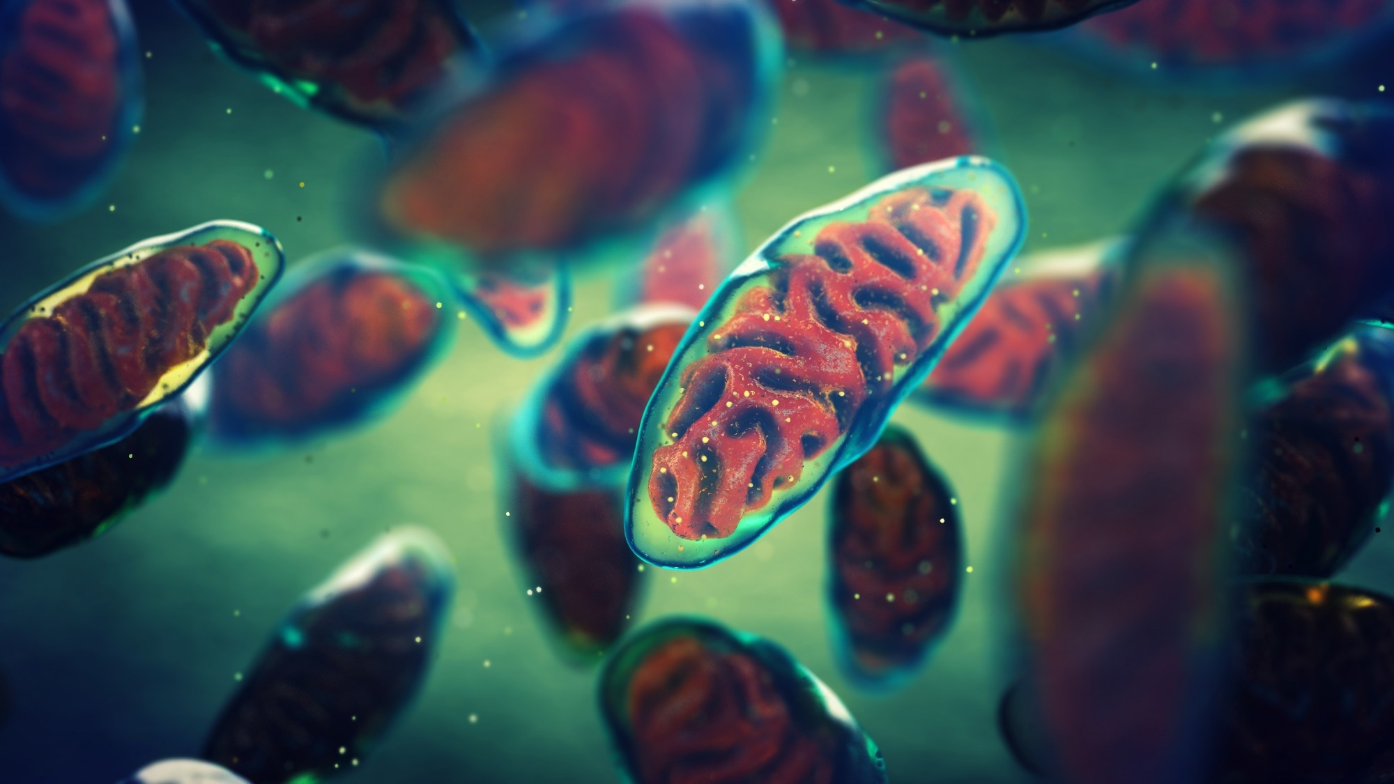
A new study reveals that vesicles packed with healthy mitochondria can supercharge tissue repair and combat chronic disease, paving the way for next-generation regenerative treatments.
Study: Harnessing tissue-derived mitochondria-rich extracellular vesicles (Ti-mitoEVs) to boost mitochondrial biogenesis for regenerative medicine. Image Credit: nobeastsofierce / Shutterstock
In a recent study in the journal Science Advances, researchers report a novel mechanism wherein mitochondria-rich extracellular vesicles (named “Ti-mitoEVs”) transfer their functional mitochondrial cargo to damaged cells, potentially reversing injury and disease in energy-demanding tissue. The study also demonstrated that high-intensity interval training (HIIT) in mice increases Ti-mitoEV secretion, indicating a physiological role for these vesicles in tissue homeostasis and repair.
Study findings revealed that murine skeletal muscle-derived Ti-mitoEVs significantly reduced inflammation and promoted tissue repair in acute muscle injury and chronic kidney disease models. Mechanistic investigations suggest these benefits are due to boosted mitochondrial biogenesis, establishing a promising and novel therapeutic platform for regenerative medicine. While non-mitoEVs (vesicles with lower mitochondrial content) also showed some benefit in reducing inflammation, Ti-mitoEVs were markedly more effective in promoting tissue repair and restoring mitochondrial function.
Background
Mitochondria (mt) are the powerhouses of our cells, generating all the energy required for normal physiological functioning. Mitochondrial damage triggers a cellular energy scarcity with severe heart, kidney, and muscle tissue injury repercussions. Restoring mitochondrial function represents an imperative and time-sensitive priority, but decades of research have still not yielded a reliable therapeutic means.
Current clinical policy involves the use of small-molecule drugs, such as Q10 (an antioxidant coenzyme) and resveratrol (a Sirt1 activator), to mitigate detrimental outcomes, including oxidative stress via mitochondrial reactive oxygen species (mtROS). Still, these drugs lack tissue specificity and often demonstrate poor bioavailability. While mitochondrial transplantation sounds like a sound and obvious treatment modality, it remains too logistically and immunologically complex for current medical systems to implement.
Recent regenerative medicine research aims to meet the need of mitochondrial function restoration while bypassing the limitations of mitochondrial transplantation. A growing body of evidence is investigating the potential of extracellular vesicles (EVs), minuscule, membrane-bound sacs that cells naturally use to shuttle molecular cargo, as special drug delivery systems. The study further showed that muscle-derived Ti-mitoEVs have a much higher yield and improved functionality compared to mesenchymal stromal cell (MSC)-derived EVs.
About the study
The present study hypothesized that if EVs packed with healthy, functional mitochondria isolated directly from an energy-demanding source like skeletal muscle could be transferred to sites of mitochondrial injury, the EVs might create a potent, natural tool for tissue repair.
To test this hypothesis and isolate mitochondrial-rich EVs, researchers harvested skeletal muscle from healthy C57BL/6 mice (male, age = 6-8 weeks). These muscles were digested to break down the extracellular matrix and release vesicles using collagenase IV and dispase enzymes. A multi-step differential ultracentrifugation protocol was then leveraged to separate (pellet) vesicle fractions based on their relative weights.
Additionally, size-exclusion chromatography (SEC) purification was employed in part of the experiments to ensure the specificity of isolated Ti-mitoEVs and to rule out the effects of co-isolated contaminants.
Transmission electron microscopy (TEM) was used to visually identify fractions containing high “tissue-derived mitochondria-rich extracellular vesicles (Ti-mitoEVs)” concentrations. Mitochondrial protein concentrations were quantified using liquid chromatography-tandem mass spectrometry (LC-MS/MS). Long-range polymerase chain reaction (PCR) analysis and MitoTracker Deep Red dye assays were performed to determine the amount and coverage of mtDNA available and assess its functional integrity.
Finally, the therapeutic effects were estimated in vitro using human kidney (HK-2) cells and in vivo using C57BL/6 mice. Specifically, Seahorse XF Analyzer (a measure of cellular oxygen consumption) was used to estimate the efficacy of Ti-mitoEVs on HK-2 cells, while acute muscle injury (induced via cardiotoxin injection) and chronic kidney disease (CKD; induced by folic acid) murine models of disease were monitored for their health changes following vesicle administration. Biodistribution studies showed that systemically administered Ti-mitoEVs homed to injured kidneys and colocalized with renal cells.
Study findings
The differential ultracentrifugation protocol identified a low-g-force fraction (2,000g to 30,000g) that was highly enriched in Ti-mitoEVs. TEM images confirmed these findings, noting that this fraction had substantially more mitochondria-containing vesicles than any other separated fraction. Impressively, LC-MS/MS assays in tandem with long-range PCR revealed that Ti-mitoEVs carried ~663-fold more mtDNA than higher centrifugal force isolates, and most importantly, demonstrated complete mtDNA genome coverage.
In vitro experiments revealed that Ti-mitoEVs successfully delivered their mitochondrial cargo to damaged human kidney cells, with confocal microscopy visualizing the process in real time (vesicles fusing with recipient cells and releasing their mtDNA). Notably, Seahorse XF assays revealed that this “donation” (transfer) significantly boosted the recipient cells’ bioenergetic capacity, elevating both maximal and ATP-linked respiration (p < 0.05). Ti-mitoEV treatment was critically observed to help restore the damage to mtDNA levels caused by hydrogen peroxide-induced oxidative stress.
Results were even more striking in animal experiments, with cardiotoxin-induced acute muscle injury models demonstrating significantly reduced tissue damage and immune cell infiltration, and CKD models demonstrating significantly less renal fibrosis and inflammation.
Immunohistochemistry staining confirmed these findings, with treated muscles exhibiting significantly higher levels of the mitochondrial biogenesis markers (TFAM and TOM20) compared to untreated controls, indicating the active rebuilding of the muscle’s energy machinery. Damaged kidney cells also exhibited restored mitochondrial mass and function.
The authors emphasize that for clinical translation, future work should clarify the specific cellular origin of Ti-mitoEVs, further optimize isolation and storage protocols, and rigorously ensure biosafety and quality assurance.
Conclusions
The present study demonstrates a novel regenerative medicine approach: using tissue-derived vesicles to deliver healthy, functional mitochondria to injured cells. It validates the process as feasible and effective in reversing acute muscle injury and CKD across both in vitro (human) and in vivo (murine) models.
This work represents a robust proof-of-concept for a new class of therapeutics that leverages the body’s intercellular delivery systems to heal from within. Future studies should identify the specific cellular sources of these potent vesicles and confirm the therapy’s efficacy in larger animal models before its application in human regenerative medicine.
Journal reference:
- Lou, P., Zhou, X., Zhang, Y., Xie, Y., Wang, Y., Wang, C., Liu, S., Wan, M., Lu, Y., & Liu, J. (2025). Harnessing tissue-derived mitochondria-rich extracellular vesicles (Ti-mitoEVs) to boost mitochondrial biogenesis for regenerative medicine. Science Advances, 11(29). DOI — 10.1126/sciadv.adt1318. https://www.science.org/doi/10.1126/sciadv.adt1318
