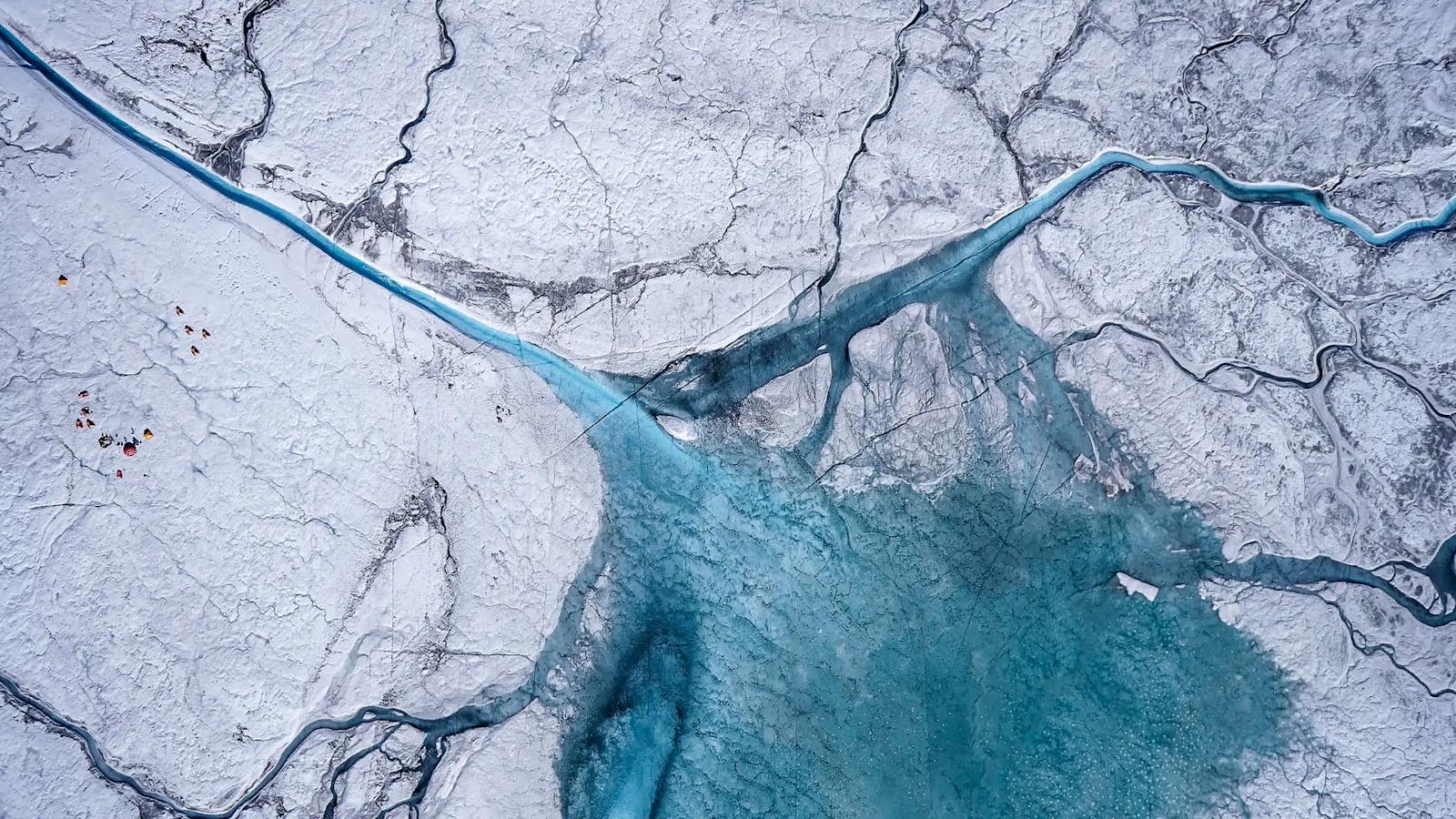
Map-making neurons change even in familiar settings, study finds – News Bureau

CHAMPAIGN, Ill. — Neurobiologists at Northwestern University and the University of Illinois Urbana-Champaign found the brain’s internal GPS changes each time mice navigate a familiar, static environment.

Even when a mouse walks the same path every day — and the path and surrounding conditions remain identical — each journey activates different “map-making” neurons in the brain.

This finding illuminates the fundamental mystery of how the brain processes and stores spatial memories, with implications for scientists’ understanding of memory, learning and even aging.

The researchers published their findings in the journal Nature.

“Our study confirms that spatial memories in the brain aren’t stable and fixed,” said Northwestern neurobiology professor Daniel Dombeck, the study’s senior author. “You can’t point to one group of neurons in the brain and say, ‘That memory is stored right there.’ Instead, we’re finding that memories are passed among neurons. The exact same experience will involve different neurons every time. It’s not a sudden change, but it slowly evolves.”

Located deep within the brain’s temporal lobe, the hippocampus stores memories related to spatial navigation. For decades, neurobiologists thought the same hippocampal neurons encoded memories of the same places, Dombeck said. Thus, the path someone might take from their bedroom to their kitchen should activate the exact same sequence of neurons during each midnight walk for a glass of water.

About 10 years ago, however, scientists imaged mouse brains as the animals ran through a maze. Even as the mice ran through the same maze day after day, different neurons fired during each run. Scientists wondered if the results were a fluke — maybe the rodents’ experience of the maze changed, with differences in speed, smell or something subtle in the environment.

To probe these questions, the researchers designed an experiment that gave them unprecedented control over the mice’s sensory input. The mice ran through the virtual maze on treadmills, ensuring precise measurement of speed. The maze was presented on a multisensory virtual reality system previously developed in Dombeck’s laboratory. This not only controlled what the animals saw, but a cone on the nose of the mice provided identical smells for every session, controlling for every possible environmental variable.

After running the experiment several times, the results were clear: Even in a highly reproducible virtual world, a different group of neurons activated each time. The finding confirmed that the brain’s spatial maps are inherently dynamic, constantly updating regardless of how static a space might be.

Understanding the fluidity of memory has implications for human disease as well as AI learning, says molecular and integrative biology professor Jason Climer. Photo by Anne Freeman

“This evidence suggests that memories are fluid. This could be related to deeper questions of why the brain can do things modern artificial intelligence struggles with, things like learning new things continuously,” said U. of I. molecular and integrative biology professor Jason Climer, the co-first author of the study. Climer performed the study while a postdoctoral researcher in Dombeck’s group. “It also may play a role in natural forgetting — an active process, often overlooked, but essential for healthy memory function.” 

Although few patterns arose throughout the course of the experiment, the researchers did notice one consistent factor: The most excitable neurons, which were more easily activated, maintained more stable spatial memories throughout multiple runs through the virtual maze. Since neuron excitability decreases with age, the finding could help scientists understand the role of aging and disease as it relates to the brain’s ability to encode new memories.

“The small core of neurons that are stable are special, and better understanding what makes them special could lead to new treatments for memory dysfunction,” Climer said. “Memory deficits are the hallmark of Alzheimer’s disease and are also a major barrier for patients suffering from a range of neuropsychiatric disorders such as schizophrenia. By better understanding fundamental aspects of memory like the changes over time we report in our paper, we are providing new targets for understanding differences in these patients’ brains and novel strategies for treatments. Understanding how the brain handles the problem of memory also has a lot to teach us about how computers and AI might be improved.”

The National Institutes of Health supported this work. Dombeck lab members Heydar Davoudi and Jun Young Oh are also co-first authors of the paper, along with Climer.

This news release is adapted from content provided by Northwestern University. 

Continue Reading