Sample Page
LHC bans commercial activities on Sundays amid smog crisis
Written by
admin
in
1. Pakistan
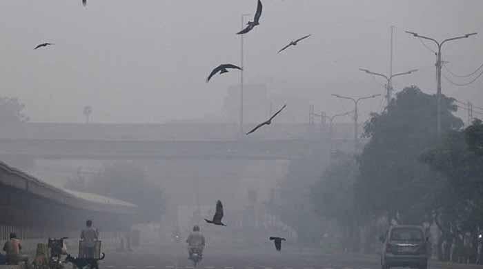
Birds fly past on a street amid dense smog in Lahore, Pakistan, on November 1, 2025. — AFP
LHC Justice Shahid Karim hears pleas on environmental issues.
Court orders strict enforcement of 10pm closure for marriage…
Continue Reading
←
Best Beats Studio Buds deal: Save $70.01 at Amazon
One million people in Gaza receive WFP food parcels but more crossings needed for continued scale-up | World Food Programme – ReliefWeb
→
More posts
Canon honored for Excellent Production Support at TSMC 2025 Excellent Performance Awards
December 24, 2025
Summer awareness campaign to help Western Australians stay safe in the sun
December 24, 2025
Softball To Host First-Ever “Pre-Season Pa‘ina” Fundraiser
December 24, 2025
SCIRP Open Access
December 24, 2025