Mapping transcription factor (TF) binding sites across the genome is key to understanding how cells control gene expression, yet pinpointing their footprints across the genome—especially in single cells or small samples—has remained a technical bottleneck in the fields of molecular biology, genetics, and epigenetics. A new technique called DynaTag may be a promising new approach to understanding TF landscapes.
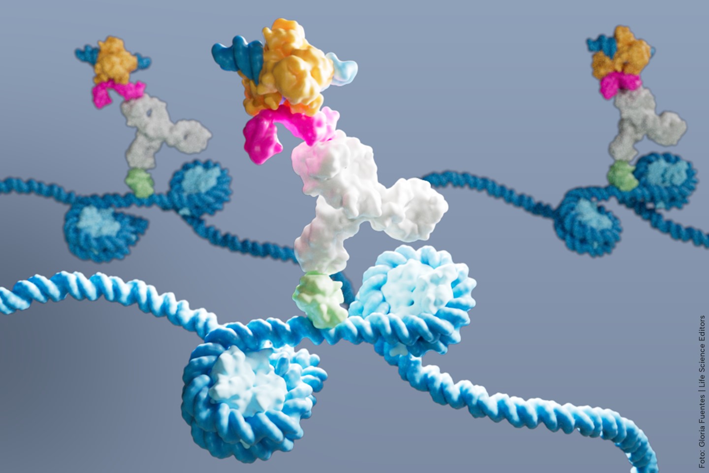
In a study titled “DynaTag for efficient mapping of transcription factors in low-input samples and at single-cell resolution,” published in Nature Communications, DynaTag (cleavage under Dynamic targets and Tagmentation) enables high-resolution mapping of transcription factor binding sites—even from low-input or single-cell samples. The method, developed by researchers at the University of Cologne, relies on a physiological salt buffer to preserve TF–DNA interactions that are typically lost during sample processing with standard high-salt protocols. “TF–DNA interactions are sensitive to these stringent conditions, causing their dissociation from chromatin and incompatibility with these tagmentation-based technologies,” reported the researchers. DynaTag utilizes a physiological intracellular salt solution throughout all nuclei handling steps to retain specific interactions while suppressing nonspecific protein–DNA interactions, added the research team.
“DynaTag outperforms existing methods such as ChIP-seq and CUT&RUN in terms of sensitivity and resolution,” said senior author Robert Hänsel-Hertsch, PhD, principal investigator at the Center for Molecular Medicine Cologne (CMMC). “The new method delivers high-resolution mapping of the DNA regions where transcription factors bind.”
Using DynaTag, the team profiled 15 transcription factors in both stem cell and cancer models. In embryonic stem cells, the method revealed dynamic binding patterns for factors such as NANOG, MYC, and OCT4 as cells transitioned to an epiblast-like state. Single-nuclei DynaTag further showed that distinct TF occupancy patterns were enough to distinguish cell states, highlighting its utility in developmental biology and heterogeneous tissue analysis.
The authors also applied DynaTag to small cell lung cancer (SCLC), a fast-growing cancer type notorious for its resistance to therapy. In a patient-derived mouse model, the researchers used DynaTag to compare TF binding before and after chemotherapy. They found that the transcription factors FOXA1 and MYC showed increased occupancy in chemotherapy-resistant tumors, while factors like ASCL1 and POU2F3 declined. Likewise, the results further showed significantly increased FOXA1 and MYC occupancies at genes involved in several metabolic pathways, such as fatty acid metabolism.
Intriguingly, the method also suggested a gain-of-function for a mutant p53 variant (R248Q), which became more active at genes linked to epithelial-mesenchymal transition (EMT)—a potential route for cancer cells to escape treatment. These insights, the researchers say, were not detectable using alternative methods like ATAC-seq footprinting.
“It had already been established that certain signaling pathways promoting resistance or metastasis are activated after chemotherapy in small cell lung cancer. However, it was not known which transcription factors regulate these signaling pathways,” said Hänsel-Hertsch. “With the help of DynaTag, we were able to identify specific transcription factors that show increased binding to genes belonging to these signaling pathways after chemotherapy and are likely to promote further tumor growth.”