Thank you. Listen to this article using the player above. ✖
Want to listen to this article for FREE?
Complete the form below to unlock access to ALL audio articles.
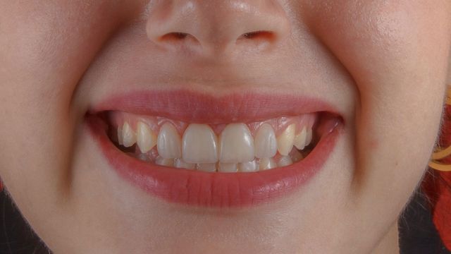
Until now the sensory neurons inside the tooth were primarily thought to send pain signals to the brain, but a new study shows those neurons are multitaskers that also trigger a jaw-opening reflex that almost instantaneously prevents damage and further injury to teeth.
The reflex that pops open the lower jaw was a widely known craniofacial reflex, but until this study the cellular origins of this phenomenon were not known.
University of Michigan researchers in sensory neuroscience, dentistry and mechanical engineering found the origin using special live imaging, behavior-tracking tools and mice molars to uncover the neurons’ additional role of monitoring the inner tooth and outer enamel.
The discovery and understanding of this additional role shows how important healthy, active nerves are for preserving teeth.
“We suspected there was a more fundamental role for tooth nerves,” said Joshua Emrick, senior author of the study and assistant professor at the U-M School of Dentistry. “When we consider regenerating a tooth pulp, we need to bring back the nerves.”
Emrick’s research team looked at how nerve cells reacted to stimulation of the molar teeth of mice in real time. Their experiments revealed a newly defined, protective role for intradental High-Threshhold Mechanoreceptors, highly specialized sensory neurons that respond to tooth damage. These HTMRs detect dangerous threats and send the message rapidly to the brain for instantaneous action.
“Our study challenges the prior assumption that nerves inside the tooth primarily function to elicit pain and force us straight to the dentist for help,” Emrick said. “If you’ve ever accidentally bitten down on your fork, you’ve probably experienced a startling jolt, but also stopped short of fracturing your teeth. You may thank these intradental HTMRs for that.”
The reflex is really about self-preservation.
“We think protection of the teeth through this jaw-opening reflex is highly conserved among mammals that haven’t developed the ability to replace teeth—like humans or in the molar teeth of mice,” Emrick said. “Our work reports an ability to use these neurons to also elicit pain which will open up possibilities for developing new methods for relieving toothache at the dentist’s office.”
To break it down further, the study, published in Cell Reports, showed that when enamel or dentin is damaged, the neurons fire a response. Follow-up experiments determined what happened after the HTMRs were activated. As previously known, the group identified that they trigger acute pain, but more surprisingly they also witnessed a rapid jaw-opening reflex within 5 to 15 milliseconds of the activation.
While the authors focused their work on understanding how the HTMRs function within the tooth, this important subclass of sensory neurons may protect other oral and body structures from damage.
Elizabeth Ronan, postdoctoral fellow at the School of Dentistry and lead author of the work, said the findings are the start of a deeper understanding.
“While we typically think of sensation as giving rise to our perceived external experience of the world, sensory neurons are equally essential in protecting and maintaining our tissues throughout life,” she said. “Much remains to be discovered regarding how sensory neurons function within individual tissues, especially internal ones such as the teeth.”
Reference: Ronan EA, Gandhi AR, Koecklin KHU, et al. Intradental mechano-nociceptors serve as sentinels that prevent tooth damage. Cell Reports. 2025;44(8). doi: 10.1016/j.celrep.2025.116017
This article has been republished from the following materials. Note: material may have been edited for length and content. For further information, please contact the cited source. Our press release publishing policy can be accessed here.