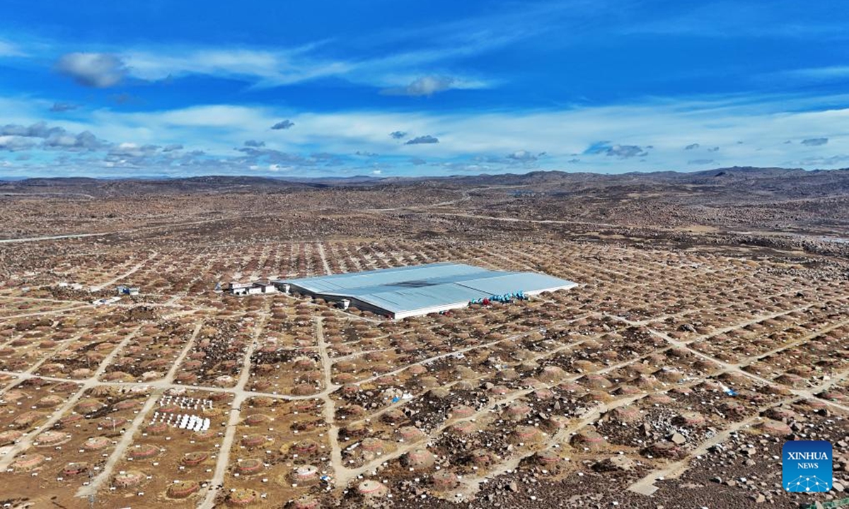
Introduction
Bloodstream infection (BSI) is a severe and often fatal complication linked to accelerated morbidity and mortality, with an annual incidence ranging from 150 to 309 cases per 100,000 population and a mortality rate ranging from 12.5% to 22.7.1–3 Neonates, in particular, face even higher risks and mortality rates of BSIs, with a mortality rate ranging from 19.7% to 30%.4–6 Escherichia coli (E. coli) and Klebsiella pneumonia (K. pneumoniae) stand as prominent pathogens responsible for neonatal BSIs, with significant mortality rates linked to their infectious impact, presenting a formidable obstacle to anti-infective treatment.6,7 Therefore, it is crucial in clinical practice to identify predictors associated with poor prognosis in E. coli and K. pneumoniae neonatal BSIs.
The emergence of antimicrobial resistance (AMR), particularly in extended-spectrum β-lactamase (ESBL)-producing bacteria, constitutes a significant global health challenge. This is primarily due to its frequent association with the failure of empirical antibiotic therapy, leading to elevated morbidity and mortality rates.8 In 2019, an estimated 4.95 million deaths were associated with bacterial AMR, among which 1.27 million were attributable to bacterial AMR.9 ESBL is a Gram-negative bacterium of the Enterobacteriaceae family, which harbors ESBL genes either within its plasmids or integrated into its chromosomal DNA.10 It produces β-lactam hydrolyzing enzymes, providing resistance to penicillin, aztreonam, and first-, second-, and third-generation cephalosporins, while lacking the ability to hydrolyze carbapenems or cephamycin.11 Among a wide range of Gram-negative bacteria harboring ESBL genes, E. coli (ESBL-EC) and K. pneumoniae (ESBL-KP) are the most common hosts, causing infections such as BSIs, urinary tract infections, and diarrhea, particularly among neonates, in both hospital and community settings.12–14 Although our previous research demonstrated a notably high prevalence of K. pneumoniae infections among preterm neonates, coupled with suboptimal treatment outcomes and elevated antimicrobial resistance patterns,15 the existing literature on ESBL-EC and ESBL-KP neonatal bloodstream infections remains scarce. Significant knowledge gaps persist regarding their clinical manifestations, epidemiological characteristics, and antimicrobial resistance profiles. Addressing these gaps is critical for developing targeted strategies to mitigate the impact of ESBL-EC and ESBL-KP neonatal BSIs.
The present study aims to bridge this knowledge gap by systematically investigating the clinical features and antimicrobial resistance profiles of ESBL-EC and ESBL-KP. Through analyzing clinical data, AMR patterns, and the production of ESBL, this research seeks to enhance our understanding of these pathogens, and facilitate the development of evidence-based containment strategies and optimized therapeutic interventions.
Methods
Study Setting and Patients
We conducted a retrospective cohort study from January 2017 to December 2023 at the Maternal and Child Health Hospital of Guangxi Zhuang Autonomous Region, which is the largest children’s hospital in the region. During the study period, 180,297 blood culture tests were conducted, with a positivity rate of 1.88% (3394/180,297). In this study we focused on cases of BSIs caused by E. coli and K. pneumoniae in diagnosed patients. For each patient, only the first E. coli or K. pneumoniae BSI isolate was selected for analysis. The inclusion criteria were defined as follows: (a) patients aged 28 days or younger, (b) meets criteria indicative of neonatal bloodstream infections,16 (c) a positive blood culture for E. coli or K. pneumonia, (d) hospitalization with complete clinical data available. Conversely, the analysis systematically excluded outpatients with incomplete or unavailable medical records. The protocol for this study was approved by the research administration of Medical Ethics Committee of Maternal and Child Health Hospital of Guangxi Zhuang Autonomous Region (No. 2023-3-42). This study used an anonymous method to protect the participants and obtained their permission.
Data Collection and Definitions
Various clinical features of the enrolled patients were collected from the hospital’s electronic health records system. The collected data encompassed essential demographic information (gender, gestational age, and weight), medical history (underlying diseases, cesarean sections, premature ruptures of membrane), interventions (invasive procedures and devices, antibiotic exposure), bloodstream infection type and outcomes.
According to the gestational age at birth, neonates can be classified as follows: term infants, with a gestational age of 37 weeks or more to 42 weeks; late premature infants, with a gestational age between 28 weeks and 37 weeks; and extremely preterm infants, with a gestational age between 22 weeks and 28 weeks. Based on the weight at birth, newborns can be categorized as follows: extremely low birth weight (ELBW) denotes infants with a birth weight below 1000g; very low birth weight (VLBW) indicates infants with a birth weight ranging from 1000g to under 1500g; low birth weight (LBW) signifies infants with a birth weight between 1500g to less than 2500g; normal birth weight (NBW) pertains to infants with a birth weight between 2500g to 4000g; macrosomia describes infants with a birth weight exceeding 4000g. Hypoalbuminemia is generally defined as a serum albumin level of less than 35 g/L. Anemia refers to a peripheral hemoglobin (Hb) concentration of less than 120 g/L. Treatment outcomes were divided into two distinct groups based on clinical documentation. Patients who achieved clinical recovery or showed significant improvement upon discharge were classified as having a favorable prognosis, while those who died during hospitalization or were discharged in critical condition were classified as having a poor prognosis.
Microbiological Methods
In this investigation, all intentionally collected isolates were cultivated on blood agar plates at 37 °C in an incubator for 18–24 hours. The isolates were then identified using the Zhuhai DL-96IIautomatic bacterial identification drug sensitivity apparatus (Zhuhai DL Biotech Co., Ltd.,China) or the VITEK2 Compact system (BioMérieux, Marcy l’Etoile, France). The antibiotic susceptibility tests were performed on the isolates utilizing either the Kirby-Bauer methodology, the Zhuhai DL-96II automated bacterial identification drug sensitivity apparatus or the VITEK 2 Compact system. Each protocol was carried out according to the manufacturer’s guidelines. The minimum inhibitory concentrations (MICs) of common clinical antibiotics, including gentamicin, sulfamethoxazole, ciprofloxacin, ceftazidime, cefazolin, cefepime, meropenem, piperacillin-tazobactam, imipenem, amikacin, ampicillin-sulbactam, ceftriaxone, ticarcillin-clavulanic acid, chloramphenicol, amoxicillin, cefuroxime, minocycline, levofloxacin, and cefoxitin. The findings were elucidated in accordance with the protocols delineated by the Clinical and Laboratory Standards Institute M100 document, and ESBL screen was determined based on the susceptibility test for ceftriaxone and ceftazidime (MIC ≥ 2 μg/mL), excluding carbapenem-resistant strains.
The confirmation of ESBL-producing isolates was conducted using the combination broth microdilution. A comparison of the MIC was made between ceftazidime and cefotaxime alone versus those of ceftazidime and cefotaxime containing clavulanic acid. Following the designated incubation period, a reduction of ≥3-fold in the minimum inhibitory concentration (MIC) of any antimicrobial agent when tested in combination with clavulanate, compared to its MIC value when tested independently, was identified as indicative of ESBL production, in accordance with the criteria established by the Clinical and Laboratory Standards Institute (CLSI). The quality control strains employed were E. coli ATCC 25922 and K. pneumoniae ATCC 700603 (National Center for Clinical Laboratories, Beijing, China).
Statistical Analysis
Statistical analyses were performed using SPSS version 25.0. Count data were reported as percentages, and group comparisons were performed using χ2 tests or Fisher’s exact test for categorical variables. Variables that showed significance (p<0.10) in the univariate analysis, including extremely low birth weight, very premature infants, anemia, hypoalbuminemia, CRKP, mechanical ventilation, and central venous catheterization, were selected for inclusion in a logistic regression model to evaluate their association with poor Prognosis. Statistical significance was considered at p-values less than 0.05.
Results
Baseline Characteristics of 139 Neonates with Escherichia coli and Klebsiella pneumoniae Bloodstream Infections
During the study period, a total of 139 unique cases of neonatal BSIs caused by E. coli and K. pneumoniae were identified. Of these, 29 BSIs were caused by ESBL-EC (20.8%), 13 by ESBL KP (9.3%), 87 by non-ESBL strains, and 10 by CRKP (7.1%). Among the participants, 74 (53.2%) were male. The majority of neonates included in the study were term infants (50.4%) and normal birth weights (48.9%). Most of them developed late-Onset infections (66.2%) and breast-fed (70.5%). The most underlying diseases was hypoalbuminemia (35.9%), while the invasive procedures and devices was mechanical ventilation (53.2%), respectively. The predominant antibiotic exposures was β-lactam-β-lactamase inhibitors (31.6%). 26 (18.7%) neonates with poor prognosis were hospitalized after the onset of E. coli and K. pneumoniae bloodstream infections (Supplementary Table 1).
Microbiological Characteristics of Escherichia coli and Klebsiella pneumoniae Isolates
Among the 139 isolates, 95 were E. coli and 44 were K. pneumoniae. All strains have completed antimicrobial susceptibility testing, and the drug resistance profiles are shown in Table 1. Among Escherichia coli strains, the three antimicrobial agents with the highest resistance rates were ampicillin (82.1%), sulfamethoxazole (65.2%), and cefazolin (44.2%). Notably, no strains exhibited resistance to carbapenems. In comparison, the resistance rates of ampicillin (P=0.003), ceftazidime (P=0.015), ceftriaxone (P=0.000), cefepime (P=0.000), cefuroxime (P=0.000), cefazolin (P=0.000), ciprofloxacin (P=0.000), levofloxacin (P=0.01) and minocycline (P=0.031) were significantly higher in the ESBL-EC strains than non- ESBL-EC strains.
Table 1 Antibiotic Susceptibility Profiles for Escherichia coli and Klebsiella pneumoniae Isolates
|
Meanwhile, the resistance rates of K. pneumoniae to ampicillin, cefuroxime, cefazolin, and ceftriaxone are alarmingly high, standing at 100.00%, 54.5%, 54.5%, and 50.00%, respectively. Furthermore, the resistance rates for imipenem and meropenem exceed 20%, specifically standing at 22.7%. In contrast to non-ESBL-KP, ESBL-KP exhibited an elevated level of resistance towards ampicillin-sulbactam (P=0.000), ceftriaxone (P=0.000), cefepime (P=0.000), cefuroxime (P=0.000), cefazolin (P=0.000), ciprofloxacin (P=0.000), chloramphenicol (P=0.000), gentamicin (P=0.001), levofloxacin (P=0.001), sulfamethoxazole (P=0.000), and ticarcillin-clavulanic acid (P=0.000).
Risk Factors Associated with Bloodstream Infections Caused by ESBL-Producing Strains
Among 139 patients analyzed, 42 (30.2%) BSIs were attributed to ESBL strains, while 87 (62.6%) were caused by non-ESBL strains. Notably, 10 cases (7.2%) involved CRKP strains. Comparative analysis of demographic, and clinical parameters between patients with ESBL-producing and non-ESBL BSIs is presented in Table 2. The factors most significantly associated with ESBL strains bloodstream infections in neonates were late premature infant (P=0.037), very low birth weight (p=0.001), cesarean section (p=0.003), pneumonia (P=0.037), mechanical ventilation (p=0.000), and length of hospital stay >30 days (p=0.001). In contrast, term infants (p=0.010) and normal birth weight infants (P=0.021) were more prevalent in the non-ESBL strains group than in the ESBL strains group.
![]() |
Table 2 Comparison of Neonatal Variables Between Those with BSIs Caused by ESBL and Non-ESBL Strains
|
Additionally, the comparison of patient variables between individuals with BSIs due to ESBL E. coli and those with BSIs due to non-ESBL E. coli showed that cesarean section (p=0.031), pneumonia (P=0.041), mechanical ventilation (p=0.014), indwelling catheters (p=0.048) and length of hospital stay >30 days (p=0.012) were more associated with ESBL E. coli BSIs (Table 3).
![]() |
Table 3 Comparison of Neonatal Variables Between BSIs ESBL-EC and Non-ESBL-EC
|
Furthermore, the analysis of patient characteristics in individuals suffering from BSIs caused by ESBL-producing K. pneumoniae compared to those with infections caused by non-ESBL-producing K. pneumoniae revealed that late premature infants (p=0.031), very low birth weight (p=0.004), and mechanical ventilation (p=0.011) were more commonly linked to ESBL K. pneumoniae BSIs (Table 4).
![]() |
Table 4 Comparison of Neonatal Variables Between BSIs ESBL-KP and Non-ESBL-KP
|
Risk Factors for Poor Prognosis in Neonates with Escherichia coli and Klebsiella pneumoniae Bloodstream Infections
The results of the univariate and multivariate analysis were depicted in Table 5. The percentages of favorable and poor prognosis in neonates afflicted with bloodstream infections caused by E. coli and K. pneumoniae stood at 81.29% (113 out of 139) and 18.71% (26 out of 139), respectively. Univariate analysis indicated that very premature infants (p=0.000), extremely low birth weight (p=0.000), hypoalbuminemia (p=0.000), anemia (p=0.015), and isolation of CRKP (p=0.000) were significantly correlated with poor prognosis. In the multivariate analysis, hypoalbuminemia (OR: 3.922, 95% CI: 1.189–12.937, p=0.025), and isolation of CRKP (OR: 11.548, 95% CI: 1.785–74.708, p=0.010) were identified as significant predictors of poor prognosis in neonates afflicted with bloodstream infections caused by E. coli and K. pneumonia.
![]() |
Table 5 Logistic Regression Analysis for Variables Associated with Poor Prognosis of Neonates BSIs with E. coli and K. pneumonia
|
Discussion
Bloodstream infections in newborns caused by E. coli and K. pneumoniae present a formidable challenge, given the rising prevalence of ESBL and CR strains, especially in healthcare facilities, and they are two of the drivers of nosocomial and community infections globally.17,18 Between 2017 and 2023, at the Maternal and Child Health Hospital of Guangxi Zhuang Autonomous Region in Southwest China, we found that approximately one-third of the 139 BSIs caused by E. coli and K. pneumoniae were due to ESBL strains and 7.2% were due to CRKP. Among these strains, 20.8% were ESBL-EC, whereas only 9.3% were ESBL-KP. This finding was almost similar to the frequencies of ESBL producers among the neonates in the previous two studies, which reported ESBL-EC at 30.4% and ESBL-KP at 8.2%.19,20 However, a previous study about neonatal sepsis in low- and middle-income countries revealed a much higher prevalence of ESBL-EC (38%) and ESBL-KP (83%) compared to the findings in this study.21 Furthermore, previous studies conducted in china have reported even higher percentages.22,23 Consistent findings have been documented in the literature from various regions, such as Iran,24 South Africa,25 Ghana,26 and India.27 These findings could potentially be attributed to variations in phenotypic detection methods for ESBL production and differences within the study populations.
Genes encoding ESBL are predominantly located on transposons or insertion sequences within plasmids in conjunction with other resistance genes.8 Consequently, they have the potential to disseminate quickly, leading to resistance against multiple antimicrobials including aminoglycosides, cephalosporins, trimethoprim, sulfonamides, tetracyclines, chloramphenicol, and fluoroquinolones.28 The antibiotic resistance profiles from our study also showed that ESBL-EC exhibited a higher resistance rate to cephalosporins, fluoroquinolones, tetracyclines compared to non-ESBL strains. Meanwhile, ESBL-KP demonstrated elevated resistance rates to aminoglycosides, β-lactam-β-lactamase inhibitors, cephalosporins, sulfonamides, chloramphenicol, and fluoroquinolones. According to the guidelines from the Infectious Diseases Society of America, carbapenems, fluoroquinolones, and cotrimoxazole are recommended for the treatment of infections outside of the urinary tract caused by ESBL-E.29 However, high resistance to fluoroquinolones and cotrimoxazole has been found in our study, consistent with prior research.18 In contrast, no ESBL-E strains exhibited resistance to carbapenems, and ESBL-KP strains showed a resistance rate of 22.7% to imipenem and meropenem, which agrees with earlier studies in Zambia and México.30,31 Therefore, it is crucial to monitor the resistance profiles of commonly used antibiotics in the local area. Of utmost importance is the monitoring of the resistance patterns displayed by ESBL isolates and understanding their impact on patient management, particularly among neonates.
The clinical characteristics associated with neonatal BSIs caused by ESBL isolates are vital for the prevention of these infections and the effective administration. Co-morbidities like cesarean section, prematurity, very low birth weight, pulmonary disease, malnutrition, malignancy, gastroenteritis, mechanical ventilation, indwelling catheters, central venous catheterization were investigated as risk factors for neonatal BSIs due to ESBL-E isolates.32–35 In our study, patients with late premature infant, very low birth weight, cesarean section, pneumonia, and mechanical ventilation also had a higher risk of ESBL isolates bloodstream infection. Furthermore, we conducted a detailed analysis of the risk factors associated with ESBL-EC and ESBL-KP neonatal BSI. Our investigation revealed a distinct correlation between ESBL-EC infections with cesarean section, pneumonia, mechanical ventilation, and indwelling catheters, whereas ESBL-KP infections showed a notably association with the presence of late premature infants, very low birth weight, and mechanical ventilation. This finding indicates that different subtypes of Enterobacteriaceae bacteria carrying ESBL may demonstrate variations in their pathogenicity and impact on host resistance. E. coli and K. pneumoniae are the most prevalent hosts of ESBL.8 The ST131 clone is predominant in ESBL-producing E. coli, while ST11 is dominant in ESBL-producing K. pneumoniae.11,36 These strains harbor distinctive biological traits or pathogenic mechanisms that enhance their propensity to serve as risk factors for bloodstream infections. Nevertheless, studies on the virulence and molecular epidemiological characteristics have not been undertaken. Subsequent research should prioritize whole-genome sequencing and functional assessments to elucidate the molecular mechanisms that underlie these adaptations and pathogenicity, thereby informing targeted interventions.
Although we have conducted a statistical analysis on the risk factors associated with ESBL strains infections, the total number of ESBL infection cases is insufficient for a multivariate analysis. Consequently, we undertook a study analyzing the prognosis of 139 cases of neonatal bloodstream infections to identify the risk factors contributing to poor prognosis. Our study indicated that very premature infants, extremely low birth weight, hypoalbuminemia, anemia, and isolation of CRKP were significantly correlated with a poor prognosis. In the management of neonates, invasive procedures and devices such as mechanical ventilation and central venous catheterization, although crucial for life support, heighten the vulnerability to infections in newborns, especially premature infants and those with low birth weight.37 Research has confirmed that prematurity, low birth weight, hypoalbuminemia, anemia, CRKP, and invasive procedures such as mechanical ventilation are associated with a poor prognosis for neonatal BSI.38–41 Consistent with our research results, further studies are needed to explore the specific mechanisms. Importantly, isolation of CRKP and hypoalbuminemia were identified as significant predictors of poor prognosis in neonates afflicted with bloodstream infections caused by E. coli and K. pneumonia. In line with the findings of Dilan et al39 and Yasemin et al42 recent research has consistently demonstrated that the presence of carbapenem-resistant gram-negative bacteria, specifically isolation of CRKP serves as a significant predictor of unfavorable outcomes in neonatal bloodstream infections. According to the results of our search in the PubMed database, we did not retrieve any relevant literature indicating that hypoalbuminemia is an independent risk factor for poor prognosis in neonatal BSI. Proteins play vital roles in the body, contributing to immune function, nutrient metabolism, and cell structure. Newborns have an immature immune system at birth, and hypoalbuminemia could potentially compromise immune function, elevating the susceptibility to infections. In the context of neonatal BSI, hypoalbuminemia might worsen the disease severity, impacting treatment efficacy and prognosis.
Our study was subject to certain limitations. The predominant limitation pertained to its retrospective study and single-center design, encompassing a cohort of 139 neonatal patients, thereby rendering it susceptible to selection bias. Further prospective, multicenter studies are warranted. Secondly, our research exclusively targeted bloodstream infections caused by E. coli and K. pneumoniae, without encompassing other gram-negative bacteria. Furthermore, our research has focused solely on drug resistance phenotypes rather than delving into the molecular aspects such as drug resistance mechanisms, virulence, and molecular epidemiology studies. Exploring the correlation between strain molecular characteristics and clinical traits is crucial for infection prevention and control, which will be the primary focus of our future investigations. Lastly, the study design did not include a preset long-term follow-up, resulting in a lack of data on mortality within 28 days, which limits the completeness of the research conclusions and the evaluation of long-term patient outcomes. In future studies, we will consider incorporating a long-term follow-up plan to more comprehensively assess treatment efficacy and patient prognosis.
Conclusion
The predominant children’s hospital in this autonomous region exhibits a relatively low incidence of ESBL-EC and ESBL-KP. Compared to non-ESBL strains, ESBL strains demonstrated a higher resistance rate to cephalosporins, fluoroquinolones, and tetracyclines. Late-preterm infants, very low birth weight, cesarean section history, pneumonia, and mechanical ventilation were found to be associated with bloodstream infections caused by ESBL strains. The isolation of CRKP and hypoalbuminemia were identified as significant predictors of poor prognosis in neonates afflicted with bloodstream infections caused by E. coli and K. pneumonia. The implementation of infection control initiatives and antimicrobial stewardship programs is critical for controlling healthcare expenditures, reducing antimicrobial resistance rates, enhancing patient outcomes, and mitigating the adverse ecological impacts of antimicrobial overuse.
Data Sharing Statement
The datasets used and/or analysed during the current study are available from the corresponding author upon reasonable request.
Ethics Approval and Consent to Participate
This research adhered to the ethical principles outlined in the Declaration of Helsinki and followed applicable guidelines. Ethical approval was obtained from the Institutional Review Board of the Maternal and Child Health Hospital of Guangxi Zhuang Autonomous Region (Approval No. 2023-3-42). Since the study involved a retrospective analysis of de-identified medical records, participant privacy was safeguarded, and no additional risks were introduced. Due to the use of anonymized data and the study’s retrospective design, the ethics committee granted an exemption from obtaining informed consent.
Acknowledgments
We extend our sincere appreciation to the Clinical Laboratory Department of the Maternal and Child Health Hospital of Guangxi Zhuang Autonomous Region for their invaluable support. Special thanks go to the management and microbiology team for enabling efficient bacterial identification and automated susceptibility testing. We also recognize the dedicated efforts of all clinical and laboratory personnel involved in this study. Lastly, we are deeply grateful to all participants for their contribution.
Author Contributions
All authors made a significant contribution to the work reported, whether that is in the conception, study design, execution, acquisition of data, analysis and interpretation, or in all these areas; took part in drafting, revising or critically reviewing the article; gave final approval of the version to be published; have agreed on the journal to which the article has been submitted; and agree to be accountable for all aspects of the work.
Funding
This work was supported by a self-funded project by the Health Commission of Guangxi Zhuang Autonomous Region (Z-A20240291 and Z-A20220288).
Disclosure
The authors declare that they have no conflict of interest.
References
1. Amanati A, Sajedianfard S, Khajeh S, et al. Bloodstream infections in adult patients with malignancy, epidemiology, microbiology, and risk factors associated with mortality and multi-drug resistance. BMC Infect Dis. 2021;21(1):636. doi:10.1186/s12879-021-06243-z
2. Kontula KSK, Skogberg K, Ollgren J, Järvinen A, Lyytikäinen O. Population-based study of bloodstream infection incidence and mortality rates, Finland, 2004–2018. Emerg Infect Dis. 2021;27(10):2560–2569. doi:10.3201/eid2710.204826
3. Holm MKA, Jansåker F, Gradel KO, et al. Decrease in all-cause 30-day mortality after bacteraemia over a 15-year period: a population-based cohort study in Denmark in 2000–2014. Int J Environ Res Public Health. 2021;18(11):5982. doi:10.3390/ijerph18115982
4. Hadfield BR, Cantey JB. Neonatal bloodstream infections. Curr Opin Infect Dis. 2021;34(5):533–537. doi:10.1097/QCO.0000000000000764
5. Labi AK, Enweronu-Laryea CC, Nartey ET, et al. Bloodstream infections at two neonatal intensive care units in Ghana: multidrug resistant enterobacterales undermine the usefulness of standard antibiotic regimes. Pediatr Infect Dis J. 2021;40(12):1115–1121. doi:10.1097/INF.0000000000003284
6. Johnson J, Robinson ML, Rajput UC, et al. High burden of bloodstream infections associated with antimicrobial resistance and mortality in the neonatal intensive care unit in Pune, India. Clinl Infect Dis. 2021;73(2):271–280. doi:10.1093/cid/ciaa554
7. Wang J, Zhang H, Yan J, Zhang T. Literature review on the distribution characteristics and antimicrobial resistance of bacterial pathogens in neonatal sepsis. J Matern Fetal Neonatal Med. 2022;35(5):861–870. doi:10.1080/14767058.2020.1732342
8. Husna A, Rahman MM, Badruzzaman ATM, et al. Extended-Spectrum β-Lactamases (ESBL): challenges and opportunities. Biomedicines. 2023;11(11):2937. doi:10.3390/biomedicines11112937
9. Murray CJL, Ikuta KS, Sharara F. Global burden of bacterial antimicrobial resistance in 2019: a systematic analysis. Lancet. 2022;399(10325):629–655. doi:10.1016/S0140-6736(21)02724-0
10. Sobkowiak A, Scherff N, Schuler F, et al. Plasmid-encoded gene duplications of extended-spectrum β-lactamases in clinical bacterial isolates. Front Cell Infect Microbiol. 2024;14:1343858. doi:10.3389/fcimb.2024.1343858
11. Castanheira M, Simner PJ, Bradford PA. Extended-spectrum β-lactamases: an update on their characteristics, epidemiology and detection. JAC-Antimicrob Resist. 2021;3(3):dlab092. doi:10.1093/jacamr/dlab092
12. Hu Y, Feng Y, Zong Z. Global distribution of Klebsiella pneumoniae producing extended-spectrum β-lactamases in neonates. Precision Clin Med. 2024;7(4):pbae031. doi:10.1093/pcmedi/pbae031
13. Nieto-Rosado M, Sands K, Portal EAR, et al. Colonisation of hospital surfaces from low- and middle-income countries by extended spectrum β-lactamase- and carbapenemase-producing bacteria. Nat Commun. 2024;15(1):2758. doi:10.1038/s41467-024-46684-z
14. Jiménez-Rojas V, Villanueva-García D, Miranda-Vega AL, et al. Gut colonization and subsequent infection of neonates caused by extended-spectrum beta-lactamase-producing Escherichia coli and Klebsiella pneumoniae. Front Cell Infect Microbiol. 2023;13:1322874. doi:10.3389/fcimb.2023.1322874
15. Wei X, Liang J, Zhang H, et al. Clinical features and risk factors of Klebsiella pneumoniae infection in premature infants: a retrospective cohort study. BMC Infect Dis. 2024;24(1):1311. doi:10.1186/s12879-024-10201-w
16. See LL. Bloodstream infection in children. Pediatr Crit Care Med. 2005;6(3 Suppl):S42–44. doi:10.1097/01.PCC.0000161945.98871.52
17. Ojulong J, Gebru GN, Duduyemi B, et al. Prevalence of antimicrobial resistance in Klebsiella pneumoniae, Enterobacter cloacae, and Escherichia coli isolates among stillbirths and deceased under-five children in Sierra Leone: data from the child health and mortality prevention surveillance sites from 2019 to 2022. Microorganisms. 2024;12(8). doi:10.3390/microorganisms12081657
18. Akenten CW, Khan NA, Mbwana J, et al. Carriage of ESBL-producing Klebsiella pneumoniae and Escherichia coli among children in rural Ghana: a cross-sectional study. Antimicrob Resist Infect Control. 2023;12(1):60. doi:10.1186/s13756-023-01263-7
19. Mairi A, Meyer S, Tilloy V, Barraud O, Touati A. Whole genome sequencing of extended-spectrum Beta-Lactamase-producing Klebsiella pneumoniae isolated from neonatal bloodstream infections at a neonatal care unit, Algeria. Microb Drug Resist. 2022;28(8):867–876. doi:10.1089/mdr.2021.0337
20. Ding Y, Zhang J, Yao K, Gao W, Wang Y. Molecular characteristics of the new emerging global clone ST1193 among clinical isolates of Escherichia coli from neonatal invasive infections in China. Eur J Clin Microbiol Infect Dis. 2021;40(4):833–840. doi:10.1007/s10096-020-04079-0
21. Kakaraskoska Boceska B, Vilken T, Xavier BB, et al. Assessment of three antibiotic combination regimens against Gram-negative bacteria causing neonatal sepsis in low- and middle-income countries. Nat Commun. 2024;15(1):3947. doi:10.1038/s41467-024-48296-z
22. Zhu M, Wang L, Zhuge Z, et al. Risk factors associated with multi-drug resistance in neonatal sepsis caused by Escherichia coli. Infect Drug Resist. 2023;16:2097–2106. doi:10.2147/IDR.S403135
23. Zhu M, Jin Y, Duan Y, He M, Lin Z, Lin J. Multi-drug resistant Escherichia coli causing early-onset neonatal sepsis – a single center experience from China. Infect Drug Resist. 2019;12:3695–3702. doi:10.2147/IDR.S229799
24. Mansouri S, Savari M, Malakian A, Abbasi Montazeri E. High prevalence of multidrug-resistant Enterobacterales carrying extended-spectrum beta-lactamase and AmpC genes isolated from neonatal sepsis in Ahvaz, Iran. BMC Microbiol. 2024;24(1):136. doi:10.1186/s12866-024-03285-6
25. Ballot DE, Bandini R, Nana T, et al. A review of -multidrug-resistant Enterobacteriaceae in a neonatal unit in Johannesburg, South Africa. BMC Pediatric. 2019;19(1):320. doi:10.1186/s12887-019-1709-y
26. Eibach D, Belmar Campos C, Krumkamp R, et al. Extended spectrum beta-lactamase producing Enterobacteriaceae causing bloodstream infections in rural Ghana, 2007–2012. Int J Med Microbiol. 2016;306(4):249–254. doi:10.1016/j.ijmm.2016.05.006
27. Chelliah A, Thyagarajan R, Katragadda R, Leela KV, Babu RN. Isolation of MRSA, ESBL and AmpC – β -lactamases from neonatal sepsis at a tertiary care hospital. J Clin Diagn Res. 2014;8(6):DC24–27. doi:10.7860/JCDR/2014/8597.4512
28. Ghafourian S, Sadeghifard N, Soheili S, Sekawi Z. Extended spectrum beta-lactamases: definition, classification and epidemiology. Curr Issues Mol Biol. 2015;17:11–21.
29. Tamma PD, Aitken SL, Bonomo RA, Mathers AJ, van Duin D, Clancy CJ. Infectious diseases society of America 2023 guidance on the treatment of antimicrobial resistant gram-negative infections. Clinl Infect Dis. 2023:ciae403.
30. Egbe FN, Cowden C, Mwananyanda L, et al. Etiology of bacterial sepsis and isolate resistance patterns in hospitalized neonates in Zambia. Pediatr Infect Dis J. 2023;42(10):921–926. doi:10.1097/INF.0000000000004008
31. Lona-Reyes JC, Pérez-Ramírez RO, Rodríguez-Patiño V, Cordero-Zamora A, Gómez-Ruiz LM, Llamas-Ramos L. Prevalence of extended-spectrum beta-lactamases in enterobacteria of neonatal sepsis and associated factors. Revista Chilena de Infectologia. 2019;36(4):433–441. doi:10.4067/S0716-10182019000400433
32. Ndir A, Diop A, Faye PM, Cissé MF, Ndoye B, Astagneau P. Epidemiology and burden of bloodstream infections caused by extended-spectrum beta-lactamase producing Enterobacteriaceae in a pediatric hospital in Senegal. PLoS One. 2016;11(2):e0143729. doi:10.1371/journal.pone.0143729
33. Kim YK, Pai H, Lee HJ, et al. Bloodstream infections by extended-spectrum beta-lactamase-producing Escherichia coli and Klebsiella pneumoniae in children: epidemiology and clinical outcome. Antimicrob Agents Chemother. 2002;46(5):1481–1491. doi:10.1128/AAC.46.5.1481-1491.2002
34. Lukac PJ, Bonomo RA, Logan LK. Extended-spectrum β-lactamase-producing Enterobacteriaceae in children: old foe, emerging threat. Clinl Infect Dis. 2015;60(9):1389–1397. doi:10.1093/cid/civ020
35. Herindrainy P, Rabenandrasana MAN, Andrianirina ZZ, et al. Acquisition of extended spectrum beta-lactamase-producing Enterobacteriaceae in neonates: a community based cohort in Madagascar. PLoS One. 2018;13(3):e0193325. doi:10.1371/journal.pone.0193325
36. Liu J, Du SX, Zhang JN, Liu SH, Zhou YY, Wang XR. Spreading of extended-spectrum β-lactamase-producing Escherichia coli ST131 and Klebsiella pneumoniae ST11 in patients with pneumonia: a molecular epidemiological study. Chinese Med J. 2019;132(16):1894–1902. doi:10.1097/CM9.0000000000000368
37. Fernandez Colomer B, Cernada Badia M, Coto Cotallo D, Lopez Sastre J. The Spanish national network “Grupo Castrillo”: 22 years of nationwide neonatal infection surveillance. Am J Perinatol. 2020;37(S 02):S71–S75. doi:10.1055/s-0040-1714256
38. Leal YA, Álvarez-Nemegyei J, Velázquez JR, et al. Risk factors and prognosis for neonatal sepsis in southeastern Mexico: analysis of a four-year historic cohort follow-up. BMC Pregnancy Childbirth. 2012;12:48. doi:10.1186/1471-2393-12-48
39. Gumus DD, Demir OO, Aykac K, et al. Gram-negative bacteremia, the risk factors, and outcome in children. Diagn Microbiol Infect Dis. 2024;109(3):116324. doi:10.1016/j.diagmicrobio.2024.116324
40. Chen J, Yu JL. [Predictive factors for poor prognosis of neonates with early-onset sepsis]. Zhongguo dang dai er ke za zhi. 2020;22(2):146–151. doi:10.7499/j.issn.1008-8830.2020.02.012 Xhosa
41. Sahu P, Srinivasan M, Thunga G, Lewis LE, Kunhikatta V. Identification of potential risk factors for the poor prognosis of neonatal sepsis. Med Pharm Rep. 2022;95(3):282–289. doi:10.15386/mpr-2331
42. Ozsurekci Y, Aykac K, Cengiz AB, et al. Bloodstream infections in children caused by carbapenem-resistant versus carbapenem-susceptible gram-negative microorganisms: risk factors and outcome. Diagn Microbiol Infect Dis. 2017;87(4):359–364. doi:10.1016/j.diagmicrobio.2016.12.013