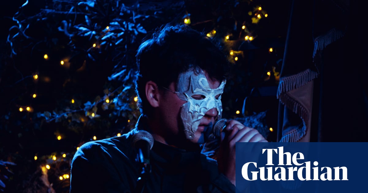
In this study, we have demonstrated a significant association between elevated GV levels and reduced LCR levels and the development and severity of brain injury in neonates with HIE following asphyxia. Furthermore, our findings suggest that the combined detection of GV and LCR holds potential predictive value for the early identification and evaluation of HIE.
The impairment of energy metabolism in cerebral cells following hypoxic-ischemia represents the initial stage in the progression of HIE. The accumulation of LAC in tissues during anaerobic metabolism, along with fluctuations in glucose levels under stress conditions, is closely associated with brain injury. Consequently, maintaining homeostasis and stabilizing glucose levels are crucial for mitigating brain injury after hypoxic-ischemia5,6,7. Research has demonstrated that the metabolic interplay between glucose and LAC is disrupted following HIE, leading to disorders in LAC and glucose metabolism20. Both hyperglycemia and hypoglycemia are prevalent among asphyxiated neonates21. While hypoglycemia is a significant risk factor for hypoxic-ischemic brain injury22hyperglycemia and erratic glucose fluctuations also impair cerebral cell energy metabolism, contributing to neurological damage23,24. Therefore, this retrospective study aims to predict the risk of HIE and evaluate its severity by monitoring changes in LAC and glucose levels.
GV represents the magnitude of glucose fluctuation within a given time period, reflecting an unstable condition where glucose levels oscillate between maximum and minimum values. Research has demonstrated that maintaining long-term stable glucose levels can decrease the incidence of brain injury and mortality risk25,26. Currently, GV has gained widespread application in adult endocrinology research; however, its utility in predicting neonatal HIE remains unreported. In this study, we found that the levels of GLU max-min, GLU SD, and GLU CV in the HIE group were significantly higher than those in the non-HIE group (P < 0.05). It is hypothesized that asphyxia-induced elevation in stress hormone secretion stimulated increased glycogenolysis and gluconeogenesis, leading to stress hyperglycemia. If hypoxia remains uncorrected, anaerobic metabolism intensifies, resulting in glycogen depletion and subsequent hypoglycemia. The significant fluctuations in glucose caused by this neuroendocrine response are closely associated with the development of hypoxic-ischemic brain injury19. Further comparative analysis of GV indexes among neonates in different clinical grading groups revealed that GLU max-min, GLU SD, and GLU CV progressively increased with the severity of HIE (P < 0.05). This observation is consistent with previous studies27,28which have demonstrated that significant fluctuations in glucose levels, particularly extreme highs and lows, can lead to abnormal cerebral blood flow and neuronal stress damage, thereby supporting our findings.
Changes in LAC levels serve as sensitive indicators of the degree of tissue and cellular hypoxia. Research has confirmed that neonates experiencing hypoxic-ischemic events exhibit increased anaerobic metabolism, which can lead to hyperlactatemia and accumulation of LAC in the brain. This process is closely associated with the onset and progression of HIE29. We observed that at 6 h post-admission, the LAC levels in the HIE group were significantly higher compared to the control group, whereas the LCR levels were notably lower. The LAC and LCR levels at 6 h post-admission were correlated with disease severity (P < 0.05), which was consistent with prior research29.
In the metabolic processes of neonatal HIE, glucose and LAC exhibit interactive relationships that collectively influence cerebral cellular energy metabolism20. This study further investigated the correlation between GV and LAC indices. The results demonstrated that no significant correlation was observed between GV and the lactate indices in the control group (p > 0.05). In contrast, within the HIE group, GLU max-min, GLU SD, and GLU CV were positively correlated with the LAC levels and negatively correlated with LCR (p < 0.05). Although the association within the HIE group was not very pronounced, potentially due to the low proportion of neonates with severe HIE, we hypothesize that this association may become stronger as the proportion of neonates with increasing HIE severity rises. It is hypothesized that following hypoxic-ischemic injury, neurons are unable to secure a stable energy supply due to erratic fluctuations in glucose levels, and LAC can serve as an alternative energy source, which has been termed “alternative brain fuel“30. Consequently, there exists a compensatory relationship between LAC and glucose in the metabolic alterations observed in HIE.
Therefore, GV and LAC levels are intricately associated with brain injury following hypoxic-ischemic events. It is crucial to maintain the homeostasis of glucose and LAC metabolism in the clinical management of asphyxiated neonates to mitigate the risk of brain injury. Multivariate logistic regression analysis revealed that increased GLU CV (OR: 4.752, 95% CI: 1.249–8.667) and decreased LCR (OR: 4.149, 95% CI: 1.378–7.382) were independent risk factors for HIE following asphyxia. Additionally, elevated GLU CV (OR: 3.718, 95% CI: 0.001–7.322) and reduced LCR (OR: 1.434, 95% CI: 1.001–1.945) were associated with an increased risk of moderate-severe HIE. These findings suggest that higher GV and lower LCR levels may play partial roles in the development and progression of HIE. However, GLU CV for moderate-severe HIE exhibit imprecision (owing to wide 95% CI), potentially influenced by the proportion of HIE severity. Consequently, these findings should be interpreted with clinical prudence and in conjunction with neuroimaging and neuroelectrophysiological data. Further analysis of the ROC curve demonstrated that the combination of GLU CV and LCR achieved the highest diagnostic efficiency for both the occurrence and severity of HIE. Specifically, the AUC for predicting HIE was 0.883, with a sensitivity of 84.2% and a specificity of 78.6%. For moderate-severe HIE, the AUC was 0.736, with a sensitivity of 90.0% and a specificity of 61.1%.
Our study has some limitations. First it is a single center study, with a relatively small sample size that precluded more in-depth analyses. Second, given that blood glucose monitoring was performed intermittently rather than continuously, the available blood glucose data fail to comprehensively capture the trends of blood glucose fluctuations following asphyxia. Furthermore, owing to the scarcity of blood glucose data within the first six hours postpartum, it was not feasible to analyze the significance of GV during the TH therapeutic intervention window for clinical decision-making purposes. Finally, this study did not evaluate the long-term neurodevelopmental outcomes in infants with HIE.