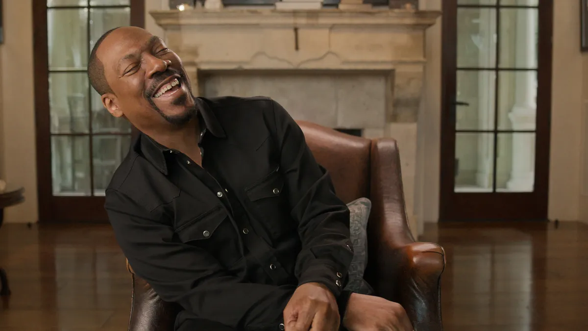
Giant unicystic ameloblastoma in a teenager of Azande tribe: a clinica

Background

Ameloblastoma, an uncommon yet locally aggressive benign odontogenic tumor of epithelial origin, ranks as the second most frequent jaw tumor, comprising roughly 1% of all such neoplasms. It predominantly affects the mandible (85%), with a predilection for posterior regions, while the maxilla is less commonly involved (15%).1,2 Individuals aged 30 to 60 are typically affected, with a slight female bias and an average diagnostic age of 35.5 years. Despite its benign nature, the tumor’s locally invasive growth carries a significant risk of recurrence.3

Globally, ameloblastoma is the most common benign odontogenic tumor, exhibiting geographical variations in incidence. It is relatively rare in non-African populations (around 0.5 per million annually) but significantly more prevalent in Nigeria and South Africa, where it constitutes a substantial proportion of odontogenic tumors. Incidence rates for Black populations in South Africa are notably higher than for White populations. While Ugandan-specific data are limited, Tanzanian studies indicate an incidence comparable to European populations (0.68 per million).4,5

Limited epidemiological data from sub-Saharan Africa may be attributed to underreporting in rural areas with poor healthcare access. Inadequate infrastructure and lack of universal health insurance in countries like Nigeria and Uganda contribute to late presentation, hindering early incidence tracking. Ameloblastoma in African individuals often presents as large, disfiguring tumors due to socioeconomic factors and limited awareness.6 Mandibular involvement, particularly in posterior regions, is common, with tumors frequently displaying cortical expansion and root resorption at diagnosis. Plexiform and follicular histological variants are more common in African populations, with fewer unicystic types.7

The higher incidence in African populations is thought to involve genetic and environmental factors, with some evidence suggesting population-specific mutations. While dietary factors have been speculated, no conclusive proof exists. This increased prevalence underscores the need for focused research and public health initiatives, potentially through collaborative networks like the proposed African Ameloblastoma Research Network.6

Clinically, ameloblastoma typically presents as a painless jaw or maxillary enlargement. Its slow but locally invasive growth can lead to root resorption, cortical perforation, and tooth displacement. Less common symptoms include pain or numbness. Diagnosis relies on histopathological examination, supplemented by radiological imaging, particularly CT scans, to differentiate it from other lesions. Accurate management necessitates integrating clinical, radiological, and histopathological findings.3,8

CT scans typically reveal a well-defined unilocular or multilocular radiolucent lesion, often causing bone expansion and tooth displacement. The multicystic variant is linked to higher recurrence rates and more aggressive behavior. Histopathologically, ameloblastoma has subtypes like follicular (most common), plexiform, and unicystic (better prognosis). The tumor features neoplastic cell nests with peripheral palisading of nuclei and loosely arranged central cells.1,9

Molecular studies have identified BRAF gene mutations in a significant proportion of mandibular ameloblastomas, offering potential for targeted therapies. Surgical excision is the primary treatment, aiming for complete removal. Approaches range from enucleation and curettage (for small, unicystic tumors, with higher recurrence risk) to more radical resections like segmental resection (lower recurrence). Reconstruction may be necessary post-surgery. Recent advances include BRAF inhibitors showing promise in neoadjuvant therapy. Metastatic ameloblastoma is rare. Recurrence rates are generally high, especially with conservative treatments and multicystic histology.1,3,10

A case of a large mandibular ameloblastoma in a young individual in a resource-limited setting highlights diagnostic and management challenges, emphasizing the need for improved awareness and multidisciplinary expertise for timely intervention in under-reported populations.

Case Presentation

A 17-year-old female of the Azande tribe, Bantu ethnicity, presented to the maxillofacial outpatient department at Kampala International University Teaching Hospital complaining for a massive swelling in the right mandibular region for the last 4 years. The swelling was a painless massive swelling not associated with any other symptoms: no dysphagia, no odynophagia, no headache. The swelling started spontaneously and gradually increased to its current size.

The patient mentioned no loss of weight, no history of chronic illness, or known familial malignancies. No history of head injury and no history of tooth extraction. A previous biopsy had been done one year ago, and the histopathology reported a diagnosis of Ameloblastoma.

On maxillofacial examination, no signs of emaciation, with normal vital signs; however, on extra-oral examination, a right-sided facial asymmetry was observed due to a marked mandibular swelling, extending from the left canine region to the right condyle, coronoid process, and sigmoid notch, significantly altering normal anatomy and preventing mouth closure. The overlying skin was stretched but retained normal coloration, with multiple linear burn scars resulting from traditional herbal therapy and a prior biopsy site. No ulceration, erythema, or signs of infection were noted. The underlying tissues were notably thinned. While the cortical bones remained partially intact, they exhibited extreme thinning. The swelling was firm with areas of fluctuance (solid-cystic consistency), non-mobile, and fixed to the underlying bone (Figure 1). On intraoral examination, buccal and lingual cortical expansion was present with intact mucosa, without ulceration or lingual involvement. The enlarged mandibular swelling displaced the tongue to the left, with a single molar and premolars supported by mucosa. The occlusal plane disruption led to displacement of adjacent teeth, contributing to the observed malocclusion. The maxillary teeth on the right side were tilted palatally. No lymph nodes were palpated.

Figure 1 Right mandibular mass deforming the face: anterior (A), right latero-posterior (B), and left lateral (C) views.

A CT Scan showed a large mass lesion (approximately 13.8 x 11.5×11 cm) arising from the body of the mandible on the right side. It was covered with bone cortex on most of its surface, except for a few defects noted. The matrix was primarily translucent and non-enhancing; nevertheless, its medial wall had an enhancing irregular mass lesion with a nodular surface. Molar and premolar teeth with no roots were observed on its medial surface. Calcifications in the matrix were absent. The rest of the mandible was normal, as the maxilla, the buccal lining and the tongue, as well. These findings were consistent with a unilocular right mandibular cystic lesion (Figure 2).

Figure 2 Unilocular translucent mass lesion arising from the body of the right mandible: axial (A), right lateral (B), and posterior (C) views.

An incisional biopsy was conducted to confirm the diagnosis, since the previous biopsy was performed out of our facility and the report was not available. During sample collection, a cloudy not foul-smelling fluid leaked from the mass. Both samples (tissue and fluid) were submitted for histopathological and cytopathological examination. Hematoxylin and Eosin stained smears showed dispersed squamous cells within an extensive mixed inflammatory background, without evidence of dyskaryosis, atypia, or malignancy. These features were those of an inflammatory cyst.

Multiple fragments of tissue, white with brown areas, firm, largest measuring 1.8×1 x 0.3 cm, and the smallest 0.5×0.4 x 0.2 cm, with a solid, homogenous white cut surface, were histologically analyzed. Sections stained with Hematoxylin and Eosin revealed odontogenic epithelial cells arranged in follicular and plexiform configurations. The columnar cells had hyperchromatic nuclei at basal layer, exhibiting peripheral palisading, and showed Vickers-Gorlin change (reverse polarization away from basement membrane) and subnuclear vacuolization. The suprabasal cells had a loose, network-like arrangement (Figure 3). These features are those of an Ameloblastoma. A chest X-ray was done with a normal result. Laboratory investigations (CBC, RFT, ESR, Coagulation profile) were unremarkable.

Figure 3 Hematoxylin and Eosin stained sections showing odontogenic epithelial cells arranged in follicular and plexiform configurations (AC: x40, x100, and x200 respectively). The columnar cells have hyperchromatic nuclei at basal layer, exhibiting peripheral palisading, and showed Vickers-Gorlin change (reverse nuclear polarity) and subnuclear vacuolization. The suprabasal cells have a loose, network-like arrangement (D: x400).

Based on the clinical presentation, radiological findings, and pathological findings, which suggested an ameloblastoma, the patient was planned for operation. It was decided that the patient should undergo an elective right total hemi-mandibulectomy (with exarticulation of the mandibular condyle and extension to the distal of the first premolar on the contralateral side). The informed consent for surgery was obtained from the patient’s mother. Under general anesthesia, an oro-tracheal intubation was done followed by open tracheostomy. A submandibular incision was made, extending from the left premolar region to the right mandibular angle, through the skin, subcutaneous tissue, and thinned muscular layer until the tumor was fully exposed. Intraorally, a perilesional incision was performed from the right retromolar region to the left second premolar, excising the extremely thin tissue in direct contact with the lesion. The mandibular bone was then sectioned and exposed up to the temporomandibular joint, followed by disarticulation of the condyle (Figure 4A and B). Intraoperative findings were consistent with a left temporomandibular joint dislocation that was corrected intraoperatively. Cosmetic surgery was used for the skin closure (Figure 5A). The patient was observed in the intensive care unit for immediate follow up. After 12 hours, the patient was able to maintain the airways but the tracheostomy was maintained for one week for prophylaxis. The feeding nasogastric tube was also maintained in situ for 2 weeks. In post-operative period, IV ceftriaxone 2g daily was given for 7 days, IV tramadol 100mg was given two times a day for 2 days and Acetaminophen 1g three times a day war given for 5 days. Wound dressing was performed every two days for 10 days.

Figure 4 Odontogenic unilocular specimen (19 x 16×10 cm), dark gray to brown, with attached skin (14 x 5 cm), with mandible, with teeth (A and B), and intraluminal growth (C). The wall is variably thickened (thickest 1cm), and the lining rough (C).

Figure 5 Cosmetic surgery was used for the skin closure (A). Patient during follow-up: one week after patient discharge (B) and one month (C) later.

The excised mass was submitted for histopathological examination. It was a single odontogenic unilocular specimen (19 x 16×10 cm), dark gray to brown, with attached skin (14 x 5 cm), with mandible, teeth, and intraluminal growth. No obvious lesion on the skin. The wall was variably thickened (thickest 1cm), and the lining rough. The sliced surface exhibited firmness with hardened regions and a gritty texture (Figure 4C). The histological features were those of Ameloblastoma, as presented above. Compiling the findings, this tumor was classified as a unicystic ameloblastoma, the intraluminal variant.

The patient was discharged two weeks post-surgery, able to take per os feeding. During the short-term follow-up after being discharged, it revealed a healed surgical site (Figure 5B and C) with no complaint of numbness of the right lower lip. Follow-up clinical and radiographic examination is essential as ameloblastoma has a high recurrence rate after 3–5 years. Depending on the follow-up findings, a wide-margin excision and reconstruction can be performed. Also, prosthetic rehabilitation is further planned for the patient to correct the surgical defect of the mandible and missing teeth.

Discussion

Ameloblastoma, a benign yet locally invasive odontogenic tumor, poses diagnostic challenges due to its resemblance to other jaw tumors. Accurate differentiation necessitates a comprehensive evaluation encompassing clinical, radiographic, and histopathological findings to distinguish it from both benign and malignant entities. Clinically, it typically manifests as a painless, slow-growing swelling in the mandible or maxilla, most commonly affecting individuals in their third and fourth decades, with a slight male predilection. However, clinical presentation alone is insufficient for diagnosis, as lesions like odontogenic keratocysts (OKCs) and dentigerous cysts (DCs) can exhibit similar symptoms.2,11

Radiographic analysis plays a crucial role in differentiating ameloblastoma. It frequently occurs in the posterior mandible and often exhibits buccal expansion. Radiographically, it may appear as unilocular or multilocular radiolucencies, sometimes with solid components. Notably, ameloblastomas are more prone to causing tooth displacement and resorption compared to OKCs and DCs. In contrast, OKCs typically present as unilocular radiolucencies and may show a dentigerous association with adjacent teeth.3,12

Histopathological examination remains the gold standard for ameloblastoma diagnosis. It is characterized by odontogenic epithelium arranged in follicular or plexiform patterns, featuring peripheral palisading and stellate reticulum-like cells. The use of lectins, such as UEA-I and BSA-I, can aid in distinguishing ameloblastoma from non-neoplastic cysts, as ameloblastomas typically show negative staining, unlike odontogenic cysts. Immunohistochemistry further assists in differentiation, with consistent calretinin expression in ameloblastoma, absent in keratocystic odontogenic tumors.13,14 The KIAA0101 marker helps differentiate ameloblastoma from its malignant counterpart, ameloblastic carcinoma. Recent advancements in deep learning applied to panoramic radiographs, utilizing convolutional neural networks like EfficientNet, have shown promising accuracy in distinguishing ameloblastoma from OKC.15,16 Cone Beam Imaging CT provides detailed information on lesion characteristics, aiding in differentiation based on features like the long-to-short length ratio and cortical expansion angle. Fine-needle aspiration offers a preliminary diagnostic tool, but its utility is limited by overlapping cytological features with other lesions, including squamous cell carcinoma in the acanthomatous variant of ameloblastoma. Molecular investigations, identifying mutations like those in the SMARCB1 gene, are increasingly used to aid in diagnosis and prognosis, potentially opening avenues for targeted therapies.17

Unicystic ameloblastoma, a variant, can mimic odontogenic cysts clinically and radiographically. Histologically, it presents as a single cystic sac lined by ameloblastomatous epithelium, often with focal intraluminal proliferation. Desmoplastic ameloblastoma, a rare variant, exhibits distinct radiographic (mixed radiolucent-radiopaque with ill-defined borders) and histological (dense desmoplastic stroma surrounding epithelial islands) features.18,19 The definitive diagnosis of unicystic ameloblastoma, particularly the intraluminal form, relies on thorough evaluation. While initial presentation might resemble other odontogenic cysts, radiographic findings of a unilocular radiolucency with bone involvement and an intraluminal nodular component raise suspicion. Critically, histopathological examination confirming ameloblastomatous epithelium with characteristic patterns within a unicystic structure definitively classifies it, excluding other similar lesions.8

Unicystic ameloblastoma is a specific entity characterized by its monocystic nature and unique histological features, believed to originate from epithelial remnants of Malassez. Considered a neoplasm with proliferative and invasive potential, it typically appears as a well-circumscribed, unilocular radiolucency. While often asymptomatic, it can cause swelling and facial asymmetry. It is more common in males and typically affects individuals in their second and third decades. Histologically, it is categorized into luminal, intraluminal, and mural subtypes.1,8 Our case was an intraluminal subtype. The proliferative potential of unicystic ameloblastoma is higher than conventional types, indicated by PCNA and Ki-67 marker expression, with a higher recurrence risk associated with tumors invading the cyst wall. Histology involves a cystic lesion lined by ameloblastomatous epithelium, potentially exhibiting follicular, plexiform, and acanthomatous changes, along with rete processes and inflammatory cells in the connective tissue. Atypical histological variations, such as mucous cell and granular cell differentiation, have been documented.20

Histological diagnosis can be challenging due to similarities with other odontogenic cysts. The presence of ameloblastomatous lining and tumor growth within the cyst is crucial for differentiation. Unicystic ameloblastoma is more prevalent in males and commonly occurs in the posterior mandible, with a mean age of onset in the mid-twenties. Management typically involves surgical resection with adequate margins, and long-term follow-up is essential due to the potential for delayed recurrence, influenced by histological subtype and invasion of the cyst wall.21

The identified histological characteristics, such as ameloblastomatous epithelium with specific arrangements, serve as definitive diagnostic indicators, differentiating unicystic ameloblastoma from simple odontogenic cysts. The unilocular appearance, while radiographically similar to other cysts, denotes the unicystic variant. The observed intraluminal nodular growth suggests a higher proliferative capacity and recurrence risk, necessitating careful surgical excision and extended follow-up.22,23

Unicystic ameloblastoma generally has a favorable prognosis, especially with prompt identification and management, exhibiting lower recurrence rates than solid or multicystic types with adequate surgery. Prognosis is influenced by histological subtype, tumor size, and invasive features, with the mural subtype showing a higher recurrence rate. Large tumors, root resorption, and cortical perforation are significant predictors of recurrence. Studies indicate relatively low recurrence rates with appropriate follow-up, including regular clinical and radiographic assessments for at least 5–10 years.24,25 Patient education on recurrence signs is crucial for early detection. Delayed presentation, as seen in the described case, can be influenced by socioeconomic factors and cultural beliefs prevalent in resource-limited settings, posing challenges for healthcare delivery, including access to advanced diagnostics, specialized personnel, and consistent follow-up care. Sustainable healthcare infrastructure and culturally sensitive approaches are essential for managing such cases effectively.25,26

Conclusion

This case of a giant unicystic ameloblastoma in a 17-year-old Azande female from Uganda underscores the challenges of managing significant odontogenic tumors in resource-limited settings. The delayed presentation, resulting in a massive lesion, highlights the impact of limited healthcare access and awareness in sub-Saharan Africa. Successful management through radical surgical resection (hemi-mandibulectomy) was achieved, emphasizing the necessity of definitive surgical intervention for such aggressive benign tumors. This report contributes to the limited literature on ameloblastoma in this region and emphasizes the critical need for improved diagnostic pathways, increased awareness, and accessible specialized care to facilitate earlier intervention and potentially less morbid outcomes for patients in similar situations. Long-term follow-up remains crucial to monitor for recurrence, and further reconstructive efforts are planned to address the functional and aesthetic deficits.

Data Sharing Statement

The data presented in this study are available on request from the corresponding author. The data are not publicly available due to patient confidentiality pertaining to medical electronic health records from which our data were collected.

Ethics Approval and Consent to Participate

Institutional ethical approval was not required to publish the case details.

Consent for Publication

Written informed consent was obtained from the mother of the patient for publication of this case report and any accompanying images. A copy of the written consent is available for review by the Editor-in-Chief of this journal.

Author Contributions

All authors made a significant contribution to the work reported; took part in drafting and reviewing the article; gave final approval of the version to be published; have agreed on the journal to which the article has been submitted; and agree to be accountable for all aspects of the work.

Funding

The authors declare that no funds, grants, or other support were received during the preparation of this manuscript.

Disclosure

The authors declare that they have no conflicts of interest in this work.

References

1. Singh RK, Jha S, Mala M, Kumar A, Goit S. Massive mandibular ameloblastoma masquerading malignancy: a unique case. IP J Otorhinolaryngol Allied Sci. 2024;7:16–19. doi:10.18231/j.ijoas.2024.005

2. Walia C, Al Rashidi AA, Mohammed SWA, Roy S. Ameloblatoma: an aggressive odontogenic neoplasm with altered vickers and gorlin criteria. J Oral Med Oral Surgery, Oral Pathol Oral Radiol. 2024;10:145–148. doi:10.18231/j.jooo.2024.027

3. jaya NS, Chacko D. Clinicopathologic profile and surgical modalities in mandibular ameloblastoma: a descriptive study. J Craniofac Surg. 2024;35:158–162. doi:10.1097/SCS.0000000000009706

4. Odukoya O. Odontogenic tumors: analysis of 289 Nigerian cases. J Oral Pathol Med. 1995;24:454–457. doi:10.1111/j.1600-0714.1995.tb01133.x

5. Shear M, Singh S. Age-Standardized Incidence Rates of Ameloblastoma and Dentigerous Cyst on the Witwatersrand. South Africa; 1978.

6. Arotiba G, Hille J, Adeola H, Odhiambo W. Ameloblastoma in Black Africans the need for multi-national collaborative research. 2017.

7. Ranchod S, Titinchi F, Behardien N, Morkel J. Ameloblastoma of the mandible: analysis of radiographic and histopathological features. J Oral Med Oral Surg. 2021;27. doi:10.1051/mbcb/2020051

8. Sebastián Pinos Pinos L, Ortiz JAR, Fernanda Torres Calle M, Mendoza FMV. Unicystic ameloblastoma: literature review. World J Adv Res Rev. 2021;10:95–103. doi:10.30574/wjarr.2021.10.3.0240

9. Merbold L, Smit C, Uys A. Maxillofacial Radiology. South African Dent J. 2023;78:278–288. doi:10.17159/sadj.v78i05.16871

10. Cunha LR de A, Franco AG, Minervino VD, et al. Analysis of surgical approaches for the treatment of ameloblastoma: an integrative review. Cent Pesqui Avançadas Em Qual Vida. 2024. doi:10.36692/v16n1-49r

11. Boffano P, Cavarra F, Tricarico G, et al. The epidemiology and management of ameloblastomas: a European multicenter study. J Cranio-Maxillofacial Surg. 2021;49:1107–1112. doi:10.1016/j.jcms.2021.09.007

12. Meng Y, Zhang Y-Q, Xin Y, Zhao Y-N, Chen Y, Liu D-G. Imaging analysis of ameloblastoma, odontogenic keratocyst and dentigerous cyst in the maxilla using spiral CT and cone beam CT. Chin J Stomatol. 2018;53.

13. Ghai S. Ameloblastoma: an updated narrative review of an enigmatic tumor. Cureus. 2022. doi:10.7759/cureus.27734

14. kuda singappulige niluka darshani gunawardhana, primali rukmal jayasooriya, wanninayake mudiyanselage tilakaratne. diagnostic dilemma of unicystic ameloblastoma: novel parameters to differentiate unicystic ameloblastoma from common odontogenic cysts. J Investig Clin Dent. 2013.

15. Nassef AH, Fouad HMA, Raslan HS, Zahran AM, Hussein AM. Kiaa0101 immunohistochemical expression in diagnostic dilemma between ameloblastoma and ameloblastic carcinoma (In Vitro Study). Alexandria Dent J. 2024. doi:10.21608/adjalexu.2023.244645.1427

16. Min Li CCM, Zhang J-Y, Gang L. Application of deep learning in differential diagnosis of ameloblastoma and odontogenic keratocyst based on panoramic radiographs. Acta Acad Med Sin. 2023;45:273–279.

17. Ariji Y, Morita M, Katsumata A, et al. Imaging features contributing to the diagnosis of ameloblastomas and keratocystic odontogenic tumours: logistic regression analysis. Dentomaxillofacial Radiol. 2011;40:133–140. doi:10.1259/dmfr/24726112

18. Doddawad VG, Shivananda S, Sunita S, Sowmya S. A rare variant of ameloblastoma of the oral cavity: a review and case report. J Datta Meghe Inst Med Sci Univ. 2024;19:173–176. doi:10.4103/jdmimsu.jdmimsu_110_17

19. Pandarinath BG, Kushalvasanth S, Dinkar D. A mixed radiopaque-radiolucent lesion in the anterior mandible associated with multiple impacted teeth. A radiodiagnostic challenge? J Cancer Res Ther. 2019;15:700–703. doi:10.4103/jcrt.JCRT_721_16

20. Jain K, Sharma G, Kardam P, Mehendiratta M. Unicystic ameloblastoma of mandible with an unusual diverse histopathology: a rare case report. J Clin Diagn Res. 2017;11:4–5. doi:10.7860/JCDR/2017/24192.9615

21. Anand R, Narwal A, Pandiar D, et al. clinicopathological profile of 80 cases of unicystic ameloblastoma aided by a histopathological comparison using modified Philipsen–Reichart classification and marx–stern classification. Head Neck Pathol. 2021;15:875–881. doi:10.1007/s12105-021-01307-7

22. Sah P, Menon A, Kamath A, Chandrashekar C, Carnelio S, Radhakrishnan R. Role of immunomarkers in the clinicopathological analysis of unicystic ameloblastoma. Dis Markers. 2013;35:481–488. doi:10.1155/2013/517834

23. Saravanakumar B, Parthiban J, Aarthi Nisha V, Sarumathi T, Prakash CA. Unicystic ameloblastoma of the mandible – report of two cases with review of literature. J Clin Diagn Res. 2014;8:7–9. doi:10.7860/JCDR/2014/8921.4238

24. Isolan CP, Moreira AG, Edges A, Post LK, Aitken-Saavedra JP. Successful conservative treatment of a mandibular unicystic ameloblastoma: 13-year follow-up. J Clin Exp Dent. 2018;10:1123–1126. doi:10.4317/jced.54897

25. Zheng CY, Cao R, Hong WS, Sheng MC, Hu YJ. Marsupialisation for the treatment of unicystic ameloblastoma of the mandible: a long-term follow up of 116 cases. Br J Oral Maxillofac Surg. 2019;57:655–662. doi:10.1016/j.bjoms.2019.06.002

26. Titinchi F, Brennan PA. Unicystic ameloblastoma: analysis of surgical management and recurrence risk factors. Br J Oral Maxillofac Surg. 2022;60:337–342. doi:10.1016/j.bjoms.2021.07.022

Continue Reading