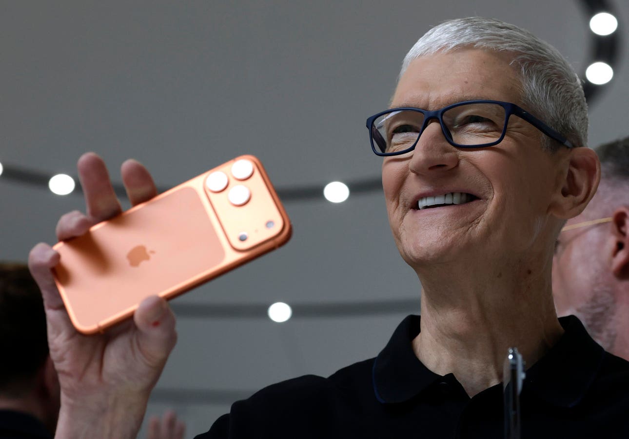
Introduction
Low back pain (LBP) is a type of pain and discomfort that affects the lumbar region of the spine. It is a widespread health concern globally, with an annual prevalence of 38%, and is linked to a significant socioeconomic burden.1 Facet cyst is one of the causes of low back pain. Facet cysts, also known as zygapophysial joint cysts, were first described in 1877 as a cystic formation involving the zygapophysial joint in the epidural, foraminal, or paravertebral area.2,3 While the exact cause of the cyst formation remains unknown, it is thought to be related to a complex degeneration process of the facet joints, especially of the mobile spine, in addition to intervertebral disc degeneration.4 Facet cysts, once thought to be a rare cause of symptomatic nerve compression with an incidence around 6.5%, are actually more common in older age groups, affecting up to 10% of patients over 70 who experience lumbar or radicular pain.3,5,6 Furthermore, facet cysts can cause significant spinal pain and nerve dysfunction and are associated with significant morbidity and reduction in quality of life if not managed properly.7,8
Facet cysts may be managed conservatively or surgically, depending on the degree of nerve compression and symptom severity. Recent study found percutaneous procedures to have a 55.8% success rate, though surgical management was still required for 38.7% of the cases for continued symptom relief.9 This technical note aims to provide a detailed, step-by-step description of the Biportal Endoscopic Spine Surgery (BESS) technique using both interlaminar ipsilateral and contralateral approaches. The goal is to offer practical surgical guidance and emphasize the advantages of Biportal Endoscopic Spine Surgery (BESS) as a minimally invasive alternative to traditional open spine surgery, especially for lumbar spine decompression.
Case Report
Case 1
A 63-year-old female presented with lumbar back pain radiating to the left feet along the L4-L5 distribution that had been present for 10 days. The pain is felt in all position and is very severe until the patient required the use of wheelchair. There are no history of trauma or precipitating events. Prior to assessment, patient has received pain medications, ie, NSAIDs and tramadol, the patient reported that pain was slightly relieved by pain medication consumption, however the pain immediately return when medication was stopped. The patient then underwent diagnostic testing and MRI result showed left L5-S1 facet cysts with associated nerve compression (Figure 1). An interlaminar Biportal Endoscopic Spine Surgery (BESS) with contralateral approach were then performed on the patient. After the procedure, the patient was in a stable condition and felt significant improvement of her symptoms. No post-operative complication was observed in the patient.
Figure 1 Preoperative magnetic resonance imaging (MRI) (A) sagittal and (B) axial view showing a facet cyst in level L4-5 with associated nerve compression.
|
Case 2
A 58-year-old male presented with severe left-leg pain that originated from his lower back for 5 weeks. The pain was especially felt in along the anterior thigh, down to the medial part of the lower left leg, reaching the thumb, along the L4 distribution. The patient experienced no sensoric or motoric deficits. History of trauma or precipitating events was denied. Prior to assessment, patient has received NSAIDs and opioids which only offers temporary relief. Patient then underwent MRI testing, showing an L3-L4 facet cyst compressing the associated nerve (Figure 2). No other significant medical history was present. Patient then underwent ipsilateral Biportal Endoscopic Spine Surgery (BESS) with immediate symptomatic improvement following the procedure. No post-operative complication was observed in the patient.
![]() |
Figure 2 Preoperative magnetic resonance imaging (MRI) showing a facet cyst in level L4-L5 (A) sagittal view. The white dot line serves as a marker indicating the L4 vertebral level (B) axial view.
|
Materials and Methods
Surgical Technique
All procedures were performed under general anesthesia with fluoroscopic guidance to confirm the spinal level. Preoperative MRI and intraoperative fluoroscopy were used to determine the optimal approach (ipsilateral or contralateral) based on the location and extent of the facet cyst, its relationship to the dura, and suspected adhesions. An ipsilateral approach was preferred for laterally located cysts, whereas a contralateral approach was favored for medially located cysts.
Portal Placement and Visualization
The procedure utilized the Joimax TESSYS biportal endoscopic system. The patient was placed in the prone position, and fluoroscopic guidance was used to localize the correct spinal level and determine optimal portal positioning. Initial skin markings were made under both anteroposterior (AP) and lateral fluoroscopic views to identify the anatomical landmarks necessary for accurate access.
In the AP view, a midline skin marking was created by connecting the medial borders of the pedicles of the vertebrae above and below the targeted lesion. This line served as a reference for the interlaminar window and helped determine whether the ipsilateral or contralateral side would be used, depending on the preoperative surgical plan. In the lateral view, a second marking was made that passed through the facet joint, the intervertebral disc space, and the interlaminar window, oriented according to the specific location of the facet cyst (Figure 3).
![]() |
Figure 3 Marking was made for endoscopic portals insertion.
|
The intersection of these two lines defines the anatomical center of the working zone. Two portals were created with skin incisions approximately 7–15 mm in size, placed at superior and inferior points relative to the intersection point (Figure 4). A blunt dilator was inserted into the lateral edge of the interlaminar space, followed by an obturator sheath with a beveled opening directed toward the ligamentum flavum.
![]() |
Figure 4 Two metal trocars were inserted through a small incision—one for the endoscope providing real-time visualization and the other as a working instrument channel.
|
For the left-sided portal placement, the endoscope was inserted through the superior portal and held in the surgeon’s left hand, while the inferior portal was used for instrumentation and held in the surgeon’s right hand. In a right-sided placement, the configuration was reversed. Proper portal positioning was confirmed by visualizing the working instruments through the endoscope, ensuring they shared the same target zone. The contralateral approach provides a more horizontal working trajectory, allowing better visualization of the cyst, especially when it extends medially or under the dura. Meanwhile, the ipsilateral approach may have a steeper angle, making it harder to access deeper cysts without excessive bone removal.
Flavectomy and Laminotomy
Continuous saline irrigation was maintained to ensure clear endoscopic visualization and prevent tissue stagnation. Soft tissue dissection and exposure of the ligamentum flavum were achieved using endoscopic burrs and radiofrequency probes.
The endoscope was initially docked on the ipsilateral spinolaminar junction. Contralateral sublaminar drilling was performed to create a working corridor extending toward the medial portion of the contralateral foramen. A partial laminotomy is performed using burr or Kerrison rongeur. The middle portion of the lamina and the spinous process were then partially removed using a Kerrison rongeur and drill, which allows for the visualization of the ipsilateral and contralateral ligamentum flavum. The thickened ligamentum flavum was then partially resected and removed using endoscopic forces to enable the visualization of the cyst, dura, and contralateral nerve decompression (Figure 5).
![]() |
Figure 5 Endoscopic images using a contralateral approach in the lumbar site level L4-L5 showing a thickened flavum ligament covering the cyst were detected, and removal of flavum ligament was performed.
|
Cyst Removal
As the foramen was expanded, the endoscope was advanced further through the caudal-ventral region of the foramen to examine the extraforaminal cyst. Once the facet cyst is visualized, a fine dissector is used to dissect the cyst from surrounding tissues. If adherent to the dura or nerve root, microscissors may be used to perform adhesiolysis.
The cyst may be removed in piecemeal fashion to avoid excessive nerve root manipulation or in-toto using a Kerrison rongeur (Figure 6). After cyst excision, the nerve root was carefully examined to ensure full decompression and structural integrity. The contralateral approach proved advantageous by avoiding nerve retraction, as the working corridor passed beneath the nerve root. In contrast, the ipsilateral approach often required retraction of the nerve to access the cyst, increasing the potential for iatrogenic injury (Figure 7). Before closure, hemostasis is ensured using a radiofrequency device, and a drainage catheter is inserted to prevent hematoma formation.
![]() |
Figure 6 Endoscopic image using contralateral approach in lumbar site level L4-L5 showing (A) Adhesion site between the cyst and dura mater was identified (B & (C) Cyst was carefully dissected with respect to the dura mater.
|
![]() |
Figure 7 The comparison trajectory (red arrow) of contralateral and ipsilateral on axial lumbar MRI images. (A) The contralateral approach passes under the spinous process to reach the opposite side, allowing access to the lateral recess and foramen while preserving the facet joint on the working side. (B) The ipsilateral approach enters directly through the same-side interlaminar space, giving straight access to the target area but coming closer to the facet joint and exiting nerve root.
|
Discussion
Synovial facet cysts are degenerative lesions of the lumbar spine that can contribute to lumbar spinal stenosis, often resulting in lower back pain and radicular symptoms. Diagnosis is primarily established through MRI studies of the lumbar spine, which can reveal the presence and extent of the cyst. Facet cysts can develop throughout the spine, from C1-C2 to the lumbosacral junction, with approximately 90% occurring in the lumbar region.10 Lumbar facet cysts are known to contribute to nerve root dysfunction and pain. Initial treatment typically involves nonoperative management, including NSAIDs and physical therapy, to alleviate symptoms and improve function. Some studies have indicated that percutaneous procedures, such as steroid injections or cyst aspiration, are generally ineffective and surgical intervention is considered for patients experiencing progressive, disabling pain unresponsive to conservative treatment or those with worsening neurological deficits.11–14 The endoscopic approach has several advantages, including smaller laminotomy, less muscle damage, and smaller incision, which preserves as much anatomical structure as possible, minimizes postoperative instability, has a lower infection rate due to being a minimally invasive approach and does not require general anesthesia.15,16 This advantage is particularly significant in symptomatic facet cyst cases, given that the prevalence of facet cysts increases linearly with age and is more common in elderly patients who may have multiple comorbidities.3 The safety profile and tolerability of endoscopic procedures for facet cyst removal in the elderly population have also been reported by other studies.16,17
Facet cysts can be removed using either a contralateral or ipsilateral biportal endoscopic approach, each offering specific advantages and limitations depending on the cyst’s characteristics and location. The contralateral approach accesses the cyst from the opposite side, crossing the midline to enhance visualization of medially located lesions or those extending beneath the dura. This technique reduces nerve root retraction, thereby lowering the risk of postoperative radiculopathy, and helps preserve spinal stability by minimizing facet joint resection. Additionally, it provides a broader surgical corridor via an extended laminotomy, typically sparing the ipsilateral facet joint. By limiting facet disruption, this approach mitigates the risk of long-term spinal instability and degenerative changes, while also allowing for more complete cyst excision, which may decrease recurrence rates.
In contrast, the ipsilateral approach offers a more direct route to the lesion, making it more practical for superficially located or lateral cysts with limited dural involvement. However, it often requires more extensive facet joint removal and greater nerve root manipulation, which can increase the potential for neural irritation and postoperative instability. Moreover, identifying the cyst capsule, particularly when it adheres to the dura, is more challenging through this trajectory, complicating the dissection and necessitating broader facet resection for adequate access.
The contralateral technique also facilitates clearer visualization of the normal dura and the adhesion plane between the dura and cyst, simplifying the dissection process. This benefit is supported by Heo et al, who favored the contralateral approach for more meticulous separation of adherent lesions.15 Furthermore, contralateral decompression has been shown to better preserve the facet joint. Matsumura et al reported higher facet preservation rates with the contralateral technique compared to the ipsilateral one (95% vs 80%).18 Similarly, a recent study found that the contralateral interlaminar approach in biportal endoscopic decompression resulted in superior lateral recess clearance and greater facet preservation than the ipsilateral approach (92% vs 84%).18 These outcomes are likely attributable to the oblique trajectory of the contralateral approach, which aligns more naturally with the anatomical orientation of the recess roof. In contrast, the ipsilateral route often necessitates more aggressive facet removal to achieve comparable exposure. Additionally, surgeon hand dominance and lesion laterality may influence the technical complexity of the procedure. Bone work typically begins at the interlaminar space and proceeds cranially, with the dominant hand controlling instruments. Right-handed surgeons may find the left-sided approach more ergonomic, whereas left-handed surgeons may prefer the right.19
A comparative summary of the advantages and limitations of both approaches is presented in Table 1. As this study does not include statistical analysis, interpretation of the results should be approached with careful clinical judgment. A longer cohort study with longer follow-up is necessary to validate the broader applicability of this technique.
![]() |
Table 1 Comparison Between Contralateral and Ipsilateral Approach for Biportal Endoscopic Facet Cyst Removal
|
Conclusion
Both the contralateral and ipsilateral approaches in biportal endoscopic facet cyst removal offer distinct advantages, making the choice highly dependent on cyst location and anatomical considerations. The contralateral approach provides a better visualization, minimizes nerve root retraction, and enhances structural preservation, making it the preferred technique for medially located, adherent, or recurrent cysts in this scenario. In contrast, the ipsilateral approach offers a more direct pathway for laterally positioned cysts but may require greater facet joint removal and nerve root manipulation. Given these considerations, a tailored surgical approach is essential, balancing cyst accessibility, nerve safety, and spinal stability to optimize clinical outcomes.
Ethics Approval and Informed Consent
This study has been approved by The Research Ethics Commission of Fakultas Kedokteran Universitas Trisakti (registration number 005/KER/FK/02/2025) and informed consent was obtained from the patients. Institutional approval was obtained prior to the publication of this case report.
Consent for Publication
The patients have obtained consent for publication.
Author Contributions
All authors made a significant contribution to the work reported, whether that is in the conception, study design, execution, acquisition of data, analysis and interpretation, or in all these areas; took part in drafting, revising or critically reviewing the article; gave final approval of the version to be published; have agreed on the journal to which the article has been submitted; and agree to be accountable for all aspects of the work.
Funding
The authors received no financial support for this article’s research, authorship, and/or publication.
Disclosure
The authors report no conflicts of interest in this work.
References
1. Özüdoğru A, Canlı M, Ceylan İ, Kuzu Ş, Alkan H, Karaçay BÇ. Five times sit-to-stand test in people with non-specific chronic low back pain—a cross-sectional test–retest reliability study. Ir J Med Sci. 2023;192(4):1903–1908. doi:10.1007/s11845-022-03223-3
2. Rosenstock T, Vajkoczy P. New classification of facet joint synovial cysts. Acta Neurochir. 2020;162(4):929–936. doi:10.1007/s00701-020-04264-2
3. Janssen SJ, Ogink PT, Schwab JH. The prevalence of incidental and symptomatic lumbar synovial facet cysts. Clin Spine Surg. 2018;31(5):E296–E301. doi:10.1097/BSD.0000000000000648
4. Bydon A, Xu R, Parker SL, et al. Recurrent back and leg pain and cyst reformation after surgical resection of spinal synovial cysts: systematic review of reported postoperative outcomes. Spine J. 2010;10(9):820–826. doi:10.1016/j.spinee.2010.04.010
5. Boody BS, Savage JW. Evaluation and treatment of lumbar facet cysts. J Am Academy Orthopaedic Surg. 2016;24(12):829–842. doi:10.5435/JAAOS-D-14-00461
6. Lalanne LB, Carmona RL, Cirillo Totera JI, Lemos FA, Wilson JTM, Beaulieu Montoya AM. Surgically managed symptomatic intraspinal lumbar facet synovial cyst outcome of surgical treatment with resection and instrumented posterolateral fusion, a case series. BMC Surg. 2022;22(1). doi:10.1186/s12893-022-01712-x
7. Kursumovic A, Bostelmann R, Gollwitzer M, Rath S, Steiger HJ, Petridis AK. Intraspinal lumbar juxtaarticular cyst treatment through ct-guided percutaneous induced rupture results in a favorable patient outcome. Clin Pract. 2016;6(4):866. doi:10.4081/cp.2016.866
8. Mobbs R, Campbell R, Phan K. NeuroSpine Surgery Research Group (NSURG) classification system for grading lumbar synovial cysts. Orthop Surg. 2018;10(1):3–7. doi:10.1111/os.12363
9. Campbell RJ, Mobbs RJ, Phan K. Percutaneous resolution of lumbar facet joint cysts as an alternative treatment to surgery: a meta-analysis. J Spine Surg. 2016;2(1):85–86. doi:10.21037/jss.2016.01.03
10. Choudhri HF, Perling LH. Diagnosis and management of juxtafacet cysts. Neurosurg Focus. 2006;20(3):1–4. doi:10.3171/foc.2006.20.3.2
11. Hsu KY, Zucherman JF, Shea WJ, Jeffrey RA. Lumbar intraspinal synovial and ganglion cysts (Facet cysts): ten-year experience in evaluation and treatment. Spine (Phila Pa 1976). 1995;20(1):80–89. doi:10.1097/00007632-199501000-00015
12. Yarde WL, Arnold PM, Kepes JJ, O’Boynick PL, Wilkinson SB, Batnitzky S. Synovial cysts of the lumbar spine: diagnosis, surgical management, and pathogenesis. Report of eight cases. Surg Neurol. 1995;43(5):459–465. doi:10.1016/0090-3019(95)80090-4
13. Lyons MK, Atkinson JLD, Wharen RE, Deen HG, Zimmerman RS, Lemens SM. Surgical evaluation and management of lumbar synovial cysts: the Mayo Clinic experience. J Neurosurg. 2000;93(1 SUPPL):53–57. doi:10.3171/spi.2000.93.1.0053
14. Sabo RA, Tracy PT, Weinger JM. A series of 60 juxtafacet cysts: clinical presentation, the role of spinal instability, and treatment. J Neurosurg. 1996;85(4):560–565. doi:10.3171/jns.1996.85.4.0560
15. Heo DH, Kim JS, Park CW, Quillo-Olvera J, Park CK. Contralateral sublaminar endoscopic approach for removal of lumbar juxtafacet cysts using percutaneous biportal endoscopic surgery: technical report and preliminary results. World Neurosurg. 2019;122. doi:10.1016/j.wneu.2018.11.072
16. Hagan MJ, Telfeian AE, Sastry R, et al. Awake transforaminal endoscopic lumbar facet cyst resection: technical note and case series. J Neurosurg Spine. 2022;37(6):843–850. doi:10.3171/2022.6.SPINE22451
17. Krzok G, Telfeian AE, Wagner R, Iprenburg M. Transpedicular endoscopic surgery for lumbar spinal synovial cyst—report of two cases. J Spine Surg. 2016;2(4):310–313. doi:10.21037/jss.2016.09.02
18. Matsumura A, Namikawa T, Terai H, et al. The influence of approach side on facet preservation in microscopic bilateral decompression via a unilateral approach for degenerative lumbar scoliosis: clinical article. J Neurosurg Spine. 2010;13(6):758–765. doi:10.3171/2010.5.SPINE091001
19. kan YY, Woong PC, Jun SG, Hoon PJ. Tse AC Yeung. comparative cohort study for expansion of lateral recess and facet joint injury after biportal endoscopic ipsilateral decompression and contralateral decompression. Asian Spine J. 2022;16(4). doi:10.31616/asj.2020.0656