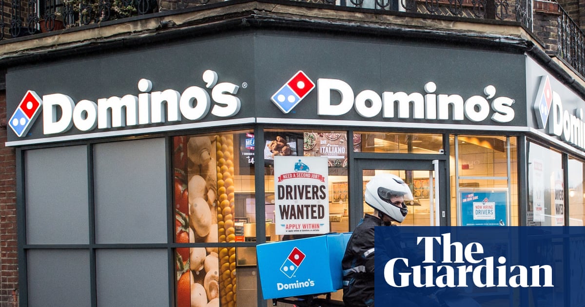
Introduction
Serratia marcescens a facultative anaerobic Gram-negative bacillus belonging to the Serratia genus within the Enterobacteriaceae family, is a significant opportunistic pathogen in hospitals. Widely distributed in hospital environments, S. marcescens is hard to fully eradicate via physical or chemical disinfection. Its high virulence enables it to cause respiratory, urinary tract, bloodstream infections, meningitis, and surgical site infections, especially in patients undergoing invasive procedures, surgeries, with weakened immunity, or experiencing trauma.1 It spreads rapidly among hospitalized patients and has triggered numerous nosocomial outbreaks.2,3 According to CHINET data,4,5 the detection rate of S. marcescens increased from 0.99% (2483 strains) of all clinically isolated strains in 2019 to 1.15% (5120 strains) in 2023, marking a 16.16% rise. In recent years, the emergence of multidrug-resistant S. marcescens, including those resistant to carbapenems,6,7 has made detecting and controlling its spread a tough challenge.
Currently, international data on the evolution of resistance in S. marcescens relative to antimicrobial exposure are limited, and this is also a relatively unexplored area in the research of S. marcescens. Antimicrobial selective pressure is a key driver of bacterial resistance. Our research endeavors to innovatively explore the macro-level quantitative relationship between the two. Theoretically, antimicrobial use precedes bacterial resistance.8,9 This study will explore the correlation between S. marcescens resistance rates and antimicrobial Defined Daily Doses (DDDs) from the previous quarter, aiming to inform strategies for curbing S. marcescens resistance and optimizing antimicrobial use in clinical practice.
Materials and Methods
Strain Source
S. marcescens strains isolated from patients (2021–2024) were collected under strict aseptic conditions, including sputum, throat swabs, bronchoalveolar lavage fluid, blood, urine, pleural effusion, peritoneal effusion, and drainage fluid, and cultured according to the National Clinical Laboratory Procedures (4th edition).10 For identical strains isolated multiple times from a single patient sample, only the initial susceptibility result per drug was retained. Colonizing or contaminating strains were excluded to avoid confounding true pathogens, based on at least two of the following clinical-microbiological discordance criteria: (1) Absence of infection-related symptoms (eg, fever, purulent discharge, leukocytosis, or imaging findings); (2) Clinical improvement despite in vitro resistance to empiric antibiotics; (3) Spontaneous resolution without antibiotic therapy. The quality control strains used were Escherichia coli ATCC 25922 and Pseudomonas aeruginosa ATCC 27853, provided by the Clinical Laboratory Center of the National Health and Family Planning Commission. The study was approved by the Hospital Ethics Committee (NO. 2025-L104) (In Q1 2021 and Q3 2023, our hospital did not perform statistical analysis due to insufficient S. marcescens detections).
Strain Identification and Antimicrobial Susceptibility Testing
Strain identification and susceptibility testing were performed using the VITEK-2 system (BioMerieux, France) and validated by the Kirby-Bauer disk diffusion method. Quality control included: (1) daily calibration with control strains (Escherichia coli ATCC 25922 and Pseudomonas aeruginosa ATCC 27853); (2) retesting ≥5% of strains per batch (minimum 5 strains); (3) recalibration and data review if results deviated from CLSI 2024 standards.11 Results were interpreted as susceptible, intermediate, or resistant, with resistance rate calculated as the percentage of resistant strains among total strains. Testing was conducted by our hospital’s microbiology lab, with data managed by the Infection Control Department.
Antibacterial Drug Usage Data
The hospital HIS rational drug use system collects data on the use of antibacterial drugs. The WHO-defined daily dose (DDD) serves as the standard reference for calculating antimicrobial agents’ consumption in DDD units. DDDs quantify prescribing frequency, with higher values indicating stronger clinical preference for an agent. The calculation formula is as follows:
DDDs = Annual drug total consumption/Drug DDD value.
Data Analysis
Data analysis used SPSS 27.0 and R 4.5.1: (1) Pearson correlation analysis (significance level α=0.05) was conducted to assess the relationship between the resistance rate of S. marcescens and DDDs of antimicrobials. (2) Multiple linear regression was used to explore independent, linear correlations. The model used S. marcescens resistance rate as the dependent variable and statistically significant antimicrobial DDDs from the previous quarter as independent variables. The stepwise method was applied for variable selection. It requires linear relationships, independent and normally distributed residuals, homoscedasticity, and no multicollinearity among independent variables. (3) The final regression model is subjected to threefold validation. Bayesian analysis used Markov Chain Monte Carlo (MCMC) simulation (10,000 iterations; weakly informative priors) in SPSS 27.0. Significance required: 95% credible interval excluding zero and posterior probability >0.95. Bootstrap Validation: Conduct 3000 BCa resamples; require original coefficient within 95% CI and CI not crossing zero. Winsorized Regression: Apply 1% bilateral winsorization, re-model. Accept if |Δβ%|<10% and p<0.05 remains significant (Δβ% = [(β_winsorized − β_original)/β_original] × 100%) and the significance level (p<0.05).
Results
Isolation Rate of S. marcescens
In the past four years, 16190 strains of pathogens were detected in our hospital, among which 522 strains of S. marcescens were isolated, accounting for 3.22% of the total number of bacteria. The total χ² value was 16.365, less than the critical value (21.026), P>0.05, and there was no significant difference in the isolation rate of S. marcescens. The total number of pathogens and S. marcescens detected in each quarter are shown in Table 1. It can be seen that the detection rate of S. marcescens is 1.96%–4.35%. Of the 522 isolates of S. marcescens, 452 (86.59%) came from respiratory tract, 31 (5.94%) from blood flow, 29 (5.56%) from urine, and the remaining 10 (1.91%) from other sources. According to the statistics of the departments submitted for examination, the top 3 departments with the number of isolates of S. marcescens were 265 (50.77%) in surgery, 139 (26.63%) in ICU and 85 (16.28%) in internal medicine.
Table 1 Detection of Serratia marcescens
|
Usage of Antibacterial Drugs
The DDDs of commonly used antimicrobial agents in the past four years are shown in Table 2.
![]() |
Table 2 The DDDs of Commonly Used Antimicrobial Agents
|
β-lactam antibiotics continue to dominate, with high DDDs for piperacillin tazobactam, cefoperazone-sulbactam, and levofloxacin. Over the four years, the usage of cefuroxime, piperacillin-tazobactam, and gentamicin showed a significant upward trend.
Drug Resistance of S. marcescens
The isolates of S. marcescens were highly susceptible to amikacin and tigecycline, with resistance rates below 5%. They also showed susceptibility to piperacillin-tazobactam, ceftazidime, ertapenem, imipenem, meropenem, trimethoprim-sulfamethoxazole, and tigecycline, with resistance rates below 10%. Resistance to cefuroxime exceeded 90%, while resistance to amoxicillin-clavulanate remained above 60%. From 2021 to 2024, resistance rates to ceftriaxone, ceftazidime, cefoxitin, imipenem, meropenem, ertapenem, and ceftazidime decreased overall, while resistance to levofloxacin increased. For more details, as shown in Table 3.
![]() |
Table 3 Drug Resistance of Serratia marcescens
|
Pearson Correlation Analysis
Pearson correlation analysis showed that S. marcescens resistance to ceftazidime, cefoperazone-sulbactam, and imipenem correlated with the DDDs of multiple antibiotics. In contrast, resistance to amoxicillin-clavulanic acid, cefuroxime, cefoxitin, cefepime, amikacin, and levofloxacin each correlated with only one antibiotic’s DDD (Table 4).
![]() |
Table 4 Correlation of Serratia marcescens Resistance Rates (Columns) with Antibiotic DDDs (Rows)
|
![]() |
Table 5 Linear Regression Analysis of Serratia marcescens Resistance and DDDs
|
Correlation Analysis
To assess independent linear correlations, we utilized multiple linear regression models. Models 1–3 included multiple independent variables, while Models 4–9 incorporated single independent variables. Table 5 shows the key parameters and test values for each regression model: regression coefficients (β) quantify the impact of independent variables on the dependent variable (p<0.05 indicates significance), the F-value assesses overall model significance, VIF<5 demonstrates low multicollinearity, R² represents explanatory power, and a Durbin-Watson value near 2 confirms residual independence. Diagnostic plots for Models 1–3 are shown in Figures 1–3. The standardized residual histograms (Figures 1a, 2a and 3a) approximate a normal distribution across all groups. The normal probability (P-P) plots (Figures 1b, 2b and 3b) demonstrate points clustering closely along the diagonal line, confirming the normality of residuals. The residual scatter plots (Figures 1c, 2c and 3c) show evenly distributed points within the ±3 range for all groups, indicating homoscedasticity and absence of influential outliers.
![]() |
Figure 1 Regression diagnostic plots for Model 1. (a). Histogram of standardised residuals (normality assessment), (b). Normal Q-Q plot (residual normality verification), (c). Scatterplot of Residuals versus fitted values (homoscedasticity assessment).
|
![]() |
Figure 2 Regression diagnostic plots for Model 2. (a). Histogram of standardised residuals (normality assessment), (b). Normal Q-Q plot (residual normality verification), (c). Scatterplot of Residuals versus fitted values (homoscedasticity assessment).
|
![]() |
Figure 3 Regression diagnostic plots for Model 3. (a). Histogram of standardised residuals (normality assessment), (b). Normal Q-Q plot (residual normality verification), (c). Scatterplot of Residuals versus fitted values (homoscedasticity assessment).
|
Robustness Verification
The final regression model is subjected to threefold validation (Table 6). Multiple robustness validations consistently demonstrated negative correlations for IPM-RR/GEN, CFS-RR/GEN, CFX-RR/TZP, and FEP-RR/CXM. Bayesian posterior means were less than zero (95% HPD excluding zero), Bootstrap confidence intervals were entirely negative, and Winsorized regression coefficients were highly consistent with the original model, with minimal changes such as +0.41% for IPM-RR/GEN.
![]() |
Table 6 Robustness Verification
|
The robustness validation for the AMC-RR/MEM and LVX-RR/GEN groups supports the original positive trend. Bayesian analysis shows posterior means significantly greater than zero, and Winsorized regression reinforces this with positive coefficients and confidence intervals excluding zero. Bootstrap results are borderline, primarily reflecting insufficient estimation precision rather than a change in effect direction. All three validation methods have consistent positive point estimates, with effect sizes exceeding the clinical threshold (|β| > 0.02), confirming the biological validity of the positive trend.
The triple validation showed consistent negative trends for CAZ-RR/TZP, CXM-RR/TZP, and AMK-RR/TZP. Bayesian and Winsorized regression results were statistically significant, while Bootstrap analysis results were borderline, reflecting limited estimation accuracy due to the sample size. The effect sizes were small (|β| < 0.005) and did not reach the clinical threshold (|β| > 0.02), but the consistent trends warrant validation through enhanced analytical approaches in future investigations.
Discussion
S. marcescens, first identified in 1819 by Venetian pharmacist Bartolomeo Bizio, was initially seen as a low-pathogenicity environmental saprophyte. However, its pathogenic potential was revealed in the 1950s through fatal urinary tract infections. It can cause infections in multiple systems, including respiratory, neurological, abdominal, cardiovascular, musculoskeletal, and ocular.12 Recently, S. marcescens has gained attention as an emerging pathogen worldwide, provoking infections and outbreaks in debilitated individuals, particularly newborns and patients in ICU.13 S. marcescens resists disinfectants, persists in the environment, and colonizes medical devices and surfaces. Its invasiveness and antimicrobial resistance make it a major hospital-acquired pathogen.
In the past four years, a total of 522 strains of S. marcescens were detected in our hospital, accounting for 3.22% of the total bacterial count in the hospital. The clinical isolated samples of S. marcescens mainly come from the respiratory tract, accounting for 86.59%, and the proportion of samples from blood, urine, secretions, ducts, etc. is relatively small. This suggests that when the immune system is weakened, the respiratory tract is a common site for S. marcescens infection, calling for clinical prevention and control. This finding aligns with relevant literature.14,15 The presence of S. marcescens can exacerbate the condition of patients with compromised immune systems.16 Furthermore, studies have indicated that this bacterium may lead to severe acute infections, resulting in respiratory distress even in individuals with normal immune function.17 S. marcescens is mainly found in surgical and intensive care unit (ICU), emphasizing the need for better infection control. This aligns with Chen Jian et al’s findings.18 Major risk factors include serious underlying diseases, major surgeries, ventilator use, tracheostomies, invasive exams, and prolonged high-dose antibiotics, which are common among ICU and surgical patients.
S. Marcescens contains R factors or R plasmids that can carry multiple resistance genes, leading to its natural resistance to first generation cephalosporins, ampicillin, and macrolides.18 The study showed that S. marcescens had a resistance rate of over 90% to cefuroxime and a high resistance rate to amoxicillin-clavulanic, consistent with Li Jin et al.19 The isolated strains of S. marcescens showed sensitivity to cefoperazone-sulbactam, cefepime, ertapenem, imipenem, meropenem, amikacin, trimethoprim-sulfamethoxazole, and tigecycline, with a resistance rate of less than 10%, consistent with previous reports.20 S. marcescens shows high sensitivity to amikacin and tigecycline, with resistance rates below 5%. Compared with the national data of bacterial resistance monitoring in CHINET in 2023,4 the resistance rate of 522 strains of S. marcescens to cefuroxime is slightly higher than the national average level. The drug sensitivity results to ceftriaxone, ceftazidime, levofloxacin, amikacin, trimethoprim-sulfamethoxazole, and tigecycline are relatively consistent with the national data, while the resistance rate to cefepime, cefoxitin, cefoperazone-sulbactam, ertapenem, imipenem and meropenem is lower than the national level. Clinically, third-generation cephalosporins, aminoglycosides, and quinolones treat S. marcescens infections, with carbapenems for severe MDR strains. Overuse of carbapenems has coincided with an increasing detection rate of carbapenem-resistant S. marcescens, hindering control and treatment.21 Notably, studies demonstrate that S. marcescens exhibits intrinsic resistance to polymyxins. Studies show S. marcescens is naturally resistant to polymyxins,6 if it also produces carbapenemases, treatment gets harder.22 Clinicians should choose antibiotics wisely to control and prevent outbreaks of S. marcescens infections.
Reports indicate that developing a new antimicrobial drug from research to clinical use generally takes 5 to 10 years. In contrast, bacteria can develop resistance in as few as 2 years.23 Thus, slowing bacterial resistance is just as critical as enhancing antimicrobial drug efficacy. The study showed resistance to cefoperazone-sulbactam and imipenem had independent negative linear relationships with gentamicin DDDs; resistance to cefoxitin correlated negatively with piperacillin-tazobactam DDDs; resistance to cefepime showed a negative association with cefuroxime DDDs. These four relationships were strongly supported by consistent results from Bayesian, Bootstrap, and Winsorized regression. In addition, amoxicillin-clavulanic resistance positively correlated with meropenem DDDs, and levofloxacin resistance positively correlated with gentamicin DDDs. These positive trends were supported by triple robustness testing. Concurrently, analyses of S. marcescens isolates revealed negative linear correlations of resistance to ceftazidime, cefuroxime, and amikacin with piperacillin-tazobactam DDDs. Although these associations were directionally consistent across all robustness validation methods, their effect sizes fell below prespecified clinical thresholds, warranting further studies to explore these potential relationships. The recurrent involvement of piperacillin-tazobactam in resistance patterns warrants attention. Existing studies have shown that S. marcescens has the ability to form biofilms, which can hinder antibiotic penetration, alter bacterial metabolic states, and thus enhance bacterial drug resistance.24 Although research on S. marcescens is limited, studies on other Gram-negative bacteria can offer insights. Research has demonstrated that use of piperacillin-tazobactam reduces rates of ceftazidime-resistant Klebsiella pneumoniae, which is similarly a member of the Enterobacteriaceae family.25 The mechanism may be related to biofilm inhibition. Studies have shown that sub-minimal inhibitory concentration (sub-MIC) concentrations of piperacillin-tazobactam can effectively reduce biofilm formation by inhibiting the adhesiveness and motility of Escherichia coli,26 and reduce Pseudomonas aeruginosa biofilm formation in a dose-dependent manner, with the strongest inhibitory effect at MIC/2 concentration.27 Whether increased use of piperacillin-tazobactam reduces S. marcescens resistance to ceftazidime by inhibiting biofilm-mediated resistance barriers remains unclear, and relevant mechanisms require further research through constructing in vitro biofilm models and conducting in vivo antibacterial experiments. Zhao Ningqiu et al28 found that different antimicrobial agents may share resistance mechanisms, such as overexpression of efflux pumps, changes in cell membrane permeability, inactivation of antimicrobial compounds, and modification of their targets. These mechanisms can cause cross-resistance or co-resistance between drugs.
From a biological perspective, these associations may involve complex mechanisms. Negative correlations may reveal potentialistic synerg inhibitory effects between antimicrobial drugs, suggesting that increased use of certain drugs can suppress the rise in resistance to target drugs, indicating that rational combination or rotation of antimicrobial drugs can effectively reduce resistance risks. Positive correlations suggest the presence of cross-resistance or co-resistance mechanisms, implying that increased use of one class of antimicrobial drugs may simultaneously or subsequently lead to increased resistance to other structurally or functionally similar drugs. Hospitals should optimize antibiotic management strategies, including targeted antibiotic rotation, limiting high-risk drug use, and adhering to data-driven guidelines. These measures can improve patient care, curb the spread of S. marcescens resistance, and preserve the long-term efficacy of essential antimicrobials.
The resistance mechanisms of S. marcescens are complex, as it inherently exhibits resistance to multiple antibiotics, such as polymyxin and cephalothin. It can produce specific β-lactamases induced by drugs, include cephalosporinase (AmpC), extended-spectrum β-lactamases (ESBLs), carbapenemases, etc. These enzymes are capable of hydrolyzing the β-lactam ring of β-lactam antibiotics, rendering the antibiotics inactive and thus enabling the bacteria to develop resistance to the corresponding antibiotics. Research on carbapenem resistance mainly focuses on serine carbapenemases, such as KPC enzyme, OXA-48, and metallo-enzymes.29 The resistance genes encoding carbapenemases in S. marcescens are mainly KPC-2,30 NDM-1,31 IMP-1, IMP-10, and it may carry multiple resistance genes, presenting a multi-drug-resistant phenotype.32 Relevant reports have shown that the synergistic effect of high-yield chromosome-mediated loss of AmpC enzyme and porin resulting in decreased permeability can also cause drug resistance to carbapenilic antibiotics.33 Moreover, reduced outer membrane permeability and overactive efflux pumps in the inner membrane are key contributors to increased drug resistance in S. marcescens.34
Currently, there is a scarcity of global data regarding how S. marcescens develops resistance in response to antimicrobial exposure. This aspect remains a relatively unexplored area within S. marcescens research. Our study used multiple linear regression to analyze the independent, linear correlation between S. marcescens resistance rates and antibiotic use one quarter in advance. We also validated our results using three methods: Bayesian, which enhances reliability; Bootstrap, which improves model adaptability; Winsorized regression, which controls the impact of outliers. Our quarterly time-series design (longitudinal study) is more effective at capturing macro trends compared to cross-sectional studies.9,35 However, the limited observation period restricts the number of data points in similar longitudinal studies.36,37 To compensate, we optimized our methodology through triple validation to ensure model stability. Future research could further investigate the impact of continuous antibiotic use on resistance through randomized controlled trials and explore resistance mechanisms. We hope this study offers a unique perspective to guide antibiotic management.
The rapid emergence and spread of resistant bacteria in hospitals are due to multiple factors, including improper antimicrobial use creating high selective pressure, inconsistent infection control measures leading to patient-to-patient transmission, inter-hospital transfer of resistance, and complex links between resistance and antimicrobial use.38 Clinicians should strictly control antimicrobial use, standardize dosages and durations, and avoid misuse that can trigger resistance. Tailoring treatments based on infection type and susceptibility results can reduce resistance risks. Healthcare facilities need to enhance resistance monitoring, strengthen multidisciplinary collaboration, and enforce strict antimicrobial use regulations. Regular analysis of resistance data can guide rational prescribing. Infection control measures, including “one use, one disinfection”, isolating patients with resistant infections, and using dedicated equipment, are essential. Hand hygiene training and rapid response to clustered cases are crucial to break transmission chains. Understanding the correlation between S. marcescens resistance rates and antimicrobial use is crucial for clinical resistance control.
Conclusion
Over the past four years, our hospital identified 522 S. marcescens strains (3.22% of all isolates), with 86.59% from respiratory samples. This indicates that the respiratory tract is a common site for S. marcescens infection, necessitating clinical prevention and control measures. Most strains were sensitive to cefoperazone-sulbactam, cefepime, ertapenem, imipenem, meropenem, amikacin, trimethoprim-sulfamethoxazole, and tigecycline, with resistance rates <10%. Our study found significant correlations between S. marcescens resistance and antibiotic usage. Negative correlations showed that strategic antimicrobial use can reduce resistance rates, while positive correlations indicated higher co-resistance risks. These results highlight the need for hospitals to implement antibiotic rotation, limit high-risk drug use, and enhance supervision to improve patient outcomes and curb resistance.
Highlights
- This study is the first to deeply investigate the macro-level quantitative relationship between S. marcescens drug resistance and antibiotic use, filling a research gap in this area.
- Using multiple linear regression models, we identified independent linear correlations between S. marcescens resistance rates and the use of various antibiotics.
- Our findings provide a scientific basis for rational and optimized antibiotic use in clinical practice, helping to delay the emergence of resistant bacteria and combat the threat of antibiotic resistance.
Ethics Approval
This study was reviewed and approved by the Ethics Committee of Wenzhou Hospital of Integrated Traditional Chinese and Western Medicine (NO. 2025-L104). Informed consent was obtained from the patient.
Acknowledgments
We are grateful to Dr Ren-ai Xu for his invaluable guidance and support throughout this research. We also thank Ms Leping Liu for her helpful comments and suggestions.
Disclosure
The authors report no conflicts of interest in this work.
References
1. Duan XK, Liu WF, Xiao YY, et al. Exploration of the feasibility of clinical application of phage treatment for multidrug-resistant Serratia marcescens-induced pulmonary infection. Emerg Microbes Infect. 2025;14(1):2451048. doi:10.1080/22221751.2025.2451048
2. Hanczvikkel A, Tóth Á, Kopcsóné Németh IA, et al. Nosocomial outbreak caused by disinfectant-resistant Serratia marcescens in an adult intensive care unit, Hungary, February to March 2022. Euro Surveill. 2024;29(26):2300492. doi:10.2807/1560-7917.ES.2024.29.26.2300492
3. Muyldermans A, Crombé F, Bosmans P, et al. Serratia marcescens outbreak in a neonatal intensive care unit and the potential of whole-genome sequencing. J Hosp Infect. 2021;111:148–154. doi:10.1016/j.jhin.2021.02.006
4. Guo Y, Hu FP, Zhu DM, et al. CHINET surveillance of bacterial resistance in China: 2023 report. Chin J Infect Chemother. 2024;24(6):627–637.
5. Hu FP, Guo Y, Zhu DM, et al. CHINET surveillance of bacterial resistance in China: 2019 report. Chin J Infect Chemother. 2020;20(3):233–243.
6. Ferreira RL, Rezende GS, Damas MSF, et al. Characterization of KPC-producing Serratia marcescens in an intensive care unit of a Brazilian tertiary hospital. Front Microbiol. 2020;11:956. doi:10.3389/fmicb.2020.00956
7. Bolourchi N, Noori Goodarzi N, Giske CG, et al. Comprehensive pan-genomic, resistome and virulome analysis of clinical OXA-48 producing carbapenem-resistant Serratia marcescens strains. Gene. 2022;822:146355. doi:10.1016/j.gene.2022.146355
8. Yan Y, Sun WG. Impact of antimicrobial consumption frequency on Staphylococcus aureus resistance rates in the following quarter. Chin J Pharmacoepidemiol. 2018;27(6):382.
9. Bronzwaer SL, Cars O, Buchholz U, et al. A European study on the relationship between antimicrobial use and antimicrobial resistance. Emerg Infect Dis. 2002;8(3):278–282. doi:10.3201/eid0803.010192
10. Shang H, Wang YS, Shen ZY. National Guide to Clinical Laboratory Procedures.
11. Clinical and Laboratory Standards Institute. Performance standards for antimicrobial susceptibility testing: thirty-fourth informational supplement. In: Document. Wayne, PA: CLSI; 2024:M100–S34.
12. Iovene MR, Pota V, Galdiero M, et al. First Italian outbreak of VIM-producing Serratia marcescens in an adult polyvalent intensive care unit, August-October 2018: a case report and literature review. J Hosp Infect. 2019;101(4):431–437. doi:10.12998/wjcc.v7.i21.3535
13. Tavares-Carreon F, De Anda-Mora K, Rojas-Barrera IC, et al. Serratia marcescens antibiotic resistance mechanisms of an opportunistic pathogen: a literature review. PeerJ. 2023;11:e14399. doi:10.7717/peerj.14399
14. Zhang Y, Zhou WQ, Li J, et al. Resistance phenotype analysis of 53 carbapenem-resistant Serratia marcescens isolates. J Clin Transfus Lab Med. 2021;23(1):87–90.
15. Simsek MD. Determination of the antibiotic resistance rates of Serratia marcescens isolates obtained from various clinical specimens. Niger J Clin Pract. 2019;22(1):125–130. doi:10.4103/njcp.njcp_362_18
16. Muyldermans A, Crombé F, Bosmans P, et al. Serratia marcescens outbreak in a neonatal intensive care unit and the potential of whole-genome sequencing. J Hosp Infect. 2020;104(4):438–445. doi:10.1016/j.jhin.2021.02.006
17. Ruiz-Sada P, Escalante M, Lizarralde E. Severe acute infection due to Serratia marcescens causing respiratory distress in an immunocompetent adult. Rom J Intern Med. 2016;54(2):134–136. doi:10.1515/rjim-2016-0013
18. Chen J, Xu CQ, Tu HX. Drug resistance analysis of Serratia marcescens in hospital infections. Chin J Health Lab Technol. 2009;19(12):2889–2990.
19. Li J, Zhang XY. Epidemiological characteristics and antimicrobial resistance of Serratia marcescens in a tertiary hospital from 2017 to 2023. Chin J Infect Control. 2025;24(1):52–57.
20. Zhang ZX, Wu WH. Distribution and drug resistance analysis of 56 Serratia marcescens isolates. Chin J Disinfect. 2013;30(12):1134–1137.
21. Yuan Y, Chen JZ, Pan YP, et al. Carbapenem-resistant Serratia marcescens: resistance genes and molecular epidemiology. Acta Univ Med Anhui. 2019;54(1):33–38.
22. Ma JL, Liu ZZ, Li DJ, et al. Molecular epidemiology of carbapenemase-producing Serratia marcescens infections in a neurosurgical ICU. Chin J Nosocomiol. 2020;30(3):393–397.
23. Zou J, Li N. Bacterial resistance mechanisms and control strategies. Basic Med Forum. 2006;10(1):1052–1053.
24. Erdal B, Yalınay M, Elmas Ç, et al. Investigation of pseudomonas aeruginosa biofilm formation and quorum sensing genes in piperacillin/tazobactam and ciprofloxacin sub-minimal inhibitory concentrations. Mikrobiyol Bul. 2020;54(4):547–558. doi:10.5578/mb.70087
25. Hsueh PR, Chen WH, Luh KT. Relationships between antimicrobial use and antimicrobial resistance in gram-negative bacteria causing nosocomial infections from 1991–2003 at a university hospital in Taiwan. Int J Antimicrob Agents. 2005;26(6):463–472. doi:10.1016/j.ijantimicag.2005.08.016
26. Song J, Hong H, Feng W, et al. Effect of subinhibitory concentrations of piperacillin/tazobactam on biofilm formation ability of Escherichia coli. Chongqing Med. 2018;47(3):368–370.
27. Ray C, Shenoy AT, Orihuela CJ, et al. Killing of Serratia marcescens biofilms with chloramphenicol. Ann Clin Microbiol Antimicrob. 2017;16(1):19. doi:10.1186/s12941-017-0192-2
28. Zhao NQ, Liu C, Xue YX, et al. Cross-resistance and co-resistance of bacteria to antimicrobial compounds. Chin J Antibiot. 2021;46(1):11–19.
29. Wu HO, Zeng WL, Dai YY, et al. Molecular mechanisms of carbapenemase-producing Serratia marcescens. Chin J Health Lab Technol. 2022;32(2):170–173.
30. Rodriguez C, Brengi S, Caceres MA, et al. Polyclonal dissemination of KPC-2 in Serratia marcescens, including a clone with epidemic behavior in the nosocomial niche. Int J Antimicrob Agents. 2017;49(5):657–658. doi:10.1016/j.ijantimicag.2017.03.003
31. Phan HTT, Stoesser N, Maciuca IE, et al. Illumina short-read and MinION long-read WGS to characterize the molecular epidemiology of an NDM-1 Serratia marcescens outbreak in Romania. J Antimicrob Chemother. 2018;73(3):672–679. doi:10.1093/jac/dkx456
32. Wang JM, Zhang HQ, Zhang YH, et al. Resistance mechanisms and homology analysis of carbapenem-resistant Serratia marcescens in 12 burn patients. Chin J Health Lab Technol. 2017;27(9):1354–1356,1359.
33. Guo P, Qiao Y, Zhang HT, et al. Drug resistance and imipenem resistance mechanisms of clinical Serratia marcescens isolates. Chin J Infect Chemother. 2017;17(2):187–191.
34. Montagnani C, Cocchi P, Lega L, et al. Serratia marcescens outbreak in a neonatal intensive care unit: crucial role of implementing hand hygiene among external consultants. BMC Infect Dis. 2015;15:11. doi:10.1186/s12879-014-0734-6
35. Olesen SW, Barnett ML, MacFadden DR, et al. The distribution of antibiotic use and its association with antibiotic resistance. Elife. 2018;18(7):e39435. doi:10.7554/eLife.39435
36. Bruyndonckx R, Hens N, Aerts M, et al. Exploring the association between resistance and outpatient antibiotic use expressed as DDDs or packages. J Antimicrob Chemother. 2015;70(4):1241–1244. doi:10.1093/jac/dku525
37. Meyer E, Gastmeier P, Deja M, et al. Antibiotic consumption and resistance: data from Europe and Germany. Int J Med Microbiol. 2013;303(6–7):388–395. doi:10.1016/j.ijmm.2013.04.004
38. White RL, Friedrich LV, Mihm LB, et al. Assessment of the relationship between antimicrobial usage and susceptibility: differences between the hospital and specific patient-care areas. Clin Infect Dis. 2000;31(1):16–23. doi:10.1086/313916