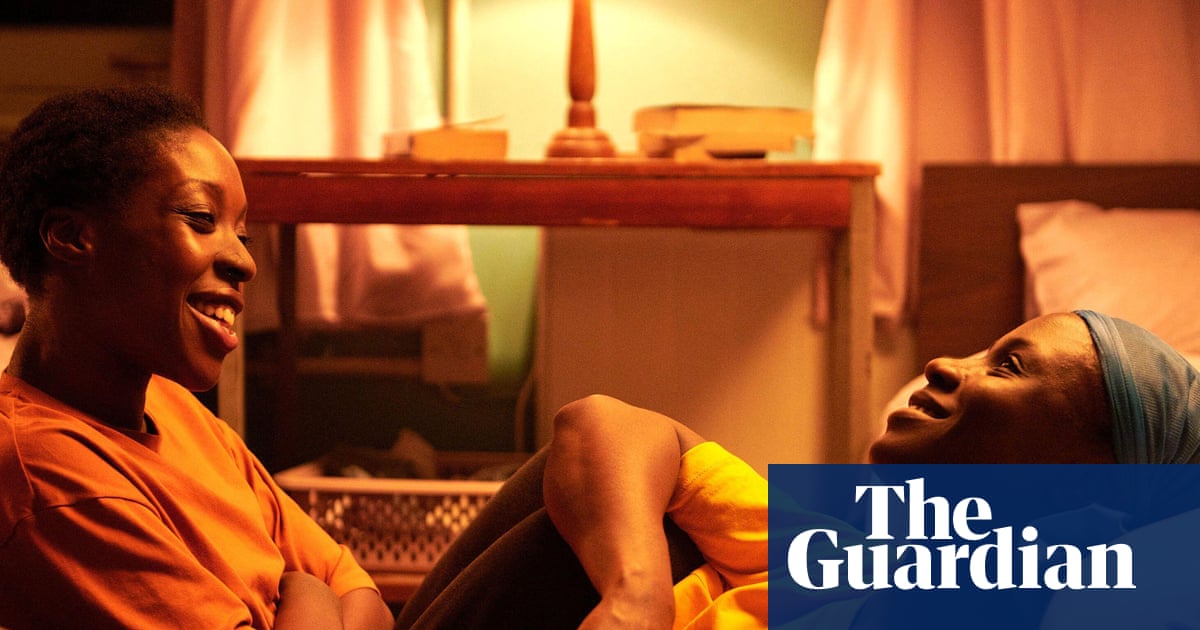
Participation of host cell proteins in inclusion bodies of non-segmented RNA virus infected cells: a molecular insight | Virology Journal

Rhabdoviridae is a family of viruses within the order Mononegavirales, characterized by their bullet-shaped structure. The Vesicular Stomatitis Virus (VSV) and the Rabies Virus (RABV) serve as prototypical members of this family and are commonly utilized for research in this field.

Rabies virus (RABV)

RABV is the most medically significant virus in the family. RABV causes an acute infection of the central nervous system, progressing through five general stages in humans: incubation, prodrome, acute neurologic period, coma, and death. The incubation period is highly variable, ranging from less than 10 days to over 2 years, but typically lasts 1–3 months [28].

For the RABV, IBs are known as Negri bodies (NBs), named after Adelchi Negri who discovered NBs in the cerebellum of RABV-infected animals and humans [29]. These NBs are a hallmark of RABV infection and were previously used as a diagnostic marker. These are typically spherical structures that measure 2 to 10 micrometres in diameter. Electron microscopy revealed that NBs consist of a granular or filamentous matrix made up of viral nucleoprotein, with viral particles observed budding from them [29]. Cytological staining indicated the presence of nucleic acids, suggesting they may function as replication complexes [5, 29].

A recent study demonstrated the NBs formation through LLPS, evidenced by their spherical shape, ability to fuse into larger droplets, and dissolution upon osmotic shock [30, 31]. These NBs primarily consist of two proteins: the RNA-binding Nucleoprotein (N) and the intrinsically disordered Phosphoprotein (P). A crucial part of P, specifically the amino-terminal section of its second intrinsically disordered domain (IDD2), plays a key role in phase separation [30]. Electron microscopy studies have revealed that RABV-IBs typically contain cavities and lack membranes for up to 12 h post-infection but gradually acquire a granular double membrane, suggesting that these structures may derive membrane from the endoplasmic reticulum (ER) [5]. In situ hybridization and short-term RNA labelling of RABV-infected cells have demonstrated the presence of all RABV mRNAs (N, P, M, G, L) within these structures. Notably, the viral genomes, antigenomes, and mRNAs synthesized inside the IBs are encased by a protective structure formed by N and P proteins, which may shield them from degradation [5, 32]. Cellular stress granules (SGs) also exhibit liquid-like properties and are located near NBs but do not merge with them. These SGs increase in number, grow in size through fusion events, and remain in close proximity to NBs. During RABV infection, mRNAs produced in NBs accumulate in SGs, suggesting a complex interplay between viral replication and the host’s stress response mechanisms [33]. Nonetheless, several host cellular proteins are also known to play vital roles in the formation and regulation of NBs.

Toll-like receptor 3 (TLR3) is one such critical host protein involved in this process, particularly in the context of RABV infection [34]. TLR3 is an essential innate immune receptor that detects viral double-stranded RNA (dsRNA), primarily localizing to endolysosomal compartments within cells. Upon recognition of dsRNA, it triggers the activation of immune pathways, leading to the release of type I interferons and pro-inflammatory cytokines, which orchestrate a protective immune response. TLR3 signals through the TRIF (Toll/interleukin-1 Receptor domain-containing adaptor protein inducing interferon beta) adaptor protein, activating key transcription factors such as Interferon Regulatory factor 3 (IRF3) and Nuclear Factor Kappa B (NF-κB), which drive the expression of antiviral genes [35]. Structurally, TLR3 is a horseshoe-shaped solenoid protein characterized by 23 leucine-rich repeats (LRRs) in its ectodomain, a feature that may give it an inherent capacity to form aggregates [35]. During RABV infection, TLR3 is activated, contributing to the immune response and the formation of NBs, cytoplasmic inclusions that resemble aggresomes and may facilitate viral replication. Confocal microscopy and 3D imaging have shown that NBs have a highly organized structure, with TLR3 at the core surrounded by a halo of viral N and P, suggesting that the receptor plays a pivotal role in their formation, possibly initiating the aggregation process [34]. Further supporting this idea, studies on TLR3-deficient mice infected with RABV demonstrate impaired NB formation, resulting in a milder infection and reduced viral replication [36]. This suggests that TLR3 is critical for activating immune responses and is crucial in forming viral IBs like NBs. Despite these findings, the exact molecular mechanisms and interactions between TLR3 and viral proteins remain poorly understood and warrant further investigation [34].

Staufen 1 (STAU1) is a host cell SG localizing protein having dsRNA-binding domain (dsRBD), involved in the transport, localization, and regulation of mRNAs across various subcellular compartments. STAU1 is a member of the Staufen family of proteins, which are known for their involvement in post-transcriptional regulation [37]. It is characterized by multiple dsRBD, STAU1 can recognize and bind RNA molecules with double-stranded secondary structures. It is commonly associated with polysomes, where it participates in mRNA translation and regulation [38]. STAU1 plays a crucial role in the STAU1-mediated mRNA degradation (SMD) pathway, a post-transcriptional mechanism where it binds to specific sites in the 3′ untranslated region (3′UTR) of mRNAs with complex secondary or tertiary structures, leading to rapid mRNA degradation [37]. Interestingly, STAU1 is also associated with RABV-IBs, where it seems to play a dual role. Immunofluorescence Assay (IFA) has shown that STAU1 colocalizes with RABV-N protein in IBs. Downregulation of STAU1 leads to an increase in virus titer, enhanced viral replication and significantly increased IBs. STAU1 has also been shown to regulate RABV RNA levels, acting as an antagonist to the virus, like its role in SGs [38]. However, the exact function of STAU1 in the context of RABV-IBs remains poorly understood and demands further investigation.

Cytosolic chaperonin (CCT), composed of two rings with eight subunits each (CCT1–8), assists in folding various proteins, including tubulin, actin, and cyclin E, through ATP hydrolysis [39]. Mass spectrometry identified CCTγ, a TRiC/CCT subunit, as a host factor in RABV-infected cells. IFA showed that CCTγ and CCTα colocalizes with NBs, which contain viral N and P proteins. Although overexpressing CCTγ and CCTα did not significantly increase viral replication, their knockdown markedly reduced RABV replication. These findings suggest that CCTγ and CCTα are recruited by viral N and P proteins to NBs and plays a supportive role in RABV replication, though its role NB formation remain yet to be understood [39, 40].

Heat shock protein 70 (Hsp70) is another crucial molecular chaperone protein in host cells that is primarily upregulated in response to cellular stress. Its main function is to bind to protein substrates, stabilizing them to prevent denaturation or aggregation until cellular conditions improve [41]. Beyond its role in stress response, Hsp70 is also essential for various processes during normal cell growth. It aids in the folding of newly synthesized proteins, facilitates the transport of proteins and vesicles within the cell, assists in forming and dissociating protein complexes, and promotes the degradation of damaged or unwanted proteins [41]. Hsp70 was identified earlier in newly synthesized rabies virions, was also shown to accumulate within IBs alongside ubiquitinylated proteins, and has been suggested to play a proviral role during RABV infection [42, 43].

Focal adhesion kinase (FAK) is a cytoplasmic protein tyrosine kinase primarily located at focal adhesions, which are dynamic complexes that link cells to the extracellular matrix (ECM) [44]. FAK regulates key cellular processes like motility, survival, proliferation, and differentiation. It is activated by external signals such as integrin engagement, growth factors, and mechanical stress, triggering its tyrosine phosphorylation. A critical site for FAK activation is tyrosine 397 (Y397), which recruits Src family kinases via their SH2 domains. These kinases further phosphorylate FAK, amplifying the signalling cascade. Phosphorylated FAK interacts with various focal adhesion proteins, activating them directly or through additional phosphorylation. This leads to the formation of a signalling complex that transmits ECM signals into the cell, influencing important functions like migration, survival, and cell cycle progression. Overall, FAK acts as a central mediator in coordinating cell responses to changes in the extracellular environment [44].

RABV-P interacts with FAK through a specific binding region spanning residues 106 to 131 of P, which corresponds to its dimerization domain, and the C-terminal region of FAK, which contains the proline-rich regions (PRRs), PRR2 and PRR3 [45]. This interaction was further confirmed through co-immunoprecipitation (co-IP) studies in RABV-infected cells, where FAK was found to colocalize with P in NBs. The binding between P and FAK suggests a potential link between viral protein trafficking and the cellular signalling pathways regulated by FAK, particularly in the context of viral replication and host cell manipulation during infection. The association of FAK-P inside RABV-IBs suggests its role in viral replication complex but it still needs more evidence and investigations to specify its function [45].

Cellular cytoskeleton, an important component of host cells, is a network of interlinking filaments present in the cytoplasm. It consists of microtubules, actin filaments and intermediate filaments, responsible for transportation of proteins across the cytoplasm and providing shape and support to cell [46]. It is also known to play a vital role in the transport of RABV-RNPs, but it is not involved in the formation of IBs. Several studies highlight this distinction. For instance, Xavier Lahaye et al. (2009) demonstrated that inhibiting microtubules with Nocodazole during the early stages of RABV infection did not prevent IB formation. However, at later stages, microtubule disruption hindered the appearance of smaller IBs, which originate from the larger IBs, suggesting that microtubules are crucial for the transport of RNPs but not for the assembly of IBs [5]. Similarly, Jovan Nikolic et al. (2017) found that Nocodazole treatment in RABV-infected cells resulted in larger-than-usual NBs. The study also showed that depolymerizing actin filaments with cytochalasin D caused NBs to fragment, reducing their surface area [31]. Live-cell imaging revealed that RNPs were ejected from the NBs, but when microtubules were disrupted with Nocodazole, RNPs were still released, but failed to move away from the NBs and remained clustered near the NBs [9, 31]. This suggests that while microtubules are not required for RNP ejection, they are essential for the movement of RNPs away from the NBs. The role of actin in this process remains unclear due to a lack of experimental evidence [9, 31].

In summary, these studies show that the cytoskeleton, particularly microtubules, is crucial for the transport of RNPs but not for the formation of IBs. This indicates that the cytoskeleton could be important for facilitating the movement and distribution of viral components after IBs have formed, while IB assembly itself occurs independently of cytoskeletal activity [31, 46]. Therefore, the involvement of the cellular cytoskeleton in the formation of RABV-IBs is not very clear and needs more investigation.

Taken together, RABV forms cytoplasmic IBs or NBs during infection and several host proteins play crucial roles in viral IB formation and virus lifecycle. The mechanisms underlying the aggregation of these NBs and the role of host proteins in RABV replication remain inadequately understood. However, the role of viral proteins in the formation as well as the biophysical properties of RABV IBs has been studied in detail in Nikolic et al., Nature Communications 2017 [31].

Vesicular stomatitis virus (VSV)

VSV another prototype virus belonging to the Rhabdoviridae family also forms IBs during its infection cycle. Initially, after VSV infects a host cell, viral RNA synthesis occurs throughout the cytoplasm [10]. However, as protein synthesis begins, the machinery for RNA synthesis is redirected to create inclusions that become the primary sites for mRNA production [10].

During VSV infection, the formation of viral IBs is dependent on LLPS of N, P and L proteins which results in compartmentalization. These phase-separated structures are linked to proteins with low-complexity regions and those enriched in RNA Binding Domains (RBDs), such as KH (K homology) or RRM (RNA recognition motif); since RNA-protein interaction can lead to phase separation [47]. While both VSV N and L proteins can bind RNA directly, the P protein contains regions of low complexity that enhance this process. The expression of P alone does not induce viral IB formation; the presence of RNA-binding proteins (RBPs) L and N is crucial [17].

Studies have also demonstrated that VSV inclusions interact with various host cell proteins, including TIA-1 (T-cell Intracellular Antigen 1), TIAR (T-cell Intracellular Antigen 1 Related protein), and PCBP2 (poly(rC) binding protein 2).

TIA-1 (T-cell intracellular antigen-1) is a key protein involved in the formation and regulation of SGs, which are cytoplasmic aggregates of mRNA and proteins that form in response to cellular stress, such as oxidative stress, heat shock, or viral infection. TIA-1 is an RBP that plays a critical role in the regulation of mRNA stability, translation, and localization within the cell, especially under stress conditions [48]. It is characterized by its RRMs and a prion-like domain, which contribute to its ability to bind RNA, specifically to the 3′UTRs of target mRNAs. The prion-like domain, in particular, is thought to facilitate the LLPS properties of TIA-1, which are essential for SG formation [22]. This domain allows TIA-1 to undergo dynamic, reversible aggregation, a key feature in the assembly and disassembly of SGs. In response to cellular stress, TIA-1 is recruited to SGs, where it interacts with other RBPs and translational regulators. These SGs serve as sites where translation initiation is temporarily stalled, and mRNA is sequestered for either storage or degradation. TIA-1’s role in SGs involves not only binding and stabilizing RNA but also coordinating the assembly of SGs by interacting with other proteins, such as G3BP1(Ras GTPase-activating protein-binding protein 1), Caprin1, and other components of the RNA granule machinery [48,49,50]. IFA has revealed that during VSV infection, the SG protein TIA-1 co-localizes with the viral nucleocapsid protein VSV-N within cytoplasmic viral IBs. Interestingly, transient depletion of TIA-1 (via siRNA) enhances VSV replication, while permanent depletion in TIA-1 knockout MEFs inhibits replication [51]. This paradox may be due to TIA-1’s role in translational regulation and mRNA processing, where its loss could disrupt cellular factors required for VSV growth or activate antiviral responses. These findings suggest that the role of TIA-1 in VSV replication is complex and context-dependent, emphasizing the need for further research for clarification [51].

T-cell intracellular antigen 1-related (TIAR) is another prominent member of the RBP family involved in SG formation. TIAR shares many functional characteristics with its paralog TIA1, and both proteins play significant roles in RNA metabolism and regulation of mRNA stability, localization, and translation [52]. TIAR is a modular RBP consisting of three characteristic RRMs, which are highly conserved with those found in its paralog TIA1. In addition to these RRMs, TIAR contains an IDR at its carboxyl-terminal end. This C-terminal domain also includes a lysosome-targeting motif [52]. Depletion of TIAR by siRNA did not result in a significant increase in VSV replication as in the case of TIA-1. Interestingly, TIAR depletion led to an upregulation of TIA-1 levels in VSV-infected cells. This increase in TIA-1 may explain the modest, though non-significant, enhancement of VSV replication in TIAR-depleted cells. In contrast, IFA revealed that TIAR co-localizes with VSV-N in IBs, similar to TIA-1. While both proteins exhibit similar localization patterns, their roles in VSV replication appear to differ and would require more investigation to understand their role in IB formation [51].

Poly (rC)-binding protein 2 (PCBP2), another RBP characterized by its high affinity for poly(C) sequences. It contains three KH domains—KH1, KH2, and KH3—which are separated by variable-length insert sequences. Each KH domain adopts a conserved structure consisting of three α-helices and an antiparallel β-sheet, following the classic β1α1α2β2β3α3 motif [53]. The KH domains are consensus RBD, known for their ability to recognize and bind RNA, as well as C-rich single-stranded (ssDNA) and double-stranded DNA (dsDNA). This enables PCBPs to regulate various stages of RNA metabolism, including RNA splicing, stability, and translation [53]. IFA studies have revealed that PCBP2 is primarily localized to the nucleus, where it is involved in regulating RNA-related processes [53]. But live cell imaging has also revealed that during stress, PCBP2 is localized to SGs and PBs, it acts as a nucleo-cytoplasmic shuttling protein [54]. Immunoprecipitation (IP) assays have identified PCBP2 as a prominent interacting partner of VSV-P. Silencing PCBP2 with siRNA showed enhanced VSV replication while overexpressing PCBP2 in transfected cells suppressed viral growth. These results indicate that PCBP2 acts as a negative regulator of VSV replication, likely by reducing viral mRNA accumulation and inhibiting genome replication. Co-IP and IFA revealed that PCBP2 interacts with and co-localizes with the VSV P protein within virus-infected cells [55]. Additional IFA studies also showed that PCBP2 co-localizes with TIA-1 along with VSV P in viral IBs, suggesting a potential role for PCBP2 in the formation of these structures. However, the exact function of PCBP2 in the formation and maintenance of IBs remains to be fully understood, and further research is needed to clarify its precise role in VSV-IBs [51].

The siRNA-mediated knockdown of TIA-1 and PCBP2 in VSV-infected cells increases viral titers, suggesting that both proteins play an antiviral role during VSV infection. This indicates that under normal conditions, TIA-1 and PCBP2 likely act to restrict viral replication. However, their association with key viral proteins such as VSV-N and VSV-P within VSV-IBs presents a paradox [51]. While these SG proteins are generally involved in antiviral responses, their co-localization with viral components in IBs raises questions about their exact function in the context of viral replication. These proteins may have dual roles—contributing to both the host’s antiviral defence and the virus’s ability to form and maintain IBs, which may be crucial for its replication and assembly. Understanding how these SG proteins modulate the viral replication cycle, particularly within IBs, will provide valuable insights into the intricate mechanisms of viral-host interactions and may reveal new therapeutic targets for controlling VSV and similar viral infections [51].

The viral mRNAs synthesized in IBs needs to be transported outside for translation and this transportation is heavily dependent on the host cell’s microtubule network. Disruption of microtubules with nocodazole results in a noticeable accumulation of viral mRNA within the IBs, indicating the active involvement of the cytoskeleton in viral RNA trafficking [51]. However, disrupting actin filaments using cytochalasin D or tubulin with nocodazole does not significantly affect the formation of VSV-IBs, suggesting that the cytoskeleton network may not have a role in the formation of IBs but may play an important role in the transportation of viral RNA. However, the microtubule network could serve as a track for moving viral RNA and other cellular machinery, highlighting the intricate connection between viral replication and host cytoskeletal dynamics. The accumulation of viral RNA in the IBs during dysfunctional cytoskeleton suggests that while mRNAs are transported from IBs for the translation of proteins, IBs by themselves may not be the regulatory centres for RNA distribution [10, 51].

The formation of VSV-IBs is dependent on both active viral replication and continuous host cell protein synthesis. Inhibition of cellular translation using Cycloheximide (CHX) results in a marked reduction in the number of viral IBs, highlighting the essential role of host cell protein synthesis in their formation and maintenance. This suggests that active translation is not only necessary for the synthesis of viral proteins but also plays a crucial role in the structural integrity and function of IBs during VSV infection [51].

Together, these findings underscore the complex interplay between viral and host cell proteins in driving the formation and maintenance of viral IBs. The IBs serve as a space for viral replication where viruses can subvert some of the host proteins. This strategic manipulation of host proteins allows the virus to optimize the conditions necessary for efficient viral assembly and RNA synthesis. The interaction with host proteins not only supports viral replication but may also help the virus evade host immune responses, contributing to its pathogenicity and ability to persist within the host.

Chandipura virus (CHPV)

CHPV, a member of the Rhabdoviridae family, is a tropical virus first identified in 1965. Despite its long history, research on CHPV gained momentum following small regional outbreaks between 2003 and 2010, and the recent largest fatal outbreak in India in 2024 [56, 57].

CHPV has a compact genome of approximately 11 kb composed of negative-sense single-stranded RNA (ssRNA), encoding five structural proteins: Nucleoprotein (N), Phosphoprotein (P), Matrix protein (M), Glycoprotein (G), and Polymerase protein (L). When compared with other members of the family, knowledge regarding CHPV biology remains limited in various aspects of its life cycle [58].

Our recent study has revealed that CHPV forms cytoplasmic IBs, similar to those of other nsNSVs. We demonstrated that the CHPV N, P, and L proteins are components of these IBs, CHPV-N alone cannot initiate the IB formation but the underlying mechanisms of IB formation remain unclear [6]. Additionally, we have identified IDRs in CHPV proteins, with CHPV-P exhibiting the highest propensity for the disorder. These finding suggests that CHPV proteins may have the potential to phase separate and form cellular aggregates or IBs [6, 59].

It may be noted that IBs look very similar to SGs which can be induced by different types of stress such as viral infection, or Sodium arsenite treatment (Fig. 2). Interestingly, CHPV-IBs also show association with several host cell RBPs involved in SG formation including TIA-1(T-cell Intracellular Antigen 1), G3BP1 (Ras-GAP SH3-domain binding protein 1), PABP1 (Poly(A) Binding Protein 1), eIF3η (Eukaryotic Translation Initiation Factor 3 Subunit η), and Ago2 (Argonaute 2) in Vero E6 and N2a cells. Here, while TIA-1 has been already described above, it would be important to go into detail about other host proteins [6].

Fig. 2

IBs or SGs formation in Vero Cell. (a) VeroE6 cells immunostaining with anti-TIA-1 (red) and anti-PABP-1 (green) show SGs formation induced by Sodium arsenite in the lower panel, while the upper panel shows control cells. The nuclei were counterstained with Hoechst dye. The white arrow points to the SGs. Scale bar = 10 μm. (b, c) CHPV-infected cells form Viral IBs. CHPV-infected Vero E6 cells showing IBs immunostaining with anti-CHPV-N (green) and/ or with anti-CHPV-L (red) and their co-localization with SG protein TIA-1 (red). The nuclei were counterstained with Hoechst dye. The white arrow indicates IBs. The white arrow indicates IBs. Scale bar = 10 μm

G3BP1 (Ras GTPase-activating protein-binding protein 1), is a central component in the assembly of SGs, which are cytoplasmic aggregates formed during stress conditions. It is a 62 kDa protein featuring multiple domains, including a RasGAP domain, nucleic acid-binding motifs, and a SH3 domain (Src Homology 3 domain) [60]. These domains help G3BP1 interact with various proteins and RNA molecules. It has a low-complexity region that allows it to undergo LLPS, a critical feature for forming SGs in response to cellular stress [60]. G3BP1 colocalizes with CHPV-N in CHPV-IBs of infected cells along with TIA-1, but understanding its exact role in CHPV-IBs demands more investigation [6].

PABP1 (Poly A Binding Protein 1), is another crucial RBP involved in the regulation of mRNA metabolism, translation, and stability. It is a ~ 72 kDa protein with several functional domains including four RRMs that bind to the poly(A) tail of mRNA, enabling PABP1 to control mRNA stability and translation [61]. RRMs mediates PABP1’s interactions with other proteins and plays a role in forming protein complexes and its C-terminal region helps with homodimerization, allowing PABP1 to form oligomeric complexes that assist in poly(A) tail binding and function. It also prevents mRNA degradation and enhances the recruitment of translation initiation factors. Under stress conditions, PABP1 can also be involved in the formation of SG, where mRNAs are stored temporarily during cellular stress, and its interactions with other proteins influence mRNA fate [6, 61].

IFA has revealed that RBPs, G3BP1 and PABP1 colocalize with CHPV-IBs formed during CHPV infection. However, their specific contributions to the formation of these IBs are still not fully understood. In contrast, TIA-1, another RBP that associates with CHPV-induced IBs, has been identified as playing a crucial proviral role in the CHPV infection process. Studies using siRNA-mediated silencing of TIA-1 have demonstrated a marked reduction in several key viral processes. Specifically, the depletion of TIA-1 resulted in a significant decrease in CHPV-N protein, a reduced formation of viral IBs, a substantial drop in CHPV-N specific RNA levels, and a notable decline in virion production. These findings suggest that TIA-1 could play an essential role during CHPV replication, and its silencing could disrupt multiple stages of the viral life cycle [6].

AGO2 (Argonaute 2), is a key protein in the RNA interference (RNAi) pathway and plays a central role in gene silencing and post-transcriptional regulation of gene expression. By guiding RNA-induced silencing complex (RISC) to mRNA targets, Ago2 plays a key role in silencing specific genes at the post-transcriptional level, making it essential for processes like viral defence, developmental regulation, and cellular homeostasis [62]. Whether the association of CHPV-IBs with Ago2 inhibits its suppressive role in gene silencing through microRNAs, remains unknown [6].

Though IFA can only conclude a co-localization of various host factors with CHPV-N in CHPV-IBs, their role in CHPV-IB formation remains unclear. Yet it suggests that CHPV could subvert host proteins into the IBs, which could help the virus to survive inside cells and escape cellular defences. The mechanism of IB formation remains yet to be understood in detail [6].

Additionally, our study also reveals a novel role for the protein kinase R (PKR), traditionally recognized as an antiviral molecule; our findings suggest that PKR acts as a pro-viral factor during CHPV infection.

Protein kinase R (PKR) is a key intracellular sensor of stress, particularly in response to viral infections. It is activated by dsRNA produced during viral replication, as well as by other ligands such as protein activator of the interferon-induced protein kinase PKR (PACT) and heparin [63]. Upon activation, PKR inhibits protein synthesis by phosphorylating the α subunit of the eukaryotic translation initiation factor 2 (eIF2α), halting translation as part of the host’s antiviral defence [63]. Human PKR is a 551-amino-acid protein composed of two main domains: an N-terminal regulatory dsRBD and a C-terminal catalytic kinase domain. The dsRBD contains two dsRNA-binding motifs (RBMs) that recognize a specific higher-order structure of dsRNA, rather than its nucleotide sequence, which allows PKR to detect viral infections. PKR is typically maintained in an inactive monomeric form. The formation of an active, phosphorylated dimer dissociates from the activating ligand and phosphorylates downstream targets, such as eIF2α, to block protein synthesis which could lead to SG formation [63].

Interestingly, silencing PKR resulted in a significant decrease in CHPV-N protein, decreased viral IBs, diminished CHPV-N RNA and reduced virion production. IFA also confirmed the co-localization of PKR with CHPV-IBs, further making a distinction in the architecture of CHPV-IBs from canonical SGs. However, the mechanism and precise role of PKR remain largely unclear and yet to be understood. While these SG proteins co-localize with CHPV-IBs, their dynamics differ significantly from those of canonical SGs [6].

Overall, much more research is needed to elucidate the involvement of host cellular proteins in the formation of IBs, their physical interactions with CHPV proteins, and their roles in the virus lifecycle. A Summary of host and viral factors participating in Rhabdovirus IBs formation is shown in Fig. 3.

Fig. 3
figure 3

Summary of host and viral factors involved in Rhabdovirus inclusion bodies formation

Continue Reading