A stabilising sheath of interlocking polymers enables coacervate microdroplets to more closely mimic natural cells than ever before. The researchers behind the work showed how the protected coacervates can form part of a chemical signalling cascade with natural cells, and suggest that the technique could have applications in drug delivery and tissue engineering.
Artificial cells provide a window into the complex chemistry underpinning life. These simplified systems help us to understand cellular functions and provide insight into how the very first living systems may have emerged. However, developing a model that replicates the intricacy of natural cells remains a substantial challenge.
Coacervates are one potential candidate – these dense liquid microdroplets form when two charged components in a mixture spontaneously separate into different phases, creating a membrane-free compartment rich in biomolecules. But using these compartments as synthetic cells is plagued with difficulties explains Evan Spruijt, an artificial cell researcher at Radboud University in the Netherlands. ‘A typical problem is that they are liquid droplets and they will fuse as soon as they meet,’ he says. ‘They also wet all kinds of surfaces and get stuck there.’
While simple membranes can provide a supportive barrier, they fail to protect coacervates from higher salt concentrations, which can dissolve these droplets completely.
Now, Eduardo Fernandez-Megia’s team the University of Santiago de Compostela in Spain has developed a particularly stable variety of coacervate using a type of branched polymer, known as a dendrimer. Forming both a component of the droplet structure and a highly interconnected protective layer across the surface, the dendrimer molecules provided an immediate boost in stability. The membrane-bound unit can survive several days in aqueous solution, compared with just a few minutes for a membrane-free system. The particle also remains unperturbed by increasing salt concentrations, tolerating more than twice the ionic strength of unprotected systems.
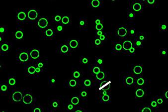
The team also showed how the system could facilitate cell-like behaviour, replicating a simple enzymatic cascade by adding fluorescently labelled glucose oxidase (GOX) and horseradish peroxidase (HRP) to the coacervates’ interior. When glucose diffuses through the membrane, the GOX enzyme oxidises it, producing hydrogen peroxide as a byproduct. The HRP then uses this peroxide to oxidise another organic molecule that triggers a fluorescent signal upon successful reaction.
The fluorescent marker produced by the GOX–HRP cascade could also successfully diffuse from the coacervate into neighbouring natural cells, which the researchers describe as a form of chemical communication. Writing in the Journal of the American Chemical Society, the team noted that this compatibility with genuine biological samples makes the membranised coacervates ‘a promising platform for the selective delivery of therapeutic enzymes’ and could also enable their use in tissue engineering.
Looking forwards, the clear stabilising potential of dendrimer membranes could become a key tool for other groups struggling with similar protocell units says Spruijt. ‘I think the most exciting part is that this all works together: the stability is just right to do these experiments with living cells and the encapsulated enzymes stay stable and active,’ he adds. ‘Next, I would really like to see some selectivity. It would be really nice if these synthetic cells could be made to interact specifically with, let’s say cancer cells, in a medium with different cell types – that would be really exciting.’