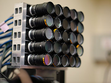
Researchers developed a new microscope that combines diffractive optics with 25 tiny cameras to capture simultaneous images at multiple depths. [Image: Eduardo Hirata Miyasaki]
Sometimes it takes 25 “eyes” to see tiny, moving organisms or complicated cellular structures.
Researchers in the United States have built a 25-camera multifocus microscope that captures 3D volumes at high speeds (Optica, doi:10.1364/OPTICA.563617 ). Team members put the device through tests of tracking particle and microorganism dynamics, and they say that the optical microscope could have many applications in biomedical imaging.
Challenges of fast 3D imaging
Biologists usually want to study 3D specimens and, if the subjects are alive, their movements. However, optical microscopes create 2D images, and time-consuming tricks are needed to compile the pictures to replicate the third dimension. Sequential acquisition of a stack of 2D images is too slow to catch molecular diffusion or motions on the millisecond time scale. Faster techniques, such as light-sheet fluorescence microscopy, sacrifice spatial or temporal resolution or both.
High-numerical-aperture microscope lenses introduce spherical aberrations away from the focal plane. An aberration-corrected refocusing mechanism lessens the aberrations but requires a separate module for fixing the chromatic dispersion, thus introducing even more complexity to the system.
An array of cameras
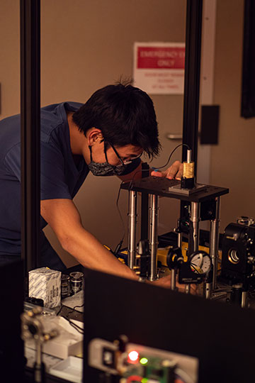
Eduardo Hirata Miyasaki building the new microscope. [Image: Sara Abrahamsson]
In the microscope developed by scientists at the University of California Santa Cruz (UCSC) and several other US institutions, a 25-plane multifocus grating changes the single image beam from a conventional wide-field microscope into a 5 × 5 array of images derived from the zeroth, first and second diffractive orders of the grating. At the receiving end of the light path, the team equipped 24 of the 25 cameras (excepting the center camera) with chromatic correction gratings to remove chromatic dispersion caused by the diffractive Fourier optics of the multifocus grating. A custom-built aluminum frame held the camera array in position. Capturing accurate color rendition is critical when performing fluorescence microscopy on living tissue.
“Employing an array of 25 cameras in a single microscope would have been a major investment and undertaking two decades ago but is today quite practical due to the availability of fast, sensitive and affordable machine vision cameras,” write the researchers, led by Sara Abrahamsson of UCSC and Eduardo Hirata Miyasaki, formerly of UCSC and now with the Chan Zuckerberg Biohub Network, USA. The team adds that the device architecture could be easily adapted to larger array sizes to provide greater imaging depth for bigger specimens.
After initial tests of fluorescent beads, the scientists imaged a 50 × 50 × 50 μm3 volume of fruit-fly larvae in both bright-field and fluorescent-imaging modes. The team could view a larva’s heart beating at about 2 Hz. Functional neuronal imaging of C. elegans (roundworms) nematodes over a larger volume (180 × 180 × 50 μm3) captured the animals’ motions as well as calcium channel activity in their neurons.