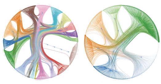
When scientists produced the first map of all synaptic connections in the roundworm Caenorhabditis elegans in 1986, many hailed it as a blueprint for the flow of brain signals. As it turned out, though, models of neuronal activity based on this wiring diagram bore little resemblance to the functional maps of brain activity measured in living worms.
This disconnect isn’t limited to worms. Mice, for instance, appear to have widespread silent synapses—wired connections that don’t send signals—and the actual responses of some cells in the fruit fly’s visual system do not match the responses the connectome predicts.
A new preprint helps to explain why: Most network features, in C. elegans at least, are not conserved between the anatomical and functional connectomes. Yet the anatomical connectome can still forecast—albeit in a complex way—observed neuronal activity in the worms, according to a second preprint by the same team, because “most signaling is happening along the wires,” says Andrew Leifer, associate professor of physics and neuroscience at Princeton University and principal investigator on both preprints.
The findings begin to address the long-standing challenge of reconciling structure and function, and show that “we weren’t entirely wrong” about the importance of synaptic connectivity, says Jihong Bai, professor of basic sciences at the Fred Hutchinson Cancer Center, who was not involved in the work.
The debut of a color-coded map of cell types in the worm brain in 2021 split the neuroscience community. It made it possible to identify individual neurons in whole-brain recordings and compare annotated recordings with the connectome—an exercise that revealed no correlation between the two.
“At the time, this finding was very controversial,” says Eviatar Yemini, assistant professor of neurobiology at the UMass Chan Medical School, who led the 2021 study but was not involved in the preprints. Researchers were divided “between those who couldn’t believe that this was the case and others who viewed this as a natural distinction between hardware and software,” he says.
An independent team replicated the findings the following year, and Yemini says he and his collaborators now have unpublished data showing that connectivity differences between male and hermaphrodite worms don’t match the two worms’ differences in brain activity.
Meanwhile, the publication of a “wireless” C. elegans connectome in 2023 revealed a dense signaling network, with all neurons forming, on average, more neuropeptidergic connections than synaptic links.
S
till, the studies that have looked for correlations between connectivity and functional activity in worms so far have been limited to cells that are spontaneously active, Leifer says.
Instead, he and his colleagues opted to systematically stimulate every neuron in the nematode brain, one at a time, and track the effects on the remaining neurons, covering two-thirds of all possible neuron pairs. Averaging the responses from 113 genetically identical worms, the team produced the first “signal propagation” atlas of C. elegans brain activity in 2023.
Neurons with direct synaptic connections are likely to respond to each other, with the response rate diminishing among neurons that have more synapses separating them, Leifer’s team found when they compared their functional atlas to the C. elegans connectome.
But when they trained a model to predict neuronal responses using the connectome, it was unable to reproduce the patterns of information flow seen in worms, the group reported in their 2023 paper. In fact, neuronal perturbation often prompted responses from cells that had no anatomical connections to the target cell, likely through neuropeptide signaling. Among neuronal pairs identified as likely candidates for extrasynaptic signaling, functional responses fell in mutant worms unable to release neuropeptide vesicles, the study found.
Despite that disparity, anatomical and functional maps might better align in terms of their broader organization, Leifer says. “Network science focuses on this implicit assumption that properties of the network, like the architecture, are informative and helpful,” he says.
Across most of the brain, the subnetworks of neurons—or modules—that are physically connected to one another do not overlap with the modules that show similar activity when another neuron is stimulated, Leifer’s team reported in one of the two new preprints, which was posted on arXiv last December and accepted for publication in PRX Life earlier this month. The only exception is the pharynx, a simple neuromuscular organ that contains a community of neurons separate from other regions.
And classifying “rich club” neurons—those that have a wealth of connections—revealed two, mostly distinct, lists of cells, the study found. AVEL and AVER, a pair of neurons involved in backward locomotion, buck that trend by having a multitude of both anatomical and functional connections.
For the remaining cells, being physically connected to several neurons doesn’t translate to having multiple functional connections, “perhaps because they might only be transiently active in certain contexts or internal states,” says study investigator Sophie Dvali, a graduate student in Leifer’s lab.
That lack of correspondence adds to mounting evidence that researchers cannot infer information flow using the synaptic connectome alone.
I
n the other preprint, posted on bioRxiv last November, Leifer and his colleagues took a different approach—they used a portion of the neuronal activity data from their 2023 signal propagation atlas to train a computer model to infer the remaining activity. They constrained the model to the worm’s connectome so that it was forced to learn pathways of information flow that depend on anatomical connections.
The model captured neuronal responses among connected cells and neurons without a synaptic link. Rather than relying on direct connections, the model appears to identify complex pathways that make it possible to ferry information between far-flung neurons, says study investigator Matthew Creamer, associate research scholar at Princeton University.
The brain is a dynamic system that acts in complex ways, Creamer says. For example, a neuron could signal to its neighbor, which in turn silences the neuron that stimulated it. “That’s exactly the kinds of things we’re trying to account for in our model.”
In contrast to the 2023 findings, connectivity appears to be critical for inferring neuronal responses, the preprint shows. When Creamer used a model that was not constrained to the connectome, or used a scrambled connectivity map, the model performed poorly.
The findings “provide evidence that there is a causal relationship between the anatomical and functional connectome, which I believe could be a significant advance,” says Hannes Buelow, professor of neuroscience at the Albert Einstein College of Medicine, who was not involved in either study.
But only the model knows where that causality is coming from, Buelow says. Testing a slice of the connectome—such as the sensory system, which contains signaling pathways that are relatively well understood—could reveal causal interactions, he says.
It’s unclear, however, the extent to which extrasynaptic signaling contributes to neuronal responses. That the model could replicate most signals using the connectome implies that wireless communication constitutes only a fraction of neuronal responses.
Although wireless signaling might not account for most neuronal communication in worms, “for any individual connection it may be very important,” Creamer says. And variability in neuronal responses between animals could underestimate how much signaling occurs outside of synapses, he adds.
Training the model using functional data from a larger number of animals, which could better account for the variability, might reveal a more significant role for extrasynpatic signaling. For now, Leifer and his team plan to use optical sensors to measure neuropeptide flow following neuronal stimulation, he says.
And understanding that variability—why different neurons respond to stimulation of the same cell in different, but genetically identical, animals—could reveal how internal processes influence neuronal interactions. Inducing a particular internal state, such as hunger or arousal, in the worm and observing how signaling patterns change is another “exciting future direction,” Leifer says.
The team hopes that the work will provide a blueprint for similar investigations in other organisms, Leifer says. The complete fly connectome, published in 2024, could be used together with new recordings of rapid neuronal activity to model that organism’s brain function, he says.
