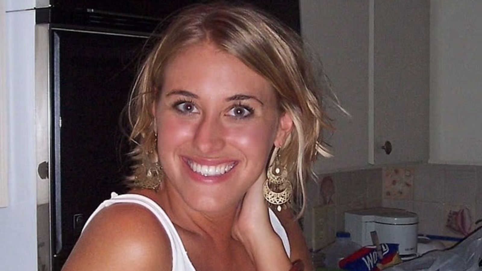
Introduction
Chronic dacryocystitis (CD), primarily resulting from primary acquired nasolacrimal duct obstruction (PANDO), is a prevalent ocular inflammatory condition and a common ophthalmological and nasal-related disorder. Symptoms of CD typically include excessive tearing, purulent discharge, skin swelling in the dacryocyst area, and pain, predominantly affecting women aged 40–60 years.1,2 These symptoms frequently impact patients’ daily lives and diminish their quality of life.
Clinically, it has been observed that patients often exhibit severe symptoms and experience disease courses that may persist for several months or even years when they seek medical treatment. Currently, the pathogenesis of PANDO remains unclear, and there is no effective pharmacological treatment available; thus, surgical intervention is the primary therapeutic approach.3,4 Presently, endoscopic dacryocystorhinostomy (En-DCR) has emerged as the preferred surgical technique for managing PANDO.5–7
Quality of life is defined as individuals’ subjective assessment of their goals, expectations, standards, and concerns within different cultural and value systems. Over the past few decades, vision-related quality of life has garnered increasing attention.8,9 Considering the essential role of vision in everyday activities,10 the symptoms of chronic dacryocystitis (especially excessive tearing) can greatly impair visual clarity and negatively impact overall quality of life. Our previous study has demonstrated that when lacrimal duct obstruction occurs, patients exhibit an excessive production of tears, known as epiphora, which disrupts the stability of the tear film on the ocular surface and may consequently impair visual acuity.11 The National Eye Institute Visual Functioning Questionnaire-25 (NEI-VFQ-25) is a widely utilized instrument for evaluating vision-related health status and quality of life in individuals with chronic ocular conditions, including cataracts, glaucoma, corneal transplantation, and various surgical interventions.12 Research has also been conducted on its applicability to macular disease, diabetic retinopathy, Graves’ ophthalmopathy, immune-mediated eye disorders, cytomegalovirus retinitis, and other chronic ocular diseases.13–15 The NEI-VFQ-25 effectively captures patients’ psychological states, social functioning, and levels of dependency, demonstrating its broad applicability. Consequently, it has been extensively translated into numerous languages and has been the subject of studies in China.16
Given that CD significantly impacts patients’ quality of life, analyzing the improvement in vision-related quality of life and associated factors before and after En-DCR surgery is of considerable importance.
Method
Participants
This study prospectively enrolled 57 patients diagnosed with CD who were scheduled to undergo En-DCR between August 2023 and December 2023. All surgeries were performed according to the standard En-DCR procedure and were performed by the same senior oculoplastic surgeon. Patients with concurrent ocular diseases affecting visual quality, significant systemic illnesses, inability to cooperate with examinations and surgical procedures, or psychiatric disorders were excluded from the study. Upon enrollment, detailed demographic and clinical information, including gender, age, and disease duration, were meticulously recorded. Comprehensive preoperative assessments were conducted at our institution. Ultimately, 54 patients completed the entire follow-up period, after accounting for those lost to follow-up. With the assistance of local community workers, individuals of the same age and gender who did not have other diseases affecting their quality of life were selected. Concurrently, a control group representative of the general population, matched for age and gender distribution, was established via a home-based questionnaire survey after obtaining informed consent.17
The main project to which this study belongs followed the tenets of the Declaration of Helsinki and was approved by the Medical Ethics Committee of Zhongshan Eye Center, Sun Yat-sen University, Guangzhou, China (2021KYPJ100), which has obtained written informed consent from the participants. The content of this study has been covered by the main project, and the collection of data through questionnaires with the patients’ consent does not impose any additional risk on the treatment of the individuals involved.
Diagnosis of Disease
CD typically arises secondary to nasolacrimal duct obstruction, leading to tear retention within the lacrimal sac and subsequent bacterial infection. The diagnosis of chronic dacryocystitis was confirmed through lacrimal duct irrigation18 and computed tomography dacryocystography (CT-DCG),19 in conjunction with clinical manifestations.
Method of Assessment of Vision-Related Quality of Life
The NEI-VFQ-25 is a widely employed, comprehensive instrument designed to assess vision-related health status and quality of life in individuals with chronic eye conditions. It consists of 25 single-choice items that address 12 dimensions of vision-related quality of life, including general health, general vision, ocular pain, near activities, distance activities, peripheral vision, social functioning, color vision, driving, role difficulties, dependency, and mental health. Each item provides 5–6 response options, corresponding to a score ranging from 0 to 100. All dimensions are equally weighted in the calculation of the overall score, with higher scores indicating an enhanced visual quality of life.20,21 The scale’s universality has facilitated its translation into multiple languages including Chinese for widespread application.
The scale’s broad applicability has enabled its translation into several languages, including Chinese, for extensive use. This study employed the NEI-VFQ-25 to evaluate vision-related quality of life in patients with CD. Assessments were conducted using questionnaires at four time points: prior to surgery, and 1, 3, and 6 months post-surgery. All questionnaires were recorded by the researchers after they asked the participants in detail. The general methodology of this study is illustrated in Figure 1.
Figure 1 This figure shows the general process of this research.
|
Evaluation of Operative Effect
The impact of the surgical intervention will be assessed six months post-operation. Currently, the efficacy of En-DCR in China is evaluated using the criteria established by Zhou Bing and Tang Xin, which categorize outcomes into three levels: cured, improved, and ineffective. The criteria for determining curative effect are as follows: 1) Cured: the patient exhibits no symptoms of excessive tearing or pus secretion; 2) Improved: the patient continues to experience epiphora but without symptoms of pus secretion; 3) Ineffective: the patient persists with symptoms of both epiphora and pus secretion.22
Statistical Analysis
Continuous variables following a normal distribution are presented as the mean ± standard deviation, while categorical variables are expressed as rates and constituent ratios. The t-test was employed to assess differences in the Visual Quality of Life (VQF) scores before and after surgery, which was normal distribution. Pearson’s correlation coefficient was utilized to examine the relationship between the duration of the disease and changes in VQF scores. A P-value of less than 0.05 was considered indicative of statistical significance. For missing data resulting from loss to follow-up, this sample will be excluded from the final statistical analysis.
Result
Basic Information
In this study, 57 patients with unilateral chronic dacryocystitis were initially enrolled. However, 3 patients were lost to follow-up, resulting in 54 patients completing all follow-up assessments. Baseline characteristics of these patients are presented in Table 1. The mean age of the patients was 51.39±9.35 years, with an average disease duration of 41.09±62.62 months. The cohort included 7 males and 47 females. Surgical outcomes were assessed at a six-month postoperative review, revealing that 34 patients were cured, 16 showed improvement, and 4 exhibited no effect. Of these, 40 patients underwent intraoperative intubation, while 14 did not. Additionally, a control group, matched for age and sex ratio, was included, consisting of 69 participants (10 males and 59 females) with a mean age of 49.26±6.01 years. Statistical analysis indicated no significant differences in age and sex ratio between the control and patient groups (p > 0.05).
![]() |
Table 1 Baseline Data of Enrolled Patients
|
Differences in Vision-Related Quality of Life Between Preoperative Patients and the Normal Population
The comparative analysis of vision-related quality of life between the preoperative patient group and the control group revealed significant differences in individual scores (p < 0.0001) (Table 2). Specifically, the patient group exhibited markedly lower scores than the control group across four domains: ocular pain (p = 0.0053), distance activities (p = 0.0251), role difficulties (p = 0.0280), and mental health (p = 0.0044).
![]() |
Table 2 Comparison of VQF Scores Between Preoperative Patient Group and Control Group
|
Differences in Vision-Related Quality of Life Before and After Surgery in Patient Group
Following a 6-month follow-up period, it was observed that the VQF scores of patients at 1, 3, and 6 months post-surgery demonstrated varying degrees of improvement compared to preoperative scores (p < 0.05), as illustrated in Figure 2. Notably, at 1 month post-surgery, patients showed significant enhancements in three areas: general health, general vision, and ocular pain. By 3 and 6 months post-surgery, improvements were also noted in general health, general vision, ocular pain, role difficulties, and mental health compared to preoperative assessments. All observed differences were statistically significant (p < 0.05) (Table 3).
![]() |
Table 3 Comparison of VQF Scores in Patients Before and After Surgery
|
![]() |
Figure 2 The variation curve of the VQF score is depicted, with the horizontal axis representing four distinct time points for patient scale evaluation and the vertical axis indicating the average VQF score of the patient cohort at these time points. Abbreviation: M AF, month after surgery.
|
Analysis of the Correlation Between the Course of Disease and the Changes in in Vision-Related Quality of Life
Correlation analysis results revealed a negative correlation between the disease course and changes in the VQF score (p < 0.0001, r = −0.6634) (Figure 3).
![]() |
Figure 3 The correlation analysis between the disease course and VQF score changes is illustrated in the figure, where the abscissa represents the disease course and the ordinate represents changes in the VQF score. #The changes in score were calculated as the VQF score at six months post-surgery minus the pre-surgery VQF score.
|
Effect of Prosthetic Tube Implantation on in Vision-Related Quality of Life
The findings indicated no significant differences in preoperative VQF scores, VQF scores at six months post-surgery, or changes in scores between the catheterized (all silicone tubing in place) and non-catheterized groups. None of these differences were statistically significant (p > 0.05) (Table 4).
![]() |
Table 4 Effect of Prosthetic Tube Implantation on VQF Score
|
Discussion
Chronic dacryocystitis (CD), as an eye condition that does not typically lead to blindness, has historically been under-researched. The etiology of CD primarily involves the obstruction or narrowing of the nasolacrimal duct, often accompanied by secondary infections. Clinical manifestations include persistent epiphora, increased ocular discharge, and purulent exudate. The prevalence of CD is notably higher among middle-aged and elderly females. In its early stages, the disease is frequently asymptomatic, lacking overt signs such as redness, swelling, or pain, which contributes to its frequent underdiagnosis.23 Nonetheless, extant research indicates that impaired lacrimal drainage in individuals with CD compromises the physiological cleaning function of the ocular surface, leading to the accumulation of deleterious substances and disruption of tear film homeostasis.24 Consequently, this may precipitate a cascade of complications, including secondary dry eye syndrome,25 infections, and potentially even blindness.3 With societal advancements and an increased emphasis on quality of life, there has been a growing recognition of CD, leading to heightened awareness and research interest.19
In clinical practice, it has been observed that the majority of patients with CD experience a prolonged disease course, with some cases persisting for months or even years. These patients frequently resort to surgical intervention once the disease significantly impairs their daily life and occupational functioning.6 Such delays in treatment not only exacerbate patient suffering but may also contribute to further disease progression. Our findings indicate that patients who adopt a proactive approach early in the disease trajectory and undergo surgical treatment tend to achieve more favorable outcomes, both in terms of prognosis and subjective quality of life. Previous research has predominantly concentrated on surgical techniques and short-term outcomes, with limited exploration of the relationship between surgical timing and improvements in patients’ quality of life. While research has suggested that surgery can improve quality of life in patients with CD,26 but the scope of assessment is not comprehensive, follow-up period is not long enough.
Furthermore, our prior study demonstrated that epiphora resulting from chronic dacryocystitis can adversely affect patients’ visual quality.11 In this study, we conducted a further examination of the results and discovered that the VQF scores for patients with CD were significantly lower than those of the control group. This finding suggests that the frequent lacrimal tearing associated with the disease adversely impacts patients’ vision-related quality of life, particularly in areas such as ocular pain, distance activities, role difficulties, and mental health.
En-DCR has emerged as a well-established surgical intervention for the treatment of CD.27 Compared to the traditional external approach, En-DCR offers advantages such as reduced surgical trauma, minimal bleeding, and the absence of facial scarring, making it a preferred option among patients. It is associated with high patient satisfaction and demonstrates a success rate exceeding 90%.28 In our study, the procedure’s efficacy rate was 92.59% (including both cured and improved cases). By establishing a channel between the lacrimal sac and the nasal cavity, the procedure facilitates the passage of tears through the lacrimal sac, bypassing the obstructed nasolacrimal duct, and allowing direct flow into the nasal cavity. This effectively alleviates symptoms such as epiphora and increased secretions.29 In the longitudinal assessment of patients following En-DCR, it was observed that postoperative VQF scores exhibited substantial enhancement. At one month post-surgery, there was a notable improvement in patients’ overall health, general vision, and ocular pain. By three and six months postoperatively, significant advancements were also observed in patients’ role-related difficulties and mental health. These findings suggest that En-DCR markedly ameliorates symptoms and enhances vision-related quality of life in individuals with CD.
Correlation analysis revealed a negative association between the duration of the disease and changes in VQF scores (p<0.0001, r=−0.6634). Patients with a prolonged disease course are at an elevated risk of infection due to prolonged obstruction of the lacrimal drainage system, which exacerbates the impact of chronic epiphora and increased secretions on their quality of life. Post-surgical intervention results in significant alleviation or even resolution of symptoms such as tearing and purulent discharge, thereby substantially improving patients’ vision-related quality of life. In certain patients with a prolonged disease course, significant ocular surface damage and severe disruption of tear film homeostasis are often observed. While En-DCR surgery effectively addresses issues related to tear overflow, it may not sufficiently mitigate other daily ocular surface function abnormalities, necessitating more comprehensive examination and treatment. Our research indicates that as the disease advances, patients experience exacerbated symptoms and a more substantial decline in vision-related quality of life. Conversely, patients who underwent surgical intervention earlier in the disease course exhibited greater postoperative improvements in vision-related quality of life, likely due to less pre-existing ocular surface damage.
For patients with anastomotic stenosis and severe lacrimal duct inflammatory reactions, lacrimal duct catheterization is adopted to support the lacrimal duct, maintain the anatomical structure of the anastomosis, and promote tear drainage. Although previous studies have demonstrated that lacrimal duct catheterization can enhance the prognosis of En-DCR,30,31 the impact of intraoperative catheterization on postoperative quality of life remains unexplored. The findings of this study indicate that for patients undergoing intraoperative catheterization (with all silicone tubing in place), the enhancement in their postoperative VQF scores was not significantly different from that of patients who did not undergo catheterization. This suggests that catheterization does not impact the improvement of vision-related quality of life. Therefore, we can decide whether to place a catheter based on the intraoperative situation without worrying about affecting the patient’s quality of life.
Furthermore, it was observed that some patients with a prolonged disease course exhibited postoperative VQF scores exceeding the average values of the normal population. This phenomenon may be attributed to the heightened subjective experiences of these patients, whose quality of life had been substantially impaired by the disease, leading to a pronounced sense of relief and well-being once symptoms were alleviated. Consequently, these patients may experience a level of satisfaction that surpasses that of the general population.
This study has some limitations that need addressing: First, patients with bilateral obstruction typically undergo En-DCR in both eyes through two separate procedures within a month, impacting the evaluation of postoperative vision-related quality of life. Consequently, patients with bilateral disease were excluded from the cohort, leading to a lack of analysis for these cases. Second, the large number of postoperative follow-up visits and questionnaire questions resulted in a slightly insufficient sample size, with some patients not complying and being lost to follow-up. This not only reduces the number of included patients, but also weakens the reliability of statistical analysis and brings about a certain degree of response bias. Larger-sample prospective studies are needed for further validation in the future. Attempting additional studies in different populations and clinical settings would enhance the generalizability of the findings.
Conclusion
CD is a condition that adversely affects patients’ vision-related quality of life, primarily manifesting in ocular pain, difficulties with distance activities, role limitations, and mental health challenges. En-DCR is an effective procedure that can significantly enhance the postoperative vision-related quality of life for these patients. In clinical diagnosis and treatment, early diagnosis and treatment should be prioritized, and medical staff should educate patients to seek timely medical attention after symptoms appear to enhance their quality of life and achieve better surgical outcomes.
Data Sharing Statement
The data used for the analysis are available from the corresponding author upon reasonable request.
Ethics Approval
This study was approved by the Institutional Review Board of the Zhongshan Ophthalmic Center, Sun Yat-sen University, China (No. 2021KYPJ100) and adhered to the tenets of the Declaration of Helsinki. Informed consent was obtained from all subjects involved in the study.
Author Contributions
All authors made a significant contribution to the work reported, whether that is in the conception, study design, execution, acquisition of data, analysis and interpretation, or in all these areas; took part in drafting, revising or critically reviewing the article; gave final approval of the version to be published; have agreed on the journal to which the article has been submitted; and agree to be accountable for all aspects of the work.
Funding
This study was partly supported by the Zhongshan Ophthalmic Center 2024 Innovations in Graduate Education Project (NO.91017-12240013).
Disclosure
The authors declare that they have no conflicts of interest in this work.
References
1. Paulsen FP, Thale AB, Maune S, Tillmann BN. New insights into the pathophysiology of primary acquired dacryostenosis. Ophthalmology. 2001;108(12):2329–2336. doi:10.1016/s0161-6420(01)00946-0
2. Yuksel D, Kosker M, Akoz I, Simsek S. Long-term results of simultaneous bilateral external dacryocystorhinostomy in cases with bilateral dacryostenosis. Seminars Ophthalmol. 2015;30(1):20–24. doi:10.3109/08820538.2013.810282
3. Chakrabarti S, Dasgupta S, Banerjee M, Pal D. Role of histomorphology and chronic inflammation score in chronic dacryocystitis. J Clin Diagn Res. 2016;10(7):EC01–EC03. doi:10.7860/JCDR/2016/19515.8089
4. Ma Y, Wang Z, Zhou G, et al. Comparison of the surgical outcomes of endoscopic dacryocystorhinostomy in chronic dacryocystitis with or without previous bicanalicular silicone tube intubation. Am J Otolaryngol. 2024;45(2):104200. doi:10.1016/j.amjoto.2023.104200
5. Watkins LM, Janfaza P, Rubin PA. The evolution of endonasal dacryocystorhinostomy. Surv Ophthalmol. 2003;48(1):73–84. doi:10.1016/s0039-6257(02)00397-1
6. Hammoudi DS, Tucker NA. Factors associated with outcome of endonasal dacryocystorhinostomy. Ophthalmic Plastic Reconstructive Surg. 2011;27(4):266–269. doi:10.1097/IOP.0b013e31820c72b7
7. Tooley AA, Klingler KN, Bartley GB, et al. Dacryocystorhinostomy for acquired nasolacrimal duct stenosis in the elderly (≥80 Years of Age). Ophthalmology. 2017;124(2):263–267. doi:10.1016/j.ophtha.2016.10.018
8. Assi L, Chamseddine F, Ibrahim P, et al. A global assessment of eye health and quality of life: a systematic review of systematic reviews. JAMA Ophthalmol. 2021;139(5):526–541. doi:10.1001/jamaophthalmol.2021.0146
9. Lin L, Yang L, Jin X, Zhao Y, Fan F. Management lacrimal sac abscesses using lacrimal probe and crawford silicon tube. BMC Ophthalmol. 2016;16(1):211. doi:10.1186/s12886-016-0378-y
10. Enander JMD, Spanne A, Mazzoni A, Bengtsson F, Oddo CM, Jörntell H. Ubiquitous neocortical decoding of tactile input patterns. Front Cell Neurosci. 2019;13:140. doi:10.3389/fncel.2019.00140
11. Meng Z, Chen R, Cao S, et al. Quality of vision in eyes with chronic dacryocystitis after endoscopic dacryocystorhinostomy. Clin Ophthalmol. 2023;17:3729–3737. doi:10.2147/OPTH.S441094
12. Wan Y, Zhao L, Huang C, et al. Validation and comparison of the National Eye Institute Visual Functioning Questionnaire-25 (NEI VFQ-25) and the Visual Function Index-14 (VF-14) in patients with cataracts: a multicentre study. Acta Ophthalmol. 2021;99(4):e480–e488. doi:10.1111/aos.14606
13. Trento M, Durando O, Lavecchia S, et al; EUROCONDOR trial investigators. Vision related quality of life in patients with type 2 diabetes in the EUROCONDOR trial. Endocrine. 2017;57(1):83–88. doi:10.1007/s12020-016-1097-0
14. Garweg JG, Stefanickova J, Hoyng C, et al; AQUA Investigators. Vision-related quality of life in patients with diabetic macular edema treated with intravitreal aflibercept: the AQUA Study. Ophthalmol Retina. 2019;3(7):567–575. doi:10.1016/j.oret.2019.03.012
15. Ng H, Vermeer KA, van Meurs JC, La Heij EC. Visual acuity inadequately reflects vision-related quality of life in patients after macula-off retinal detachment surgery. Invest Ophthalmol Visual Sci. 2020;61(10):34. doi:10.1167/iovs.61.10.34
16. Wang CW, Chan CL, Jin HY. Psychometric properties of the Chinese version of the 25-item national eye institute visual function questionnaire. Optometry Vision Sci. 2008;85(11):1091–1099. doi:10.1097/OPX.0b013e31818b9f23
17. Ezhumalai S. Outcome of camp approach in treatment of alcohol use disorder: a non-randomized controlled study. J Psychiatry Spectr. 2024;3(2):101–109. doi:10.4103/jopsys.jopsys_48_23
18. Shen X, Huang C, Wang S, Wen J. Optimisation of reduction for prolapsed silicone tube after lacrimal intubation. J Laryngology Otol. 2024;138(5):535–539. doi:10.1017/S0022215123001706
19. Eshraghi B, Abdi P, Akbari M, Fard MA. Microbiologic spectrum of acute and chronic dacryocystitis. Int J Ophthalmol. 2014;7(5):864–867. doi:10.3980/j.issn.2222-3959.2014.05.23
20. Mangione CM, Lee PP, Gutierrez PR, Spritzer K, Berry S, Hays RD; National Eye Institute Visual Function Questionnaire Field Test Investigators. Development of the 25-item National Eye Institute Visual Function Questionnaire. Arch Ophthalmol. 2001;119(7):1050–1058. doi:10.1001/archopht.119.7.1050
21. Nickels S, Schuster AK, Singer S, et al. The National Eye Institute 25-Item Visual Function Questionnaire (NEI VFQ-25) – reference data from the German population-based Gutenberg Health Study (GHS). Health Qual Life Outcomes. 2017;15(1):156. doi:10.1186/s12955-017-0732-7
22. Li M, Li W, Sha Q, Yu L, Wang X. Analysis of the safety and effectiveness of endoscopic nasal dacryocystorhinostomy in the remedy of chronic dacryocystitis. Medicine. 2024;103(3):e36934. doi:10.1097/MD.0000000000036934
23. Ali MJ, Kamal S, Gupta A, Ali MH, Naik MN. Simple vs complex congenital nasolacrimal duct obstructions: etiology, management and outcomes. Int Forum Allergy Rhinol. 2015;5(2):174–177. doi:10.1002/alr.21435
24. Marcet MM, Shtein RM, Bradley EA, et al. Safety and efficacy of lacrimal drainage system plugs for dry eye syndrome: a report by the American Academy of Ophthalmology. Ophthalmology. 2015;122(8):1681–1687. doi:10.1016/j.ophtha.2015.04.034
25. Yu X, Wang K, Hu Y, et al. Dry eye and decreased tear film stability in primary acquired nasolacrimal duct obstruction patients. Int Ophthalmol. 2024;44(1):277. doi:10.1007/s10792-024-03204-8
26. Ali MJ, Iram S, Ali MH, Naik MN. Assessing the outcomes of powered endoscopic dacryocystorhinostomy in adults using the Lacrimal Symptom (Lac-Q) questionnaire. Ophthalmic Plast Reconstr Surg. 2017;33(1):65–68. doi:10.1097/IOP.0000000000000660
27. Lueder GT. Neonatal dacryocystitis associated with nasolacrimal duct cysts. J Pediatr Ophthalmol Strabismus. 1995;32(2):102–106. doi:10.3928/0191-3913-19950301-10
28. Nowak R, Ali MJ. Experience of the first three years of independent practice following surgical training: time taken and long-term outcomes of powered endoscopic dacryocystorhinostomy. Seminars Ophthalmol. 2023;38(7):665–669. doi:10.1080/08820538.2023.2195025
29. Torabi P, Stenstrom B, Larsson AM, Bjornberg P, Svensson C, Engelsberg K. Outcomes of external and endonasal dacryocystorhinostomy according to a modified Lacrimal Symptom Questionnaire (Lac-Q). Med Hypothesis Discov Innov Ophthalmol J. 2023;12(2):55–61. doi:10.51329/mehdiophthal1470
30. Evereklioglu C. Primary probing with and without Monoka Silastic stent intubation for epiphora in older children and adults. Curr Eye Res. 2020;45(1):87–90. doi:10.1080/02713683.2019.1643026
31. Orsolini MJ, Schellini SA, Souza Meneguim RLF, Catâneo AJM. Success of endoscopic dacryocystorhinostomy with or without stents: systematic review and meta-analysis. Orbit. 2020;39(4):258–265. doi:10.1080/01676830.2019.1677726