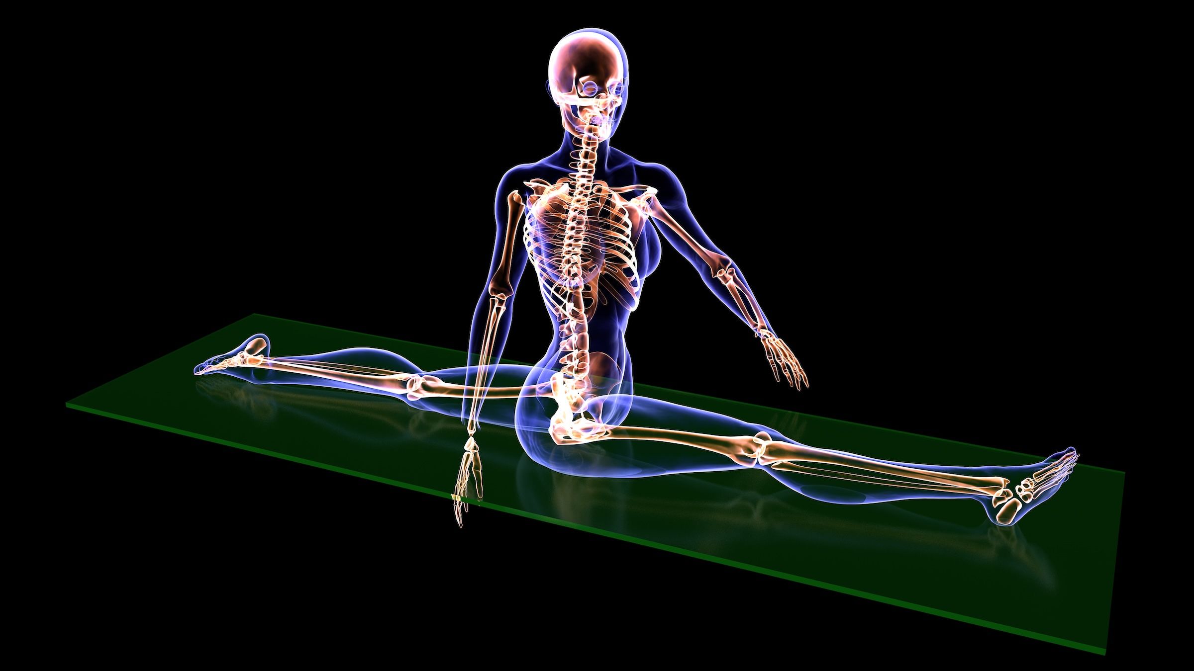
Anterior Disc Displacement of Temporomandibular Joint: Imaging diagnos

Introduction

The temporomandibular joint (TMJ) is the only bilateral joint in the human body, which is composed of mandibular condyle, articular surface of the temporal bone, articular disc and related muscles and ligaments. As a structure between the condyle and the articular surface, the articular disc plays a role in buffering occlusal force, stabilizing the joint, and regulating mandibular movement. It is also the key point for the occurrence and development of a large part of temporomandibular joint problems. Temporomandibular joint disorder (TMD) has a high incidence and involves a wide range of people.1 Anterior disc displacement (ADD) of the articular disc is the pathological basis of many TMDs. Its clinical manifestations include pain, dysfunction, and clicking or grating within the joint. These will not only affect the patient’s normal physiological functions such as chewing, swallowing, etc., but also may lead to anxiety, depression and other mental problems, damaging the patient’s health from both the physiological and the psychological level. The cause and mechanism of TMD is still unclear. Statistical data showed that it might be related to female gender, poor sleep quality, occlusion, oral parafunction, etc.2 Anxiety, depression and other psychological states were very important for the occurrence, development and treatment of TMD.3

Before the 1980s, there were many kinds of classifications of TMD in the world. In 1992, RDC/TMD was released and widely adopted.4 After 2000, the International Association for Dental Research (IADR) revised RDC/TMD and ultimately released DC/TMD in 2014.5 According to DC/TMD, TMD can be divided into two categories, pain related disorders and TMJ intra-articular disorders. The first category includes myofascial pain, myofascial pain with referral, local myalgia, arthralgia and headache attributed to TMD. The second category includes disc displacement with or without reduction, joint degenerative diseases and subluxation. The development of ADD has a certain degree of self-limitation, and some asymptomatic patients may also have structural changes. In addition to clinical symptoms and signs, imaging techniques are often used to assist in diagnosis. When treat this disease, doctors should take a combination of factors into consideration, including symptoms, clinical examinations, and the patient’s psychological state, etc. Due to the high incidence of the disease and complexity of its clinical management, it is necessary to discuss the imaging diagnosis and multidisciplinary treatment of TMD. A literature search was conducted in PubMed, CNKI, and Web of science, with articles published mostly between January, 2020 and May, 2025. To treat or not to treat? Treat now or observe for a period before intervention? Conservative treatment or surgical treatment? People often struggle, we always discuss.

Imaging Diagnosis

Magnetic Resonance Imaging (MRI)

As an effective, non-invasive and non-radiative imaging examination method, MRI is able to directly observe the position, shape, pathological features and movement during opening and closing of the mouth of temporomandibular articular disc. Currently, it is the most accurate imaging method for diagnosing ADD6,7 and the effectiveness has been tested over time in practice.8–10 With the help of MRI, many researchers have explored quantitative indicators of ADD. Zhang et al studied the relationship between the displacement distance and length of disc. Their results showed that the length of the disc decreased and the width of the disc increased as the displacement distance increased.11 The research of Shahab’s team showed that ADD with or without reduction was related to condyle horizontal angle and intercondylar angle.12 The precision of the diagnostic process is expected to further assist clinical decisions.

With the continuous development of multidisciplinary collaboration and modern technology, MRI diagnosis of TMD has intersected with the field of artificial intelligence. In the latest report, researchers constructed deep learning models based on deep learning algorithms such as convolutional neural networks to assist doctors in accurate diagnosis.13,14 Their studies demonstrated the potential application of deep learning algorithm with pre-training for automatically detecting ADD from MRI in patients with TMD.

Computed Tomography (CT)

In 1980, CT was first used for the diagnostic evaluation of TMJ.15 However, regular CT is not suitable for diagnosing soft tissues such as articular discs, muscles and ligaments. Only after injecting contrast agent into the joint can the articular disc be seen in CT scan. However, it is invasive, painful and prone to allergy, and is not widely applied.16 CT is mainly used for hard tissue in TMD.17 It can clearly show the size, shape, position, and continuity of cortical bone of the condyle, thereby evaluating the progression of the disease and providing message for further treatment decisions. Compared with CT, CBCT has higher resolution ratio, lower radiation dose and shorter scanning time,18 but its display of articular disc is still insufficient to support clinical diagnosis. Some researchers have combined the advantages of MRI and CBCT, using CBCT-MRI fused images to significantly improve the reliability of temporomandibular joint disc detection.19,20

The recent spectral CT has stronger soft tissue resolution and can relatively better display the displacement changes of the articular disc. It applies calcium inhibition technology to decalcify bones, generating virtual CT images that are not affected by calcium, thereby highlighting the articular disc.21 In the research of Zhang et al, the position of temporomandibular joint discs on calcium suppressed spectral CT images were basically consistent with those on MRI images. Calcium suppressed spectral CT imaging could accurately evaluate the position of the articular disc and the thickness of the posterior band.22 Another team also demonstrated the potential of spectral CT to perform preliminary MRI-like evaluations of articular disc, condyle bone, articular cavity effusion, etc.23 It may help patients with MRI contraindications or emergency trauma.

Ultrasonography (US)

US is convenient, low-cost, non-invasive and non-radiative. As early as 1991, researchers had conducted research on US diagnosis of the TMJ.24 At that time, US was not considered a good choice for clinical applications in TMJ. With the deepening of exploration, now many studies have shown that it is a feasible method for evaluating TMJ related disease, especially in terms of disc displacement.25–27 Although it cannot display articular discs as directly and clearly as MRI, it has its own indications such as the early screening in large population due to its low cost and convenience.28,29 Pekince et al established a quantitative US diagnostic method, which could improve the feasibility and reproducibility of US examination for ADD.30

Many factors can affect the effectiveness of US for TMD. Some researchers demonstrated that compared to ordinary linear array probes, L-shaped linear array probes can provide clearer imaging of TMJ.31 The presentation of anatomical structures of TMJ also varies with the position of the ultrasound probe.32 For ultrasound frequency, the detection effect of high-frequency US is generally better than low-frequency US.33,34 It should be noted that US depends highly on the operator.35 The operators are supposed to have rich experiences in clinical practice and image interpretation. There are many other factors that may affect the quality and interpretation of US images such as contact pressure, the type of gel or standoff pad used to scan the patient, and so on.36

The development of imaging has greatly promoted the research process of TMD. In addition to MRI, CT and US, there are other imaging techniques such as panoramic radiography, plain radiography and single photon emission computed tomography (SPECT), etc., which all have their own indications. At present, MRI is still the “gold standard” for diagnosing ADD, while CT is a nice tool for observing the bone condition of the condyle. Spectral CT and CBCT, as well as US, have great potential in observing the position of articular disc and assisting clinical diagnosis, and they are more suitable for emergency conditions and patients with MRI contraindications. The advantages, disadvantages, and indications of several imaging techniques for diagnosing ADD are shown in the Table 1.

Table 1 The Advantages, Disadvantages, and Indications of Several Imaging Techniques for Diagnosing ADD

Multidisciplinary Treatment

Reversible Conservative Treatment

Psychotherapy

Temporomandibular joint pain has gradually been recognized as a result of the interaction between physiological and psychological factors which requires multidisciplinary treatment.37 The biopsychosocial model can effectively solve many problems in the prevention and treatment of TMD, but due to various obstacles such as culture and socialites, levels of care settings, health services, etc., the clinical framework supporting this model has not been widely implemented at present.38 Psychological therapy includes cognitive-behavioral therapy, behavioral therapy, acceptance and commitment therapy, mindfulness, hypnosis, etc. For example, cognitive-behavioral therapy can correct patients’ fear of mandibular movement, enabling them to actively receive treatment and exit the cycle of erroneous cognition.39 Research has shown that cognitive-behavioral therapy, when used alone or in combination with other treatment methods such as oral appliances, pressure management, or biofeedback, is effective in most cases of maxillofacial pain.40 It is worth noting that listening to music, an intervention based on emotion regulation and attention regulation models, can even regulate TMD.41 Relaxing music and patients’ favourite music do particularly well. It is demonstrated that combining psychotherapy with other treatments together is more effective than applying it alone, and psychotherapy is expected to become a good assistant for driving away painful TMD.42

During the treatment process, it is important for the doctor to consider the patients’ chief complaints and how much impact do the symptoms have on their quality of life. Some patients may have structural abnormalities, but their subjective feelings are not obvious. Some patients’ symptoms can gradually disappear through self-compensation. In fact, ADD without reduction is common in the population. If there is no pain or restricted mouth opening, it is indicated that the tissue behind the displaced articular disc is likely to have undergone reconstruction and adaptation.43 At this point, it is recommended to observe, communicate and evaluate, applying appropriate psychological therapy rather than other interventions. Adequate patient education and psychological guidance can help patients establish a correct understanding of the disease and even affect its progression.

Physiotherapy

Physical therapy refers to the application of various non-invasive and non-pharmacological treatment methods to prevent and treat diseases. It can be divided into two categories: one mainly applies manual therapy and exercise therapy, and the other mainly applies various physical factors, including electricity, light, sound, magnetism, cold, heat, etc. Many studies have shown that manual therapy can improve joint function.44,45 A recent review demonstrated that all manual therapies, either alone or in association with other conservative interventions, can effectively reduce pain and increase degree of mouth opening.46 The effectiveness of exercise therapy has also been confirmed in many studies.47,48 Oral myofunctional therapy, as a form of exercise therapy, could improve symptoms such as swallowing difficulties caused by TMD through acting on the muscles of the neck and face.49 A study by Machado et al compared the effects of laser therapy alone and laser combined with exercise therapy, and found that adding oral exercise therapy was more effective in promoting TMD rehabilitation than using laser alone.50 In addition, research suggested that the combined use of manual therapy and exercise therapy will be more beneficial for the long-term stable rehabilitation of TMJ.51

For therapy with physical factors, electricity,52 light,53 sound,54 magnetism55 and acupuncture56 all play a role in the treatment of TMD. The efficacy of different therapies is influenced by many factors. Some may be related to psychological suggestion. Some may be related to the types of TMDs, but many samples in studies have included different types of TMDs or even mixed TMDs. In clinical practice, sometimes it is inevitable to encounter situations that we have difficulty in classifying TMDs clearly, and in such cases, symptomatic treatment may be the main solution.

Medication

There are many drugs used to treat TMD, including nonsteroidal anti-inflammatory drugs (NSAIDs), corticoids, analgesics, muscle relaxants, anxiolytics, opiates, tricyclic antidepressants, gabapentin and lidocaine patches, etc.57 Diclofenac, a commonly used NSAID, was shown to have a significant effect on early and rapid symptom relief.58 It is worth noting that taking diclofenac at a dose of 150 milligrams per day for at least 90 days has been found to be associated with an increased risk of myocardial infarction or cardiovascular death.59 Therefore, doctors should prescribe medication rigorously, and patients should strictly follow the doctors’ advice when taking medication. Analyzing the unique clinical situation of each patient is crucial for determining the optimal personalized treatment. External use, which means to locally apply the drug in the form of cream or ointment on TMJ, can relatively avoid the possible systemic adverse reactions when taking it orally. Diclofenac can achieve sufficient local concentrations,60 and there is no difference in efficacy between oral and high-dose external use.61

Arthrocentesis is an established minimally invasive method of treatment for TMJ internal derangement. Injections of drugs such as hyaluronic acid, corticosteroids, and blood products can effectively alleviate pain and increase the degree of mouth opening.62 A team has developed an optimized formulation of nanostructured lipid carriers for intra-articular administration of naproxen, a kind of NSAID. Compared with traditional form of drug delivery, this method could prolong the duration of drug action, reduce the frequency of administration, and minimize side effects.63 Ozone injection therapy, as a relatively new treatment method, is considered to have positive potential in relieving TMJ pain.64 In addition to injecting drugs into the joint cavity and capsule, muscle injection with botulinum toxin also works. However, the efficacy of injecting botulinum toxin into chewing muscles is controversial as a recent review demonstrated.65 And more research is needed to evaluate the possible negative effects of botulinum toxin. In a case report by Alvesalo et al, a patient’s masseter was temporarily paralyzed by injections of botulinum toxin A.66

Occlusal Splints

Occlusal splint is a widely used technique in TMD patients, which can help restore the relationship between the articular disc and condyle, relieve pain and get rid of mouth opening restriction. The most commonly used ones are stabilization splints and anterior repositioning splints. The stabilization splint covers the entire dental arch, and there is no relationship of intercuspation between occlusal splint and the opposite teeth in intercuspal position.67 The clinical treatment effect of stabilization splint is commendable, and most patients can benefit from it.68 However, its long-term efficacy and comparison with other treatment methods still need to be confirmed by more clinical trials. At present, research on it is continuously deepening and refining, including its hardness,69 thickness,70 and so on.

Anterior repositioning splint can guide patients to slightly extend their mandible to a comfortable position for the TMJ. It has the relationship of intercuspation, ensuring stable position of the joint during comfort occlusion.71 Research has shown that anterior repositioning splint can effectively eliminate symptoms and reposition displaced articular disc.72 Its effectiveness is superior to stabilization splint, with good short-term clinical outcomes. However, a high long-term recurrence rate is worrying.73 In recent studies, researchers have designed anterior repositioning splints using digital technology and showed that the digital process could upgrade product accuracy and clinical efficiency, alleviate patients’ discomfort symptoms effectively, and improve patients’ satisfaction significantly.74,75

Irreversible Conservative Treatment

Occlusal Adjustment

The optimum oral treatment aims to stabilize the stomatognathic system, including teeth, periodontal tissue, muscles, and temporomandibular joint.76 These elements should be viewed as a whole and we should never miss the forest for the trees. The efficacy of occlusal adjustment is influenced by the type of occlusion, and the mechanism may be related to the smoothness of occlusal contact and its regulatory effect on masticatory muscles’ activity.77 However, although occlusal adjustment can help with oral and maxillofacial pain caused by occlusal factors, it cannot solve the problem of disc displacement if it occurs.78 Even many researchers do not recommend this method as a commonly used approach for treating TMD.79,80 As an irreversible method, doctors should apply it cautiously and should not use it for preventive treatment. In addition, occlusal adjustment cannot be performed when the pain is obvious. And the teeth should be adjusted in small amounts in multiple times. During the process, continuous attention should be paid to the patients’ mental health.81

For patients with edentulism, the design of complete denture has a significant impact on TMJ. Studies by some researchers showed that lingual centralized complete denture was beneficial for maintaining the stability of complete dentures in patients with low alveolar ridge, and could help alleviate TMD symptoms in elderly edentulous patients.82,83 Recently, some researchers have been using digital technology to assist in occlusal balance. A new technology for occlusal transfer and condylar position adjustment proposed by Gong et al was easy to operate, accurate in data, and helpful for patients to restore normal relationship between articular disc and condyle.84 Researchers established a personalized reference plane to adjust jaw position, effectively relieving the symptoms of TMD.85 However, they have also indicated that position of the condyle and hinge axis obtained by digital methods may sometimes have low repeatability and high variability, and therefore lack correspondence with anatomical structure.85

Orthodontics

A certain connection may exist between TMD and malocclusion.86 Applying orthodontic treatment to address malocclusion and alleviate TMJ related symptoms to some extent has become one of the treatment options for TMD patients.87 Some studies have shown that orthodontic treatment for TMD can help alleviate clinical symptoms, improve quality of life, etc.,88,89 especially for patients with abnormal occlusion or even accompanied by mandibular adaptive deviation.90 Appropriate conservative orthodontic treatment for TMD can indeed maintain the long-term stability of patients’ TMJ function.91

It is worth noting that the relationship between orthodontics and TMD is complex and subtle. Some researchers believe that TMD may be one of the complications of orthodontic treatment. A study by Zubiate et al showed that people who received orthodontic treatment were 1.84 times more likely to suffer from TMD.92 Alam et al demonstrated that orthodontics might have negative psychological effects on patients who already had TMD, but it was not a triggering factor for the onset of the disease.93 Research of Shalish et al showed that orthodontics was not related to the diagnosis and disease characteristics of TMD.94 Controversy about the relationship between orthodontics and TMD has always existed. Recent studies suggest that before starting any orthodontic or dental treatment, a thorough examination of the function and morphology of the TMJ is necessary in order to assess treatment quality and avoid complications.95,96 Undiagnosed TMJ dysfunction may lead to further problems with the entire oral and maxillofacial system during dental treatment.

Mandibular Manipulation

If successful mandibular manipulation is performed in the early stage of acute irreversible disc displacement, the “locked” state can be quickly released, which will significantly help improve or even rebuild the normal disc-condyle relationship. The mandibular manipulation is usually treated in combination with anterior repositioning splints. Doctors first reset the “locked” TMJ manually, converting irreversible disc displacement into reversible disc displacement, and then use an anterior repositioning splint to manage reversible disc displacement.97 An 11-year MRI follow-up was conducted on patients who underwent mandibular manipulation and anterior repositioning splints treatment. The results showed that although there were slight changes in the shape and position of the articular discs, the ADD remained reversible, and the progression of TMD was successfully blocked.98

The combined effect of multiple treatments is very significant. Hao et al conducted a literature analysis and found that the combination of mandibular manipulation and occlusal splints treatment was more effective than using either mandibular manipulation or occlusal splints alone.99 It was recommended to perform intra-articular injection before mandibular manipulation, as this would help improve the intra-articular environment and reduce intra-articular viscosity.100 A study showed that intra-articular irrigation combined with mandibular manipulation and exercise therapy could help alleviate pain and promote the recovery of TMJ in patients with acute ADD without reduction.101 Some researchers combined the mandibular manipulation with various physical therapies.102

The success rate of mandibular manipulation may be related to the length of time the mandibular is locked. That is to say, the word “acute” we have mentioned earlier is very important. If the patient’s articular disc cannot be reduced for a long time, the elasticity of the tissue behind the disc will weaken and the intra-articular adhesion will be severe, which will negatively impact the success of reduction. Lei et al stated that “3 months” was probably the definition of “acute”, and this might be the key to success.97

Surgery

Minimally Invasive Surgery

In 1989, Israel and Tarro first proposed the TMJ arthroscopic disc suturing technique to treat ADD, but its success rate and long-term stability of TMJ were not satisfactory.103 In 2012, Yang’s team published an article introducing a new arthroscopic disc repositioning and suturing technique.104 They introduced a horizontal mattress suture method using 2 or 3 needles from medial to lateral to treat pure ADD. The new method stabilized the location of the disc through making the suture traction direction exactly consistent with the long axis of the disc. Their MRI evaluation demonstrated the efficiency of this method.103 The displaced articular discs can be classified mainly into pure anterior displacement and rotational displacement, the latter of which includes anteromedial displacement and anterolateral displacement. Their another publication including method introduction and efficacy evaluation of rotational displacement was published in 2019.105 Recent research reported that the clinical efficacy of Yang’s arthroscopic discopexy was highly promising.106 Researchers evaluated the growth of condylar bone after surgery, showing that after disc reduction, the condylar bone had strong ability of growing and reconstructing. In addition, the growth ability decreased with age.107 However, Yang’s arthroscopic discopexy technique also has its drawbacks. It is difficult to perform, requiring skilled TMJ arthroscopic surgeons and customized suturing equipment. In a recent study, Jerez et al designed a modification to Yang’s technique using a common, cheap, available, and disposable specimen set.108

Open Surgery

The advantage of open surgery is that it can help doctors reduce the articular disc under direct vision, thus restoring the anatomical relationship between the disc and the condyle accurately and quickly. Repositioning temporomandibular joint disc with anchors is an open surgical method for treating ADD. Wolford was the first to report the application of anchor screws for reducing articular disc.109 Compared with conservative treatment, open surgery with anchors can quickly eliminate symptoms such as pain, dysfunction, and clicking or grating within the joint. Many studies have demonstrated its effectiveness.110–112 According to a recent report, compared with traditional anchoring nails, the improved anchoring nail brought less foreign body sensation and pain, while the success rate was similar.113 Arab’s team applied umbrella perforated screw, which was first designed for orthopedic surgery, to patients with TMD.114 Their results showed that this technique could fix posterior fascia flaps easily and quickly without damaging the vascular supply to TMJ.

Removing the bilaminar zone of the temporomandibular joint disc is another open surgical method. Research demonstrated through MRI evaluation of the disc position before and after surgeries that this method was technically feasible for reducing and fixing the articular disc.115 However, the number of studies applying this method is limited, and the long-term stability of the reduced articular disc still needs further observation. In addition, it should be noted that although surgical treatment can achieve precise reduction of the articular disc and relieve symptoms, especially in the short term, there are disadvantages such as trauma, complications, and recurrence. In clinical practice, it is necessary for doctors to explain the advantages and disadvantages of various treatment methods sufficiently with patients. At present, digital technologies such as virtual surgical planning play a significant role in open surgery, especially in the management of patients with severe TMJ problems facing TMJ reconstruction. According to Del Castillo Pardo, etc., the application of virtual surgical planning can enable stock TMJ prostheses to express higher accuracy and replace custom-made prostheses in some indications, thus improving clinical efficiency and benefiting patients.116

Conclusion

TMD is a common disease affecting large population and ADD is its common pathological basis. At present, MRI is still the gold standard for diagnosing ADD, while CT can effectively observe changes in condylar bone. Different imaging techniques have their own potential applications in different conditions. The treatment of ADD is gradually developing towards multidisciplinary collaboration, and the biopsychosocial medical model is recommended. Although there are certain contradictions among researchers regarding the efficacy of various methods, especially long-term stability, most therapies can alleviate symptoms and improve patients’ life quality.

Looking forward to the future, the biopsychosocial medical model will gradually go mainstream. Diagnosis and treatment of TMD calls for an overall perspective. The oral and maxillofacial system is a whole, and as a dentist, one should pay attention to the interaction between teeth, periodontal tissue, muscles, and TMJ. When seeing a patient, symptoms, clinical signs and imaging techniques should all be taken into cautious consideration to accurately assess the stage and status of the disease. The actual needs, psychological state, etc. of him or her should be closely observed. To obtain maximum therapeutic effect with minimal invasion, dentists should first consider reversible conservative treatment, followed by irreversible conservative treatment, and surgical treatment at last.

Funding

Jingying Program of Savaid Stomatology School and Hangzhou Medical College Education Foundation.

Disclosure

The authors report no conflicts of interest in this work.

References

1. Valesan LF, Da-Cas CD, Réus JC, et al. Prevalence of temporomandibular joint disorders: a systematic review and meta-analysis. Clin Oral Investig. 2021;25(2):441–453. doi:10.1007/s00784-020-03710-w

2. Warzocha J, Gadomska-Krasny J, Mrowiec J. Etiologic factors of temporomandibular disorders: a systematic review of literature containing Diagnostic Criteria For Temporomandibular Disorders (DC/TMD) and Research Diagnostic Criteria for Temporomandibular Disorders (RDC/TMD) from 2018 to 2022. Healthcare. 2024;12(5):575. doi:10.3390/healthcare12050575

3. Florjanski W, Orzeszek S. Role of mental state in temporomandibular disorders: a review of the literature. Dent Med Probl. 2021;58(1):127–133. doi:10.17219/dmp/132978

4. Dworkin SF, LeResche L. Research diagnostic criteria for temporomandibular disorders: review, criteria, examinations and specifications, critique. 0890-2739 (Print).

5. Schiffman E, Ohrbach R, Truelove E, et al. Diagnostic Criteria for Temporomandibular Disorders (DC/TMD) for Clinical and Research Applications: recommendations of the International RDC/TMD consortium network* and orofacial pain special interest groupdagger. J Oral Facial Pain Headache. 2014;28(1):6–27. doi:10.11607/jop.1151

6. Johnson M, Sreela LS, Mathew P, Prasad TS. Actual applications of magnetic resonance imaging in dentomaxillofacial region. Oral Radiol. 2022;38(1):17–28. doi:10.1007/s11282-021-00521-x

7. Surolia P, Kumawat J, Sharma PK. Role of dynamic 3 tesla MRI in the evaluation of temporomandibular joint dysfunction. Cureus. 2023;15(3):e36681. doi:10.7759/cureus.36681

8. Somay E, Yilmaz B. Comparison of clinical and magnetic resonance imagining data of patients with temporomandibular disorders. Niger J Clin Pract. 2020;23(3):376–380. doi:10.4103/njcp.njcp_492_19

9. Lamot U, Strojan P, Surlan Popovic K. Magnetic resonance imaging of temporomandibular joint dysfunction-correlation with clinical symptoms, age, and gender. Oral Surg Oral Med Oral Pathol Oral Radiol. 2013;116(2):258–263. doi:10.1016/j.oooo.2013.04.019

10. Helms CA, Richardson ML, Moon KL, Ware WH. Nuclear magnetic resonance imaging of the temporomandibular joint: preliminary observations. J Craniomandibular Pract. 1984;2(3):219–224. doi:10.1080/07345410.1984.11677866

11. Zhang Q, Ye Z, Wu Y, et al. Nonlinear relationship between temporomandibular joint disc displacement distance and disc length: a magnetic resonance imaging analysis. J Clin Med. 2022;11(23):7160. doi:10.3390/jcm11237160

12. Shahab S, Amoozad Khalili Z, Emami Meybodi E, Banakar M. Relation between condyle horizontal angle and intercondylar angle with disc displacement in patients with temporomandibular joint disorders: an MRI evaluation. Radiol Res Pract. 2023;2023:3846525. [2090-1941 (Print]. doi:10.1155/2023/3846525

13. Lee YH, Won JH, Kim S, Auh QS, Noh YK. Advantages of deep learning with convolutional neural network in detecting disc displacement of the temporomandibular joint in magnetic resonance imaging. Sci Rep. 2022;12(1):11352. doi:10.1038/s41598-022-15231-5

14. Ito S, Mine Y, Yoshimi Y, et al. Automated segmentation of articular disc of the temporomandibular joint on magnetic resonance images using deep learning. Sci Rep. 2022;12(1):221. doi:10.1038/s41598-021-04354-w

15. Talmaceanu D, Lenghel LM, Bolog N, et al. Imaging modalities for temporomandibular joint disorders: an update. Clujul Med. 2018;91(3):280–287. doi:10.15386/cjmed-970

16. Temporomandibular joint: role of direct sagittal CT air-contrast arthrogram and MRI. (0030-6665 (Print))

17. Almashraqi AA, Sayed BA, Mokli LK, et al. Recommendations for standard criteria for the positional and morphological evaluation of temporomandibular joint osseous structures using cone-beam CT: a systematic review. Eur Radiol. 2024;34(5):3126–3140. doi:10.1007/s00330-023-10248-4

18. Dhabale GS, Bhowate RR. Cone-beam computed tomography for temporomandibular joint imaging. Cureus. 2022;14(11):e31515. doi:10.7759/cureus.31515

19. ElShennawy EM, Hamed WM, Samir SM. Diagnostic accuracy of MRI-CBCT fused images in assessment of clinically diagnosed internal derangement of the temporomandibular joint. Oral Radiol. 2024;40(2):226–233. doi:10.1007/s11282-023-00727-1

20. Wang YH, Ma RH, Li JJ, et al. Diagnostic efficacy of CBCT, MRI and CBCT-MRI fused images in determining anterior disc displacement and bone changes of temporomandibular joint. Dentomaxillofac Radiol. 2022;51(2):20210286. doi:10.1259/dmfr.20210286

21. Shim E, Kim BH, Kang WY, et al. Diagnostic performance of electron-density dual-energy CT in detection of cervical disc herniation in comparison with standard gray-scale CT and virtual non-calcium images. Eur Radiol. 2022;32(4):2209–2220. doi:10.1007/s00330-021-08374-y

22. Chen Z, Fan WP, Liu MQ, et al. Evaluation of position of articular discs in patients with temporomandibular joint disorders using calcium suppression techniques based on spectral CT. Chin J Stomatol. 2020;55(2):93–97. doi:10.3760/cma.j.issn.1002-0098.2020.02.004

23. Wang Y, Hu M, Wang Y, et al. Application of spectral CT calcium suppression imaging technology in the assessment of temporomandibular joint disorders. Chin J Stomatol. 2022;57(7):775–779. doi:10.3760/cma.j.cn112144-20220527-00282

24. Nabeih YB, Speculand B. Ultrasonography as a diagnostic aid in temporomandibular joint dysfunction. A preliminary investigation. Int J Oral Maxillofac Surg. 1991;20(3):182–186. doi:10.1016/S0901-5027(05)80013-X

25. Talmaceanu D, Lenghel LM, Bolog N, et al. High-resolution ultrasound imaging compared to magnetic resonance imaging for temporomandibular joint disorders: an in vivo study. Eur J Radiol. 2020;132:109291. (1872-7727 (Electronic). doi:10.1016/j.ejrad.2020.109291

26. Talmaceanu D, Lenghel LM, Bolog N, et al. High-resolution ultrasonography in assessing temporomandibular joint disc position. Med Ultrason. 2018;1(1):64–70. doi:10.11152/mu-1025

27. Azlag Pekince K, Caglayan F, Pekince A. The efficacy and limitations of USI for diagnosing TMJ internal derangements. Oral Radiol. 2020;36(1):32–39. doi:10.1007/s11282-019-00376-3

28. Erturk AF, Yelken Kendirci M, Ozcan I, Gokcen Rohlig B. Use of ultrasonography in the diagnosis of temporomandibular disorders: a prospective clinical study. Oral Radiol. 2023;39(2):282–291. doi:10.1007/s11282-022-00635-w

29. Friedman SN, Grushka M, Beituni HK, et al. Advanced ultrasound screening for Temporomandibular Joint (TMJ) internal derangement. Radiol Res Pract. 2020;2020:1809690. (2090-1941 (Print). doi:10.1155/2020/1809690

30. Li C, Zhou J, Shi Y, et al. Diagnostic efficacy of quantitative ultrasonography for anterior disc displacement of the temporomandibular joint. Quant Imaging Med Surg. 2023;13(10):6446–6455. doi:10.21037/qims-23-401

31. Yilmaz D, Kamburoglu K. Comparison of the effectiveness of high resolution ultrasound with MRI in patients with temporomandibular joint disorders. Dentomaxillofac Radiol. 2019;48(5):20180349. doi:10.1259/dmfr.20180349

32. Torres-Gaya J, Bosca-Ramon A, Marques-Mateo M, et al. Temporomandibular joint arthrocentesis guided by ultrasonography: an anatomical study. J Stomatol Oral Maxillofac Surg. 2021;122(4):e27–e31. doi:10.1016/j.jormas.2021.03.002

33. Habashi H, Eran A, Blumenfeld I, Gaitini D. Dynamic high-resolution sonography compared to magnetic resonance imaging for diagnosis of temporomandibular joint disk displacement. J Ultrasound Med. 2015;34(1):75–82. doi:10.7863/ultra.34.1.75

34. L KS, Zachariah GP, Chandran S. Ultrasonography: a step forward in temporomandibular joint imaging. A preliminary descriptive study. Clin Pract. 2019;9(2):1134. doi:10.4081/cp.2019.1134

35. Maranini B, Ciancio G, Mandrioli S, Galie M, Govoni M. The role of ultrasound in temporomandibular joint disorders: an update and future perspectives. Front Med. 2022;9:926573. (2296-858X (Print). doi:10.3389/fmed.2022.926573

36. Almeida FT, Pacheco-Pereira C, Flores-Mir C, et al. Diagnostic ultrasound assessment of temporomandibular joints: a systematic review and meta-analysis. Dentomaxillofac Radiol. 2019;48(2):20180144. doi:10.1259/dmfr.20180144

37. Garrigos-Pedron M, Elizagaray-Garcia I, Dominguez-Gordillo AA, Del-Castillo-Pardo-de-Vera JL, Gil-Martinez A. Temporomandibular disorders: improving outcomes using a multidisciplinary approach. J Multidiscip Healthc. 2019;12:733–747. (1178-2390 (Print). doi:10.2147/JMDH.S178507

38. Sharma S, Breckons M, Bronnimann Lambelet B, et al. Challenges in the clinical implementation of a biopsychosocial model for assessment and management of orofacial pain. J Oral Rehabil. 2020;47(1):87–100. doi:10.1111/joor.12871

39. Zhang Q, Zhang J, Ran W, Yu S, Jin Y. Effectiveness of cognitive behavioral therapy on kinesiophobia and oral health-related quality of life in patients with temporomandibular disorders, study protocol for a randomized controlled trial. Medicine. 2020;99(47):e23295. doi:10.1097/MD.0000000000023295

40. Noma N, Watanabe Y, Shimada A, et al. Effects of cognitive behavioral therapy on orofacial pain conditions. J Oral Sci. 2020;63(1):4–7. doi:10.2334/josnusd.20-0437

41. Imbriglio TV, Moayedi M, Freeman BV, et al. Music modulates awake bruxism in chronic painful temporomandibular disorders. Headache. 2020;60(10):2389–2405. doi:10.1111/head.13971

42. Christidis N, Al-Moraissi EA, Al-Ak’hali MS, et al. Psychological treatments for temporomandibular disorder pain-A systematic review. J Oral Rehabil. 2024;51(7):1320–1336. doi:10.1111/joor.13693

43. Bristela M, Schmid-Schwap M, Eder J, et al. Magnetic resonance imaging of temporomandibular joint with anterior disk dislocation without reposition – long-term results. Clin Oral Investig. 2017;21(1):237–245. doi:10.1007/s00784-016-1800-9

44. Delgado de la Serna P, Plaza-Manzano G, Cleland J, et al. Effects of cervico-mandibular manual therapy in patients with temporomandibular pain disorders and associated somatic tinnitus: a randomized clinical trial. Pain Med. 2020;21(3):613–624. doi:10.1093/pm/pnz278

45. Urbanski P, Trybulec B, Pihut M. The application of manual techniques in masticatory muscles relaxation as adjunctive therapy in the treatment of temporomandibular joint disorders. Int J Environ Res Public Health. 2021;18(24):12970. doi:10.3390/ijerph182412970

46. Arribas-Pascual M, Hernandez-Hernandez S, Jimenez-Arranz C, et al. Effects of physiotherapy on pain and mouth opening in temporomandibular disorders: an umbrella and mapping systematic review with meta-meta-analysis. J Clin Med. 2023;12(3):788. doi:10.3390/jcm12030788

47. Idanez-Robles AM, Obrero-Gaitan E, Lomas-Vega R, et al. Exercise therapy improves pain and mouth opening in temporomandibular disorders: a systematic review with meta-analysis. Clin Rehabil. 2023;37(4):443–461. doi:10.1177/02692155221133523

48. Shimada A, Ogawa T, Sammour SR, et al. Effectiveness of exercise therapy on pain relief and jaw mobility in patients with pain-related temporomandibular disorders: a systematic review. Front Oral Health. 2023;4:1170966. (2673-4842 (Electronic). doi:10.3389/froh.2023.1170966

49. Celik Guzel H, Tuncer A. The efficacy of orofacial myofunctional therapy in oral dysphagia accompanying temporomandibular dysfunction. Cranio. 2023(2151–0903 (Electronic):1–11.

50. Machado BC, Mazzetto MO, Da Silva MA, de Felicio CM. Effects of oral motor exercises and laser therapy on chronic temporomandibular disorders: a randomized study with follow-up. Lasers Med Sci. 2016;31(5):945–954. doi:10.1007/s10103-016-1935-6

51. Herrera-Valencia A, Ruiz-Munoz M, Martin-Martin J, Cuesta-Vargas A, Gonzalez-Sanchez M. Efficacy of manual therapy in temporomandibularjoint disorders and its medium-and long-termeffects on pain and maximum mouth opening: a systematic review and meta-analysis. J Clin Med. 2020;9(11):3404. doi:10.3390/jcm9113404

52. Serrano-Munoz D, Beltran-Alacreu H, Martin-Caro Alvarez D, et al. Effectiveness of different electrical stimulation modalities for pain and masticatory function in temporomandibular disorders: a systematic review and meta-analysis. J Pain. 2023;24(6):946–956. doi:10.1016/j.jpain.2023.01.016

53. Zhang Y, Qian Y, Huo K, et al. Efficacy of laser therapy for temporomandibular disorders: a systematic review and meta-analysis. Complement Ther Med. 2023;74:102945. (1873-6963 (Electronic). doi:10.1016/j.ctim.2023.102945

54. Mishra N, Barapatre P, Pandey M, et al. Data on ultrasound therapy as an adjuvant pain control method among Indian TMDS patients. Bioinformation. 2022;18(9):774–779. doi:10.6026/97320630018774

55. Jo JH, Jang Y, Chung G, Chung JW, Park JW. Long-term efficacy and patient satisfaction of pulsed radiofrequency therapy in temporomandibular disorders: a randomized controlled trial. Medicine. 2021;100(52):e28441. doi:10.1097/MD.0000000000028441

56. Liu GF, Gao Z, Liu ZN, et al. Effects of warm needle acupuncture on temporomandibular joint disorders: a systematic review and meta-analysis of randomized controlled trials. Evid Based Complement Alternat Med. 2021;2021:6868625. (1741-427X (Print). doi:10.1155/2021/6868625

57. Gil-Martinez A, Paris-Alemany A, Lopez-de-Uralde-Villanueva I, La Touche R. Management of pain in patients with temporomandibular disorder (TMD): challenges and solutions. J Pain Res. 2018;11:571–587. (1178-7090 (Print). doi:10.2147/JPR.S127950

58. Kurita Varoli F, Sucena Pita M, Sato S, et al. Analgesia evaluation of 2 NSAID drugs as adjuvant in management of chronic temporomandibular disorders. Sci World J. 2015;2015:359152. (1537-744X (Electronic). doi:10.1155/2015/359152

59. Moore N. Coronary risks associated with diclofenac and other NSAIDs: an update. Drug Saf. 2020;43(4):301–318. doi:10.1007/s40264-019-00900-8

60. Dong XD, Svensson P, Cairns BE. The analgesic action of topical diclofenac may be mediated through peripheral NMDA receptor antagonism. Pain. 2009;147(1–3):36–45. doi:10.1016/j.pain.2009.07.031

61. Businco LD, Businco AD, D’Emilia M, Lauriello M, Tirelli GC. Topical versus systemic diclofenac in the treatment of temporo-mandibular joint dysfunction symptoms. Acta Otorhinolaryngol Ital. 24(5):279–283.

62. Liapaki A, Thamm JR, Ha S, et al. Is there a difference in treatment effect of different intra-articular drugs for temporomandibular joint osteoarthritis? A systematic review of randomized controlled trials. Int J Oral Maxillofac Surg. 2021;50(9):1233–1243. doi:10.1016/j.ijom.2021.01.019

63. Guilherme VA, Ribeiro LNM, Alcantara ACS, et al. Improved efficacy of naproxen-loaded NLC for temporomandibular joint administration. Sci Rep. 2019;9(1):11160. doi:10.1038/s41598-019-47486-w

64. Haghighat S, Oshaghi S. Effectiveness of ozone injection therapy in temporomandibular disorders. Adv Biomed Res. 2020;9:73. (2277–9175 (Print)doi:10.4103/abr.abr_105_20

65. Saini RS, Ali Abdullah Almoyad M, Binduhayyim RIH, et al. The effectiveness of botulinum toxin for temporomandibular disorders: a systematic review and meta-analysis. PLoS One. 2024;19(3):e0300157. doi:10.1371/journal.pone.0300157

66. Alvesalo E, Kemppainen P, Teronen O, et al. Botulinum toxin A injections and adverse effects to mimic muscle in TMD myofascial pain treatment: a case report. J Med Case Rep. 2025;19(1):298. doi:10.1186/s13256-025-05278-z

67. Sun M, Wang JY, Liu H. Clinical efficacy observation of short-term treatment of temporomandibular joint disorder with stable occlusal splint. J Pract Oral Med. 2007;02:295–296.

68. Alajbeg I, Živković K, Gikić M. [The role of stabilization splint in the treatment of temporomandibular disorders]. Acta medica Croatica. 2015;69(1):33–43.

69. Poorna TA, John B, E KJ, Rao A. Comparison of the effectiveness of soft and hard splints in the symptomatic management of temporomandibular joint disorders: a randomized control study. Int J Rheum Dis. 2022;25(9):1053–1059. doi:10.1111/1756-185X.14379

70. Bilir H, Kurt H. Influence of stabilization splint thickness on temporomandibular disorders. Int J Prosthodont. 2022;35(2):163–173. doi:10.11607/ijp.6923

71. Wang DS, Lu HB, Wang QX. Research progress of non-surgical treatment in temporomandibular joint disorders. J Mod Oral Med. 2023;37(06):424–428.

72. Pihut M, Gorecka M, Ceranowicz P, Wieckiewicz M. The efficiency of anterior repositioning splints in the management of pain related to temporomandibular joint disc displacement with reduction. Pain Res Manag. 2018;2018:9089286. (1918–1523 (Electronic). doi:10.1155/2018/9089286

73. Chen HM, Liu MQ, Yap AU, Fu KY. Physiological effects of anterior repositioning splint on temporomandibular joint disc displacement: a quantitative analysis. J Oral Rehabil. 2017;44(9):664–672. doi:10.1111/joor.12532

74. Jin X, Chi W. Clinical effect of digitalized designed and 3D-printed repositioning splints in the treatment of anterior displacement of temporomandibular joint disc. BMC Musculoskelet Disord. 2024;25(1):348. doi:10.1186/s12891-024-07477-z

75. Sun H, Wu X, Shi T, Xing WZ. Clinical study on the effect of digital repositioning occlusal splint in the treatment of reversible anterior displacement of temporomandibular joint disc. Chin J Pract Stomatol. 2024;17(02):180–185.

76. Garcia-Cahuana LN, Martinez-Aparcana JH, Figueroa Pastrana DJ, Atoche-Socola KJ. The value of an occlusal adjustment: why?, how?, and when? case report. Rev Cient Odontol. 2022;10(3):e122.

77. Qin HY, Wang MQ, Yuan L, Zhang JH, Wang C. Preliminary study on the effect and possible mechanism of occlusal adjustment for pain caused by temporomandibular joint disorder. Oral Med Res. 2006;06:617–621.

78. Jiang H, Zhai XT, Liu HC. Summary of the seminar on clinical new concepts and development of occlusion disease and TMD. J Oral Maxillofac Rest. 2021;22(06):449–451.

79. Koh H, Robinson PG. Occlusal adjustment for treating and preventing temporomandibular joint disorders. J Oral Rehabil. 2004;31(4):287–292. doi:10.1046/j.1365-2842.2003.01257.x

80. Tsukiyama Y, Baba K, Clark GT. An evidence-based assessment of occlusal adjustment as a treatment for temporomandibular disorders. J Prosthet Dent. 2001;86(1):57–66. doi:10.1067/mpr.2001.115399

81. Liu HC, Wang L, Jiang H, et al. Exploration of indications for fine occlusal adjustment. J Oral Maxillofac Rest. 2021;22(05):350–353.

82. Cai JF, Zhang QB, Qin LM, Zhang Y. Effects of two types of complete dentures on temporomandibular joint disorders in elderly patients with edentulism. Chinese and Foreign Med Res. 2019;17(19):41–43.

83. He Y. Comparison of the therapeutic effects of lingual centralized and anatomical complete dentures in elderly TMD patients with low and flat alveolar ridges. European J Gen Dent. 2019;6(05):105–116.

84. Gong Y, Liu F, Zhu Y, et al. A proposed novel digital condylar position adjustment technique to help restore a normal disc-condyle relationship. Heliyon. 2024;10(11):e32037. doi:10.1016/j.heliyon.2024.e32037

85. Gong Y, Liu Y, Yin D. Effect of the application of digital technology-assisted optimization in the process of adjusting jaw position. Hua Xi Kou Qiang Yi Xue Za Zhi. 2024;42(2):268–276. doi:10.7518/hxkq.2024.2023327

86. Zuniga-Herrera ID, Herrera-Atoche JR, Escoffie-Ramirez M, et al. Malocclusion complexity as an associated factor for temporomandibular disorders. A case-control study. Cranio. 2023;41(5):461–466. doi:10.1080/08869634.2020.1868907

87. Gao BT, Liu Y. Research progress on orthodontic combined with multidisciplinary treatment for temporomandibular joint disorders. Chin J Pract Stomatol. 2023;16(04):398–401.

88. Zhang P, Ge Y, Zhang J, Xu W. Orthodontic treatment improves magnetic resonance imaging features and quality of life in adult patients with temporomandibular disorders. Am J Trans Res. 2023;15(5):3723–3730.

89. Penaherrera Bailon J, Tates MR, Paredes Coloma RC, Tates Almeida K, Velasquez Ron B. Damon technique for orthodontic treatment of a patient with temporomandibular joint dysfunction. J Orthod Sci. 2023;12:83. (2278–1897 (Print). doi:10.4103/jos.jos_100_22

90. Zhou Y, Xu YF, Tang XB, et al. A case of temporomandibular joint disorder with deviation deformity of mandible treated with disc reduction surgery combined with orthodontic treatment and literature review. Oral Dis Prevent Treat. 2024;07:523–531.

91. Wang H. Long term stability study of conservative orthodontic treatment for temporomandibular joint disorders. Chin J Mod Med. 2018;28(18):98–102.

92. Coronel-Zubiate FT, Marroquin-Soto C, Geraldo-Campos LA, et al. Association between orthodontic treatment and the occurrence of temporomandibular disorders: a systematic review and meta-analysis. J Clin Exp Dent. 2022;14(12):e1032–e1043. doi:10.4317/jced.59970

93. Alam MK, Abutayyem H, Alzabni KMD, et al. The impact of temporomandibular disorders on orthodontic management: a systematic review and meta-analysis. Cureus. 2023;15(8):e44243. doi:10.7759/cureus.44243

94. Shalish M, Leibovich A, Zakuto A, et al. The association between orthodontic treatment and temporomandibular disorders diagnosis and disease characteristics. J Oral Rehabil. 2024;51(3):487–499. doi:10.1111/joor.13630

95. Derwich M, Mitus-Kenig M, Pawlowska E. Interdisciplinary approach to the temporomandibular joint osteoarthritis-review of the literature. Medicina. 2020;56(5):225. doi:10.3390/medicina56050225

96. Kvaratskhelia S, Nemsadze T. The influence of the orthodontic treatment on the development of the temporomandibular joint disorder. Lit Rev. 2022;331:22–26.

97. Lei J, Yap AU, Li Y, Liu MQ, Fu KY. Clinical protocol for managing acute disc displacement without reduction: a magnetic resonance imaging evaluation. Int J Oral Maxillofac Surg. 2020;49(3):361–368. doi:10.1016/j.ijom.2019.07.005

98. Chiba M, Echigo S. Longitudinal MRI follow-up of temporomandibular joint internal derangement with closed lock after successful disk reduction with mandibular manipulation. Dentomaxillofac Radiol. 2005;34(2):106–111. doi:10.1259/dmfr/29080712

99. Hao ZW, Zhang J, Jiang XY, Li S, Wu YY. Meta analysis of manual reduction combined with occlusal splint treatment for pain and maximum mouth opening of irreversible anterior displacement of temporomandibular joint disc J Oral Maxillofac Rest. 2024;25(01):39–44.

100. Nitzan DW. Arthrocentesis—incentives for using this minimally invasive approach for temporomandibular disorders. Oral Maxillofac Surg Clin North Am. 2006;18(3):311–328. doi:10.1016/j.coms.2006.03.005

101. Yang W. Study on the therapeutic effect of joint cavity flushing combined with manual reduction and exercise on acute irreversible anterior disc displacement of temporomandibular joint. Systems Med. 2024;9(10):190–193.

102. Fang ZY, Liu LK, Cai B, Fan S, Jiang X. Observation of the recent efficacy of manual reduction combined with comprehensive physical therapy in patients with acute irreversible anterior disc displacement of temporomandibular joint. Chin J Oral Maxillofac Surg. 2018;16(06):518–522.

103. Zhang SY, Liu XM, Yang C, et al. New arthroscopic disc repositioning and suturing technique for treating internal derangement of the temporomandibular joint: part II–magnetic resonance imaging evaluation. J Oral Maxillofac Surg. 2010;68(8):1813–1817. doi:10.1016/j.joms.2009.08.012

104. Yang C, Cai XY, Chen MJ, Zhang SY. New arthroscopic disc repositioning and suturing technique for treating an anteriorly displaced disc of the temporomandibular joint: part I–technique introduction. Int J Oral Maxillofac Surg. 2012;41(9):1058–1063. doi:10.1016/j.ijom.2012.05.025

105. Liu X, Zheng J, Cai X, Abdelrehem A, Yang C. Techniques of Yang’s arthroscopic discopexy for temporomandibular joint rotational anterior disc displacement. Int J Oral Maxillofac Surg. 2019;48(6):769–778. doi:10.1016/j.ijom.2018.12.003

106. Sah MK, Abdelrehem A, Chen S, et al. Prognostic indicators of arthroscopic discopexy for management of temporomandibular joint closed lock. Sci Rep. 2022;12(1):3194. doi:10.1038/s41598-022-07014-9

107. Dong M, Jiao Z, Sun Q, et al. The magnetic resonance imaging evaluation of condylar new bone remodeling after Yang’s TMJ arthroscopic surgery. Sci Rep. 2021;11(1):5219. doi:10.1038/s41598-021-84591-1

108. Jerez D, Laissle G, Fuenzalida C, Uribe S.Modification to yang’s arthroscopic discopexy technique for temporomandibular joint disc displacement: a technical note. J Oral Maxillofac Surg. 2022;80(6):989–995. doi:10.1016/j.joms.2022.02.014

109. Wolford LM. Temporomandibular joint devices: treatment factors and outcomes. Oral Surg Oral Med Oral Pathol Oral Radiol Endod. 1997;83(1):143–149. doi:10.1016/S1079-2104(97)90105-0

110. Mehra P, Wolford LM. The Mitek mini anchor for TMJ disc repositioning: surgical technique and results. Int J Oral Maxillofac Surg. 2001;30(6):497–503. doi:10.1054/ijom.2001.0163

111. Gocmen G, Varol A, Karatas B, Basa S. Evaluation of temporomandibular joint disc-repositioning surgery with Mitek mini anchors. Natl J Maxillofac Surg. 2013;4(2):188–192. doi:10.4103/0975-5950.127649

112. Puthukkudiyil JS, Bhutia O, Roychoudhury A, et al. Does repositioning of temporomandibular joint disc with bone anchors provide better clinical outcomes than conventional disc plication procedures for anterior disc displacements without reduction in patients refractory to nonsurgical treatments? J Oral Maxillofac Surg. 2020;78(12):2160–2168. doi:10.1016/j.joms.2020.07.013

113. Mao Y, Chen X, Xie X, et al. Evaluation of an improved anchoring nail in temporomandibular joint disc repositioning surgery: a prospective study of 25 patients. J Craniomaxillofac Surg. 2021;49(10):929–934. doi:10.1016/j.jcms.2021.05.008

114. Arab K, Alfaqeeh FA, Danino MA. Internal derangement of temporomandibular joint: umbrella perforated screw technique. J Family Med Prim Care. 2021;10(2):652–656. doi:10.4103/jfmpc.jfmpc_1046_20

115. Li J, Shen D, Liu JT, Long X. MRI observation of bilaminar zone resection for the treatment of irreversible anterior displacement of temporomandibular joint disc. Oral Med Res. 2014;30(12):1186–1188.

116. Del Castillo Pardo de Vera JL, Cebrián Carretero JL, Aragón Niño Í, et al. Virtual surgical planning for temporomandibular joint reconstruction with stock TMJ prostheses: pilot study. Medicina. 2024;60(2):339. doi:10.3390/medicina60020339

Continue Reading