Imagine a medical scan that gives doctors razor-sharp images of your heart or brain in just minutes, while exposing you to less radiation and costing hospitals far less money. That vision may soon become reality, thanks to a new detector technology.
A team of researchers from Northwestern University (NWU) and Soochow University has built the first perovskite-based gamma-ray detector for nuclear medicine scans. The newly developed device captures signals with record-breaking clarity and has the potential to transform how doctors detect heart disease, cancer, and other hidden illnesses deep inside the body.
“Perovskites are a family of crystals best known for transforming the field of solar energy. Now, they are poised to do the same for nuclear medicine. This is the first clear proof that perovskite detectors can produce the kind of sharp, reliable images that doctors need to provide the best care for their patients,” Mercouri Kanatzidis, senior study author and a chemistry professor at NWU, said.
The problem with current detectors
A nuclear medicine scan involves giving the patient a very small amount of a safe, short-lived radioactive substance called a tracer. This tracer travels to the part of the body doctors want to study (like the heart, bones, or brain), and releases tiny bursts of energy called gamma rays. A special camera outside the body detects these rays and uses them to build a 3D picture on a computer.
For years, a popular nuclear medicine tool, called SPECT (single-photon emission computed tomography), has enabled physicians to track blood flow, monitor the heart’s function, or locate tumors. However, the detectors currently used in SPECT and other similar tools are far from perfect.
The most advanced ones, made of cadmium zinc telluride (CZT), are very precise but also fragile and extremely costly, sometimes running into the millions of dollars for a complete camera.
The cheaper option, sodium iodide (NaI), is more widely available but produces images that appear blurred, like a photograph taken through a foggy window. This trade-off between cost and clarity has limited access to high-quality nuclear imaging, until now.
“Our approach not only improves the performance of detectors but also could lower costs. That means more hospitals and clinics eventually could have access to the best imaging technologies,” Yihui He, one of the study authors and a professor at Soochow University, claims.
The power of perovskite detectors
During a study in 2013, the researchers discovered for the first time that apart from their use in solar cell technology, perovskites also hold promise for detecting X-rays and gamma rays.
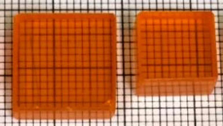
For the current study, the researchers grew large, high-quality cesium lead bromide (CsPbBr₃) crystals and engineered them into a pixelated sensor, a four-by-four array of pixels, each about seven millimeters wide and just over three millimeters thick.
The design works much like the pixels in a smartphone camera, capturing radiation in small, precise sections. They also built a custom multi-channel readout system to make sure each pixel worked seamlessly with the rest of the array. When tested, the prototype set a new benchmark for gamma-ray detection.
The perovskite-based system achieved an energy resolution of 2.5 percent at 141 kiloelectronvolts, which is the energy of the gamma rays emitted by technetium-99m, the most commonly used radiotracer in clinical practice. At higher energy levels, such as 662 kiloelectronvolts from cesium-137, the device reached a resolution of 1.0 percent.
Some individual pixels performed even better, showing a resolution as sharp as 2.2 percent at 141 kiloelectronvolts. These are the best values reported to date for perovskite detectors.
The device also excelled in imaging experiments. It could separate radioactive sources placed only a few millimeters apart, a level of detail fine enough to pick out small structures inside the body. Even faint signals were captured, and the detector remained stable and accurate over repeated use.
In other words, it was not only sharper and more precise than previous technologies but also robust enough for practical application.
“By combining high-quality perovskite crystals with a carefully optimized pixelated detector and multi-channel readout system, we were able to achieve record-breaking energy resolution and imaging capabilities. This work shows the real potential of perovskite-based detectors to transform nuclear medicine imaging.”
A practical healthcare innovation
According to the researchers, if developed into full imaging systems, perovskite detectors could make nuclear scans sharper, faster, cheaper, and safer. Patients might spend less time in scanners, need smaller doses of radiation, and receive clearer results that doctors can interpret with greater confidence.
Moreover, hospitals that cannot afford the cost of CZT-based cameras could finally access top-quality imaging, closing the gap between high-end research hospitals and everyday clinics.
“High-quality nuclear medicine shouldn’t be limited to hospitals that can afford the most expensive equipment. With perovskites, we can open the door to clearer, faster, safer scans for many more patients around the world. The ultimate goal is better scans, better diagnoses, and better care for patients,” Kanatzidis said.
The achievement is also significant for physics and materials science as it marks the first demonstration of single-photon gamma-ray imaging with perovskites, proving that the material has moved beyond the lab and into real-world applications. A NWU spinout company, Actinia Inc., is already working with medical device partners to bring the technology to hospital systems.
The study is published in the journal Nature Communications.