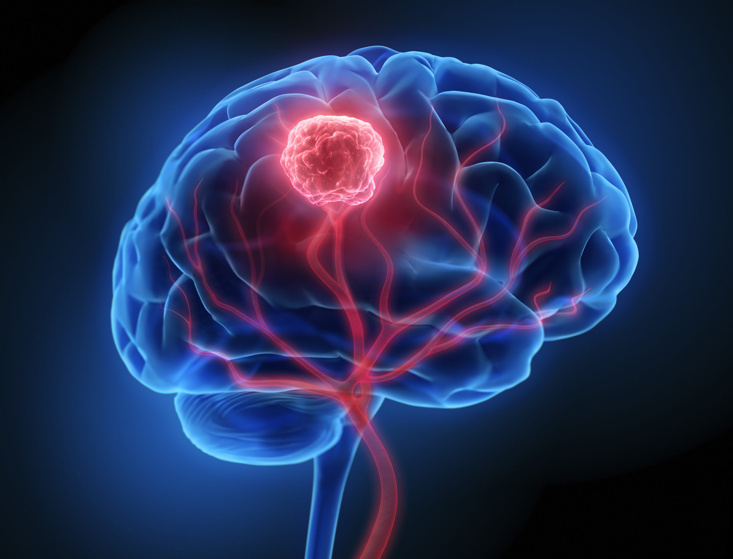
A collaboration between researchers from the University of Notre Dame, Harvard Medical School/Massachusetts General Hospital, and Boston University, have developed a new method to determine the amount of physical force (called solid stress) a brain tumor places on surrounding brain tissue. Published in the journal Clinical Cancer Research, the new method, using samples of a resected brain tumor, could eventually be used to determine the tumor type and inform post-surgical care.
“During brain tumor removal surgery, neurosurgeons take a slice of the tumor, put it on a slide and send it to a pathologist in real-time to confirm what type of tumor it is. Tumors that originally arise in the brain, like glioblastoma, are prescribed different treatments than tumors that metastasize to the brain from other organs like lung or breast, so these differences inform post-surgical care,” said co-first author Meenal Datta, PhD, an assistant professor of aerospace and mechanical engineering at Notre Dame. “By adding a two-minute step to a surgeon’s procedure, we were able to distinguish between a glioblastoma tumor versus a metastatic tumor based on mechanical force alone.”
Solid stress, unlike fluid pressure, is a mechanical force generated by tumor expansion and transmitted through solid tissue. It has been shown in previous animal studies to promote invasion, restrict blood flow, and impair treatment efficacy.
In earlier work, the same team had shown that tumors that apply solid stress to the brain can lead to more severe neurological dysfunction than tumors that infiltrate and destroy tissue. Retrospective estimates of solid stress, inferred from MRI-based growth patterns, correlated with worse neurological scores in patients. This new research was the first to attempt to provide direct, real-time measurements of solid stress.
For this research, the investigators enrolled 30 patients undergoing brain tumor resection. Prior to tumor removal, they performed intraoperative measurements of the brain surface through the craniotomy site using neuronavigation technology. This system provided a real-time, three-dimensional view of the brain, allowing the team to collect data on brain deformation caused by tumor pressure. Using these measurements and finite element modeling, they calculated tumor-induced solid stress and also estimated the percentage of brain tissue displaced or lost by the tumor.
In some cases, up to 10% of a patient’s brain volume was estimated to be replaced by the tumor. Metastatic or more nodular tumors that displaced brain tissue also exerted higher solid stress. In contrast, glioblastomas, which tend to infiltrate tissue, showed lower solid stress but greater brain tissue replacement.
To validate their method beyond their small cohort of patients, the team also tested mouse models of glioblastoma, ependymoma, and breast cancer metastasis to the brain. In the metastatic model, they observed that changes in solid stress occurred earlier than reductions in tumor size following chemotherapy. “In this model, we showed that mechanical force is a more sensitive readout of chemotherapy response than tumor size,” said Datta. “Mechanics are sort of disease-agnostic in that they can matter regardless of what tumor you are looking at.”
The researchers believe that this new approach allows for personalized assessment of both tumor behavior and disease burden. By providing insight into whether a tumor is displacing or replacing brain tissue, clinicians may better anticipate symptoms, choose therapies, and plan postoperative care.
“We present in this study a quantitative approach to intraoperatively measure solid stress in patients that can be readily adopted into standard clinical workflows,” the researchers wrote.
One limitation their work, the researchers noted, was determining the contribution of fluid pressure to tissue deformation, which they noted cannot be fully separated from solid stress. In their mouse models, the researchers were able to minimize this factor by halting blood flow prior to measurements, a technique not feasible in human subjects. In addition, current imaging tools still aren’t fully able to distinguish between fluid-filled and cancer-infiltrated edematous areas, which complicates tumor characterization.
Future studies aim to incorporate more recent imaging advances and refine fluid-versus-solid stress analysis. The team also plans prospective clinical trials to analyze how their measurements, and the actions taken based on them, influence clinical outcomes.