Since its inception in the 1930s, transmission electron microscopy (TEM) has undergone significant development. The smaller wavelength of electrons provides resolution that surpasses traditional light microscopy. Advances such as the introduction of direct electron detectors, cryo-preparation techniques, and image processing software have dramatically enhanced its resolution and versatility. These developments enable scientists to visualize specimens at atomic resolutions, which provides insights into nanostructures and, more recently, the structure and function of large biological assemblies to small proteins vital for drug discovery.
In 1933, Ernst Ruska first realized he could transmit electrons through a specimen of cotton fibers to form an image and was awarded the Nobel Prize for his discovery in 1986. In the following years, Jacques Dubochet, Joachim Franck, and Richard Henderson created a technique for generating a three-dimensional (3D) structure of the protein at an atomic level using a cryo-transmission electron microscope (cryo-TEM). Their technique taps vitrification to cool a sample to cryogenic temperatures, typically using liquid ethane around -180°C, which allows biomolecules to retain their native hydrated shape. This approach, commonly called cryo-electron microscopy (cryo-EM), earned them the Nobel Prize in Chemistry in 2017.
Detector technology resolves protein structure
During the past 30 years, every nut and bolt of the cryo-TEM has gradually been optimized. A major technical hurdle was overcome in 2010, when CMOS-direct electron detectors replaced charge-coupled device (CCD) cameras. Direct electron detectors (DEDs) have the advantage of providing immediate access to digital images and higher detective quantum efficiency (DQE) at a wide spatial frequency range, which results in images with a better signal-to-noise ratio. It enables scientists to get to high-resolution images and ultimately 3D reconstructions with fewer images, which is critical when imaging beam-sensitive biological samples.
DEDs can acquire data in movie mode to capture a series of frames at high speed during a single exposure. Further advancement came with the introduction of electron event representation (EER), which captures the position and time of each individual electron impact to enable the preservation of full temporal and spatial resolution of the data (i.e., super-resolution).
Energy filters, cold field emission guns help cross atomic barrier
In 2020, single particle analysis (SPA) cryo-EM broke the atomic resolution barrier, which allowed scientists to distinctly see atoms where blurry shapes and “blobs” once prevailed. Researchers harnessed technological improvements to determine breakthrough structure resolution of 1.2 Angstroms. For perspective, the first high-resolution structure by Henderson and colleagues was within the 10-Angstrom range attained in the early 1990s.
More recently, for the first time, researchers used the combination of a cold field emission gun electron source to lower the energy spread of the electrons, and more stable energy filters to remove inelastically scattered electrons. This combination contributed to increasing the image contrast while also boosting the resolution at the high spatial frequency range—and it allows researchers to break the atomic resolution boundary.
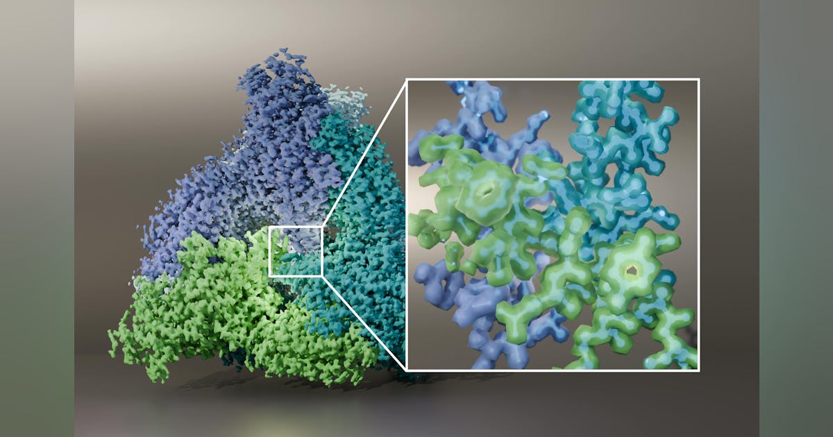
Most impressively, cryo-EM at atomic resolution enabled visualization of individual hydrogen atoms—even on water molecules inside of the protein structure. The visualization of hydrogen bonding networks inside protein structures and within drug binding pockets allows researchers to better understand how a drug interacts with its target molecule (see Fig. 1).
Autonomous microscopy and artificial intelligence (AI)
Cryo-EM innovation is reaching beyond higher resolution and contrast (see Fig. 2). Today, it’s also being adopted outside of academic labs by pharmaceutical institutions, which creates a need for automating routine workflows. Integrated software solutions offer a way to streamline data acquisition and 3D analysis with connected tools.