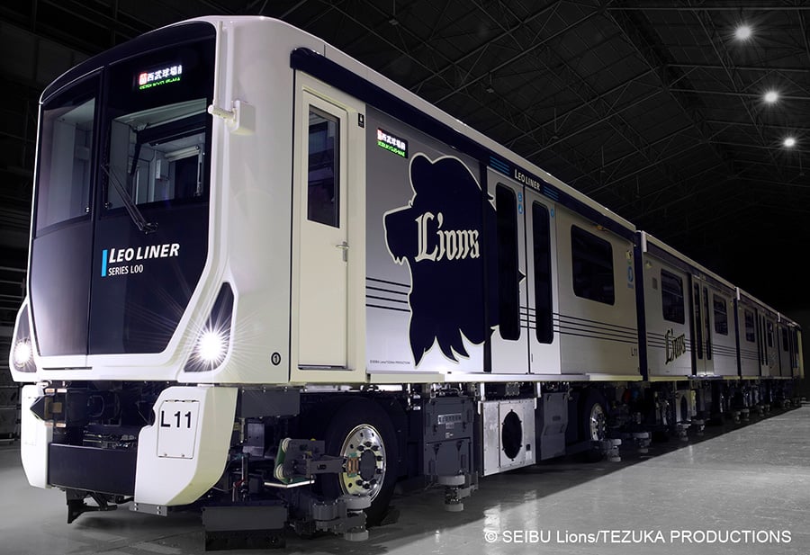
- Gaza ceasefire proposal agreed by Hamas is “almost identical” to Witkoff’s plan, Qatari official says Reuters
- LIVE: Qatar confirms Hamas ‘positive’ response to Gaza ceasefire proposal Al Jazeera
- Hamas says it accepts proposal for Gaza ceasefire and release of hostages | Israel-Gaza war The Guardian
- Hamas accepts proposed deal for ceasefire with Israel and hostage release, Egyptian source says Reuters
- Ben-Gvir says Netanyahu ‘has no mandate to pursue’ Gaza deal Dawn
Author: admin
-
Gaza ceasefire proposal agreed by Hamas is "almost identical" to Witkoff's plan, Qatari official says – Reuters
-
317 Louis Vuitton Items to Go Up for Auction at Bonhams
MONOGRAM MOMENT: Auction house Bonhams Cornette de Saint Cyr’s next online auction spans Louis Vuitton designs from the eras of Marc Jacobs, Nicolas Ghesquière, Virgil Abloh and Pharrell Williams.
“Monogram in the Spotlight” will go live on Aug. 27 until Sept. 10 with 317 pieces from a private collector including 138 handbags and small leather goods; 77 pieces of jewelry and accessories; 80 scarves, shawls and stoles; and 10 ready-to-wear pieces.
Lots start at 150 euros and are estimated to fetch up to 8,000 euros.
One of the Louis Vuitton handbags up for auction.
Thirty pieces were exhibited at Bonhams in London from July 17 to 24.
“This auction is an homage to Louis Vuitton and the exemplary single-owner collection of designs from the last ten years shows the visionary design work and groundbreaking fashion collaborations that spearheaded the brand into a new era in fashion history. The collection on offer is in pristine condition and features designs that will appeal to collectors across the globe,” said Hubert Felbacq, director of the fashion and accessories department at Bonhams Cornette de Saint Cyr in Paris.
“Monogram in the Spotlight” also includes buzzy collaborations with the likes of Tyler, The Creator, Sun Yitian and Takashi Murakami.
Louis Vuitton relaunched its seminal collaboration with Murakami last year in tandem with a campaign fronted by the brand’s ambassador Zendaya.
Key items from the auction vary from a Capucine BB Constellations bag from 2021, estimated for 4,000 to 6,000 euros; a Petite Malle bag in silver leather and padded monogram sequins, estimated for 2,500 to 3,500 euros; a bomber jacket from Ghesquière’s fall 2023 collection, estimated for 1,200 to 1,800 euros; and an Alma BB bag with a zebra head patch in collaboration with Sun Yitian, estimated for 1,000 to 1,200 euros.
Continue Reading
-
Scientists may have finally found the Universe’s missing sulfur
For decades, astrochemists have been looking for sulfur atoms in space and finding surprisingly little of the element that is a key ingredient to life. A new study could point to where it has been hiding.
An international team of researchers including Ryan Fortenberry, an astrochemist at the University of Mississippi, and Ralf Kaiser, professor of chemistry at the University of Hawaii at Mānoa and Samer Gozem, computational chemist at Georgia State University, published their research in the journal Nature Communications.
“Hydrogen sulfide is everywhere: it’s a product of coal-fired power plants, it has an effect on acid rain, it changes the pH levels of oceans and it comes out of volcanoes,” Fortenberry said. “If we gain a better understanding of what the chemistry of sulfur can do, the technological commercialization that can come from that can only be realized with a foundation of fundamental knowledge.”
Sulfur is the 10th most abundant element in the universe and is considered a vital chemical element for planets, stars and life. The lack of molecular sulfur in space has been a mystery for years.
“The observed amount of sulfur in dense molecular clouds is less – compared to predicted gas-phase abundances- by three orders of magnitude,” Kaiser said.
The answer might lie in interstellar ice.
In cold regions of space, sulfur can form two distinct, stable configurations: octasulfur crowns, which are a group of eight sulfur atoms configured in ring-like crowns, and polysulfanes, chains of sulfur atoms that are bonded by hydrogen. These molecules can form on icy dust grains, locking sulfur into solid forms.
“If you use, for instance, the James Webb Space Telescope, you get a specific signature at specific wavelengths for oxygen and carbon and nitrogen and so forth,” Fortenberry said. “But when you do that for sulfur, it’s out of whack, and we don’t know why there isn’t enough molecular sulfur.
“What this work is showing is that the most common forms of sulfur that we already know about are probably where the sulfur is hiding.”
Kaiser and Fortenberry’s research showed that these sulfur-rich molecules may be abundant in icy regions of interstellar space, giving astronomers a potential road map to solving the sulfur puzzle.
“Laboratory simulations of interstellar conditions such as this study discover possible inventories of sulfur-containing molecules that can be formed on interstellar ices,” Kaiser said. “Astronomers can then utilize the results and look for these polysulfane molecules in the interstellar medium via radio telescopes once sublimed into the gas-phase in star forming regions.”
The reason sulfur has been so difficult to find is that the bonds it forms are always changing, going from crowns to chains and a variety of other formulations.
“It never maintains the same shape,” Fortenberry said. “It’s kind of like a virus – as it moves, it changes.”
The researchers’ work identifies possible stable configurations that astronomers can search for in the universe.
“The thing that I love about astrochemistry is that it forces you to ask hard questions, then forces you to come up with creative solutions,” Fortenberry said. “And those hard questions and creative solutions can have significant, unintended positive consequences.”
Continue Reading
-
Q&A with Dr. Ahment Tutuncu
Dr. Ahmet Tutuncu
CEO
AeroRx TherapeuticsRecent advancements in COPD treatments are providing patients with potential new first-line treatments. Pharmaceutical Executive spoke with AeroRx Therapeutics’ CEO Dr. Ahmet Tutuncu about new therapies in this space, along with how big pharma companies are looking to acquire companies developing these therapies.
Pharmaceutical Executive: How are delivery methods for COPD treatments changing?
Dr. Ahmet Tutuncu: There has been a recent shift toward more personalized, convenient, and effective delivery systems including single-inhaler dry powder systems. However, nebulizers – a proven and underleveraged modality – remain essential, especially for COPD patients with physical or cognitive impairments. Novel nebulized devices will focus on enhanced drug delivery and patient usability.PE: Should the industry expect more acquisitions of companies developing COPD treatments?
Tutuncu: Yes, more acquisitions should be expected as COPD remains a high-unmet medical need and incidence continues to grow and a continued need for more effective therapies. Large pharmaceutical companies such as Merck and GSK have recognized the increasing need to deliver effective medicines to COPD patients, as demonstrated by their recent multibillion dollar COPD product acquisitions. This trend reflects the high value ascribed toward effective COPD therapy and a competitive market to bring new treatments to patients.PE: What treatments are coming next in the nebulized COPD space?
Tutuncu: New long-acting bronchodilators, such as nebulized glycopyrrolate and revefenacin, which show improved efficacy and reduced side effects, as well as Merck’s ensifentrine, a first-in-class dual-acting bronchodilator and anti-inflammatory recently approved by the FDA, have expanded treatment options. However, there remains a high-unmet need for maintenance therapy solutions for older and sicker COPD patients with persistent symptoms. AeroRx is developing a novel LABA/LAMA combination product, two proven monotherapies, to address these remaining treatment gaps for moderate-to-severe COPD patients.PE: What made AeroRX decide to come out of stealth?
Tutuncu: AeroRx recently announced Phase 2a data for its lead program AERO-007, where both low and high dose AERO-007 was well tolerated and achieved rapid and sustained 24-hour bronchodilation in COPD patients. AERO-007 is positioned to be the first LABA/LAMA combination bronchodilator drug available for nebulization delivery for moderate-to-severe COPD patients. With these exciting data, AeroRx will continue pushing forward in the clinic toward Phase 2b and beyond.Continue Reading
-
Somerset man who lost leg to smoking urges people not to start
Clara Bullock & Lili SheppardBBC News, Somerset
BBC
Cliff Hopkins had to have his leg amputated after smoking for 50 years A man who had to have his leg amputated after smoking for 50 years has said that picking up cigarettes is “never worth it”.
Cliff Hopkins, from Wells in Somerset, said he smoked two pouches of tobacco per week before deciding to quit smoking five months ago after blocked arteries led to him losing his leg.
Nearly 26,000 people in the South West have pledged to stop smoking in the past year, according to NHS figures, up from 15,000 the year before.
“It’s down to you as a person, you’ve got to want to give it up. I thought to myself, I’ve lost a leg and I don’t want to lose another one,” Mr Hopkins said.
“You think it’s not going to happen to you. But it does happen and it has for me.
“I’ve got a synthetic leg and I’m learning to walk on that. I’m getting there slowly. I’ll be back running before long, hopefully.”
He added that five months without having a cigarette has made “a massive difference, for health and wealth”.
“Don’t do it, it’s never worth it,” he said. “Think of the cost. For me, the cost is one of the major things.
“You could do so much more good for yourself with that money rather than buying cigarettes.”
Kate Anderson said some people go to food banks to fund their tobacco habit Public health specialist at Somerset Council Kate Anderson said statistics showed smokers are spending an average of £2,500 a year on cigarettes.
“Just in Somerset alone, we’ve seen people who are using food banks to fund their tobacco,” she said.
Meanwhile, she added, some people who have quit smoking have been able to use the money they have saved for things like family holidays.
But Ms Anderson also said she knows giving up smoking can be difficult.
Mr Hopkins attends a weekly meeting in Glastonbury, which is run by the council and supports people who want to quit.
“That little meeting is crucial, very much so. It is difficult to give up, it’s really hard, if you’ve smoked for as long as I have,” Mr Hopkins said.
Alison Bell, director of Public Health at Somerset Council, said she believes more people are quitting because the government is investing in local services to help them, which includes the weekly meetings Mr Hopkins attends.
“What we’ve seen in Somerset is we have the highest quit rate in the South West,” Ms Bell said.
Continue Reading
-
Each nation’s leading scorers in FIBA EuroBasket history
MUNICH (Germany) – Representing your country at a major tournament is one thing, being your nation’s leading scorer at that event is extra special.
Check out the all-time leading scorers
Top 100: Who are the all-time EuroBasket leading scorers?
But who is the top of the charts for each participating nation? We’ve got you covered with the 23 countries below, with the exception of Final Round debutants Cyprus.
Note: Active leader relates to players in the national team’s current squad for FIBA EuroBasket 2025.
Belgium
Rank
Player
Games
Points
1.
Rene Aerts
33
444
2.
John Loridon
37
339
3.
Jozef Eygel
40
270
4.
Jean Steveniers
16
244
5.
Sam Van Rossom
24
229
6.
Jonathan Tabu
28
227
7.
Eddy Terrace
9
220
8.
Francois Huysmans
14
211
9.
Maxime De Zeeuw
22
179
10.
Axel Hervelle
19
162
Active leader: Emmanuel Lecomte – 104 points
Bosnia and Herzegovina
Rank
Player
Games
Points
1
Mirza Delibasic
30
348
2
Sabahudin Bilalovic
9
217
3
Mirza Teletovic
13
195
4
Mario Primorac
12
192
5
Nenad Markovic
11
182
6
Samir Avdic
14
170
7
Gordan Firic
20
157
8
Elmedin Kikanovic
15
154
9
Jasmin Hukic
12
153
10
Nihad Djedovic
10
142
Active leader: Dzanan Musa – 107 points
Czechia
Rank
Player
Games
Points
1
Kamil Brabenec
60
918
2
Jiri Zidek
44
588
3
Frantisek Konvicka
43
496
4
Ivan Mrazek
39
483
5
Miroslav Skerik
46
461
6
Jaroslav Sip
44
453
7
Zdenek Kos
44
445
8
Jiri Baumruk
42
429
9
Jiri Pospisil
33
411
10
Jaroslav Tetiva
53
400
Active leader: Tomas Satoransky – 263 points
Estonia
Rank
Player
Games
Points
1
Sergei Babenko
17
196
2
Aivar Kuusma
8
159
3
Heino Veskila
18
157
4
Rauno Pehka
18
154
5
Priit Tomson
23
142
6
Jaak Lipso
21
140
7
Margus Metstak
12
98
8
Siim-Sander Vene
10
96
9
Joann Lossov
14
82
10
Andrus Nagel
9
75
Active leader: Siim-Sander Vene – 96 points
Finland
Rank
Player
Games
Points
1
Timo Lampen
51
621
2
Raimo Lindholm
51
489
3
Seppo Kuusela
37
417
4
Petteri Koponen
35
395
5
Kari Liimo
29
376
6
Timo Suviranta
38
366
7
Sasu Salin
35
343
8
Lauri Markkanen
13
312
9
Pertti Mutru
38
262
10
Shawn Huff
38
259
Active leader: Lauri Markkanen – 312 points
France
Rank
Player
Games
Points
1.
Tony Parker
66
1104
2.
Stephane Ostrowski
43
706
3.
Herve Dubuisson
49
657
4.
Boris Diaw
67
581
5.
Alain Gilles
39
526
6.
Richard Dacoury
31
449
7.
Antoine Rigaudeau
38
440
8.
Nicolas Batum
39
436
9.
Nando De Colo
46
419
10.
Jacques Cachemire
38
416
Active leader: Guerschon Yabusele – 118 points
Georgia
Rank
Player
Games
Points
1
Otar Korkia
36
357
2
Toko Shengelia
19
257
3
Manuchar Markoishvili
23
246
4
Giorgi Shermadini
29
227
5
Viktor Sanikidze
19
220
6
Zaza Pachulia
16
216
7
Zurab Sakandelidze
36
201
8
George Tsintsadze
27
170
9
Duda Sanadze
19
149
10
Mikhail Korkia
20
129
Active leader: Toko Shengelia – 257 points
Germany
Rank
Player
Games
Points
1
Dirk Nowitzki
49
1052
2
Dennis Schroder
20
448
3
Patrick Femerling
50
441
4
Ademola Okulaja
42
390
5
Hans-Joachim Flau
34
370
6
Herbert Kulik
39
365
7
Michael Jackel
16
347
8
Volkhardt Uhlig
33
338
9
Christian Welp
31
320
10
Detlef Schrempf
15
302
Active leader: Dennis Schroder – 448 points
Great Britain
Rank
Player
Games
Points
1
Dan Clark
20
188
2
Myles Hesson
10
132
3
Dennis Wilkinson
9
127
4
Gabe Olaseni
10
127
5
Charles Robinson
10
124
6
Luol Deng
5
123
7
Alan Bruce
9
123
8
Kyle Johnson
14
108
9
Kieron Achara
12
91
10
Andrew Lawrence
12
89
Active leader: Myles Hesson – 132 points
Greece
Rank
Player
Games
Points
1
Nikos Galis
33
1031
2
Panagiotis Giannakis
58
769
3
Giorgos Kolokithas
25
488
4
Panagiotis Fassoulas
43
473
5
Ioannis Bourousis
53
468
6
Nikos Zisis
31
449
7
Vassilis Spanoulis
38
429
8
Theofanis Christodoulou
40
379
9
Nick Calathes
41
365
10
Antonios Fotsis
44
353
Active leader: Kostas Sloukas – 277 points
Iceland
Rank
Player
Games
Points
1.
Haukur Palsson
10
123
2.
Jon Arnor Stefannson
10
119
3.
Hlynur Baeringsson
10
95
4.
Martin Hermannsson
10
87
5.
Hordud Vilhjalmsson
8
74
6.
Logi Gunnarsson
10
52
7.
Jakob Orn Sigurdarson
5
42
8.
Pavel Ermolinskij
10
37
9.
Kristofer Acox
5
35
10.
Tryggvi Hlinason
5
21
Active leader: Haukur Palsson – 123 points
Israel
Rank
Player
Games
Points
1.
Miki Berkowitz
49
917
2.
Jamchy Doron
31
659
3.
Boaz Yanay
41
472
4.
Tanhum Cohen-Mintz
35
467
5.
Lior Eliyahu
29
386
6.
Louis Grant Silver
23
335
7.
Talbut Brody
21
314
8.
Barry Leibowitz
37
292
9.
Yotam Halperin
31
281
10.
Yaniv Green
38
275
Active leader: Deni Avdija – 73 points
Italy
Rank
Player
Games
Points
1
Dino Meneghin
57
638
2
Marco Belinelli
35
528
3
Antonello Riva
24
469
4
Renato Villalta
39
448
5
Massimo Masini
40
447
6
Pierluigi Marzorati
45
392
7
Gregor Fucka
31
376
8
Carlton Myers
24
375
9
Walter Magnifico
32
373
10
Luigi Datome
32
333
Active leader: Danilo Gallinari – 219 points
Latvia
Rank
Player
Games
Points
1
Valdis Valters
32
476
2
Kristaps Janicenoks
33
302
3
Ainars Bagatskis
20
301
4
Dairis Bertans
29
298
5
Janis Blums
37
261
6
Janis Krumins
22
254
7
Roberts Stelmahers
18
237
8
Kaspars Kambala
13
221
9
Janis Strelnieks
29
220
10
Gunars Silins
20
214
Active leader: Dairis Bertans – 298 points
Lithuania
Rank
Player
Games
Points
1
Arvydas Sabonis
36
652
2
Modestas Paulauskas
39
572
3
Arturas Karnisovas
27
492
4
Jonas Valanciunas
41
475
5
Mantas Kalnietis
40
434
6
Sarunas Jasikevicius
45
405
7
Sarunas Marciulionis
20
395
8
Ramunas Siskauskas
25
347
9
Valdemaras Chomicius
38
326
10
Jonas Maciulis
41
320
Active leader: Jonas Valanciunas – 475 points
Montenegro
Rank
Player
Games
Points
1
Bojan Dubljevic
17
219
2
Nikola Vucevic
16
147
3
Predrag Drobnjak
13
139
4
Tyrese Rice
10
129
5
Vlado Scepanovic
22
107
6
Vladimir Mihailovic
17
104
7
Kendrick Perry
6
91
8
Milko Bjelica
10
72
9
Nikola Pekovic
5
65
10
Suad Sehovic
11
57
Active leader: Nikola Vucevic – 147 points
Poland
Rank
Player
Games
Points
1.
Dariusz Zelig
41
703
2.
Janusz Wichowski
45
504
3.
Mieczyslaw Mlynarski
22
482
4.
Edward Jurkiewicz
21
432
5.
Mieczyslaw Lopatka
27
412
6.
Bogdan Lipszo
33
401
7.
Eugeniusz Kijewski
22
370
8.
Andrzej Pstrokonski
44
368
9.
Jerzy Binkowski
34
364
10.
Mateusz Ponitka
25
259
Active leader: Mateusz Ponitka – 259 points
Portugal
Rank
Player
Games
Points
1
Joao Santos
11
111
2
Elvis Evora
11
73
3
Miguel Miranda
11
72
4
Mario Nogueira de Almeida
7
68
5
Carlos de Andrade
5
64
6
Joao Gomes
6
60
7
Francisco Jordao
6
58
8
Antonio Tavares
4
49
9
Paulo Cunha
4
44
10
Antonio Nogueira Cardoso
5
41
Active leader: 0
Serbia
Rank
Player
Games
Points
1
Radivoj Korac
34
844
2
Dragan Kicanovic
40
662
3
Drazen Dalipagic
43
623
4
Nenad Krstic
33
461
5
Dejan Bodiroga
37
440
6
Predrag Danilovic
33
403
7
Milos Teodosic
32
384
8
Ratko Radovanovic
35
384
9
Bogdan Bogdanovic
29
367
10
Vlade Divac
34
364
Active leader: Bogdan Bogdanovic – 367 points
Slovenia
Rank
Player
Games
Points
1.
Goran Dragic
51
696
2.
Ivo Daneu
50
515
3.
Jaka Lakovic
51
509
4.
Erazem Lorbek
35
375
5.
Rasho Nesterovic
29
327
6.
Zoran Dragic
34
331
7.
Bostjan Nachbar
30
324
8.
Luka Doncic
16
311
9.
Jure Zdovc
28
281
10.
Matjaz Smodis
31
256
Active leader: Luka Doncic – 311 points
Spain
Rank
Player
Games
Points
1
Pau Gasol
58
1183
2
Juan Antonio San Epifanio
52
894
3
Emiliano Rodriguez
53
864
4
Juan Carlos Navarro
53
768
5
Wayne Brabender
42
684
6
Francisco Buscato
54
607
7
Rudy Fernandez
61
547
8
Alberto Herreros
41
538
9
Marc Gasol
49
520
10
Andres Jimenez
36
484
Active leader: Willy Hernangomez – 245 points
Sweden
Rank
Player
Games
Points
1
Bo Widen
23
275
2
Staffan Widen
23
261
3
Jorgen Hansson
16
223
4
Erik Sahlstrom
9
144
5
Anders Gronlund
16
131
6
Ulf Lindelof
16
118
7
Sten Feldreich
7
114
8
Par Jonas Larsson
9
113
9
Hans Albertsson
16
111
10
Jeffery Taylor
5
106
Active leader: Viktor Gaddefors – 22 points
Türkiye
Rank
Player
Games
Points
1
Hidayet Turkoglu
49
607
2
Ibrahim Kutluay
40
526
3
Yalcin Granit
25
386
4
Ersan Ilyasova
32
355
5
Mirsad Turkcan
35
338
6
Altan Dincer
27
336
7
Cedi Osman
18
264
8
Zeki Tosun
21
260
9
Ender Arslan
35
246
10
Mehmet Okur
27
245
Active leader: Cedi Osman – 264 points
Related Articles
Roster tracker: Who is playing at FIBA EuroBasket 2025?
FIBA
Continue Reading
-
Netanyahu lashes out at Albanese as ‘weak politician who betrayed Israel’ as diplomatic row escalates | Australian foreign policy
Benjamin Netanyahu has launched an extraordinary broadside against Anthony Albanese, labelling Australia’s prime minister “weak” in a social media post, hours after local Jewish groups called for calm in diplomatic relations.
“History will remember Albanese for what he is: A weak politician who betrayed Israel and abandoned Australia’s Jews,” read a post on X from the account of Israel’s prime minister, on Tuesday evening Australian time.
Albanese’s office was approached for comment.
It came shortly after Australia’s peak Jewish group urged composure on Tuesday as Israel’s foreign minister warned of “additional measures” against Australia amid a diplomatic “tit-for-tat” over visa cancellations.
“There are real-life consequences here and we want to see the countries work through any issues before things get out of hand,” said Alex Ryvchin, the co-chief executive of the Executive Council of Australia Jewry.
The Israeli foreign minister, Gideon Sa’ar, accused the Albanese government of “choosing to fuel” antisemitism after cancelling the visa of the far-right Israeli politician Simcha Rothman ahead of his planned speaking tour in Sydney and Melbourne later this week.
Sa’ar also announced he would revoke the visas of Australian representatives to the Palestinian Authority, in a move labelled “unjustified” and “undermining international efforts towards peace and a two-state solution” by Australia’s foreign minister, Penny Wong.
The escalating diplomatic row followed Australia’s decision earlier this month to recognise a Palestinian state at the UN general assembly in September after new commitments by the Palestinian Authority.
Rothman’s visa was cancelled by the home affairs department on Monday, citing “an unacceptable risk” to order in Australia and concern the member of Netanyahu’s governing coalition would make “inflammatory statements to promote his controversial views and ideologies”.
Simcha Rothman (left) and Israel’s national security minister Itamar Ben-Gvir in the Knesset. Photograph: Amir Cohen/Reuters In a Hebrew-language video message shortly after his statement, Sa’ar warned Australia was “persecuting Israel” and his government would “take additional measures”.
But Jewish groups in Australia called for cooler heads to prevail in light of the countries’ “extensive economic, scientific and cultural ties”.
Ryvchin said both countries should end the “diplomatic tit-for-tat that erodes the goodwill and cooperation built up over decades”.
“Calm heads need to take control of the situation otherwise there will be a risk to some $2bn dollars in bilateral trade, extensive investment in Australian start-ups, vital security cooperation and the Israeli-made medicine and medical technology that we all rely on,” he said.
The Australia/Israel & Jewish Affairs Council said while it did not agree with many of Rothman’s views, it found the decision to cancel his visa to be a “disturbing precedent”.
“It is very disappointing that Australia and Israel have regressed from a close friendship to unproductive diplomatic jousting, which of course was started by unwarranted and hostile actions by the Australian government,” the council said in a statement.
Sign up: AU Breaking News email
The president of the Zionist Federation of Australia, Jeremy Leibler, told the Jewish Independent website on Tuesday that Rothman’s views were “out of step with mainstream Israeli opinion and with the values of most Australian Jews”.
But he criticised the decision to ban him, saying: “It is one thing to disagree strongly with an elected representative of a democracy and ally, it is quite another to deny them entry on that basis. Australia ought not to set such a precedent.”
The progressive Jewish Council of Australia supported the decision but questioned why Rothman had been granted a visa in the first place.
“As with Russia, members of Israel’s ruling coalition parties should already be subject to sanctions by the Australian government, including Rothman,” its spokesperson, Bart Shteinman, said.
“Israel’s aggressive decision to retaliate to Rothman’s visa refusal by expelling Australian diplomats to the Palestinian Authority is a major escalation in hostilities against Australia.
“If it needed any further demonstration, Israel is not interested in maintaining a relationship with the Australian government and cares little what strong words or statements it issues.”
The department cited Rothman’s previous statements as part of its decision to revoke the visa they approved on 8 August.
These included his claim in May that children within Gaza are “enemies” who should not be allowed to flee into Israel as “you don’t let them conquer your country with refugees”.
In another example cited, Rothman was reported to have said the idea of a two-state solution between Israel and Palestine had “poisoned the minds of the entire world” and was the “first step toward the destruction of the state of Israel”.
The department said that if it was publicly known that Rothman had entered Australia with the government’s permission, it “may encourage others to feel emboldened to voice any anti-Islamic sentiments”.
The right-leaning Australian Jewish Association, who invited Rothman and organised the speaking tour, said it would host an online event with Rothman on Sunday evening, stating, the “show will go on”.
Sa’ar’s decision to revoke the visas of three Australian diplomats – who are based in East Jerusalem and work in the West Bank – is expected to complicate Australia’s work with the Palestinian Authority.
The foreign affairs department is working to remove the affected representatives.
Continue Reading
-
Dare to Dream 2 | Episode 3
Episode three of Dare to Dream: The Next Move is released on Wednesday 20 August at 8pm CEST (2pm EDT), and stars double Olympic Mixed Relay medallist Morgan Pearson as the man on a mission to find clarity and calm in the wild waters of T100 racing.
WATCH ON WORLD TRIATHLON YOUTUBE AND TRIATHLONLIVE
Opening with a quick-fire tour of his Colorado home, the USA men’s number one Morgan Pearson takes a beat to assess his new-found position as one of eight Hot Shots contracted for the second T100 season. There may have been a few raised eyebrows from some of his fellow pros when he was announced for the tour, but a fastest swim and run in Vancouver showed he is well at home. As well as where he needs to make progress.
“I swam growing up. I ran growing up. I didn’t bike growing up. I didn’t get a bike until 2018 when I started triathlon. So it is pushing me to be a better biker, there’s just no doubt about that when you’re racing these guys.”
“I wasn’t really sure at first (about the Hot Shot contract)… it’s gonna be really high level racing, you know, these guys are just absolute animals. I thought about it for a few days and I was like, ‘this is what I wanna do. I want to be really out of my comfort zone’.
Pearson namechecks the achievements of the other Hot Shots, the inevitable comparisons both by himself and his peers, something that has at times hindered his ability to find consistency at the highest level. And that is in spite of being the first American male on the teamsheet for the past two Games.
“Hayden Wilde and Leo Bergere are coming off Olympic medals, so of course they definitely deserve it. And then Vince, he’s a legend of our sport, so he’s definitely someone who’s deserving. Maybe I’m a bit of the wild card or like the X factor with the Hot Shots.”
‘You know, two Olympic medals, two different Olympics. I don’t even know if that was my dream, that probably exceeds my dreams when I first started in 2018. So there’s a lot to be grateful for.”
After the impact of the passing of his brother in March 2021 ahead of the Tokyo Olympics, Pearson talks of ‘giving himself a pass’ for a disappointing individual result at Tokyo 2020, mental and physical exhaustion taking a toll. After a torrid time on the bike course, his 31st place in Paris, however, clearly cuts deeper, even having achieved that second relay silver medal.
“Tokyo was a tough pill to swallow… but I think Paris… I’ll be honest, that race will probably haunt me the rest of my life.”
So perhaps inevitably the episode keeps coming back to the immense mental and physical demands of the sport. One of the fastest runners on the WTCS and now T100 circuit, finding that rhythm at the end of 82km of swim and bike is the next challenge.
“Overthinking is a huge part of racing, especially for me. Not just the running, biking, and even swimming… I think achieving that flow state and achieving that point where you’re not really overthinking, you’re just trusting your instincts… and that those instincts are also the right decision… that’s ultimately like the place where I like to be at in a race.”
And then there is again the habit of comparing himself to the rest of the field, putting himself under pressure at the WTCS level, and a pressure that is less apparent – at leats for now – in his early T100 races.
“I can think of so many races where it’s just like, you’re running with guys and I’m like, why is this guy here? I’m just faster than him… then all of a sudden you get tight and then things feel harder than they should.’
“I also think what it might help is my mindset. When I do a WTCS I put so much pressure on myself it can be a bit crippling at times. And here I am just a beginner, racing for seventh, eighth, and not being happy, but excited with the progress and excited to see where this can go. I think that mindset is a healthy one.”
Continue Reading
-
Insights from age-stratified endoscopic detection metrics support initiating colorectal cancer screening at age 45 | BMC Gastroenterology
In recent years, the evaluation of CRC in the younger population has increased, challenging the previous notion that this disease primarily impacts older individuals [30]. This pattern emphasizes the need to revise screening guidelines to encompass younger age groups for early disease prevention and to decrease the occurrence of complex and costly advanced diagnoses [31,32,33]. Despite potential debates on the cost-effectiveness of limiting screening guideline expansions, research has shown that the overall advantages of reductions far surpass any associated healthcare expenditures in the long run [34, 35]. Timely detection not only saves lives but also minimizes expenses by reducing the necessity for extended treatments and hospitalizations in advanced CRC stages [36].
Our findings indicate a significant enhancement in colonoscopy utilization among individuals aged 55 and above compared to those in the 45–49 and 50–55 age groups. Nonetheless, there has been a noticeable increase in colonoscopy rates within the 45–49 age group over the past years, particularly evident in 2021 and 2022. Additionally, our study shows a strong link between age and polyp incidence, with individuals over 55 consistently displaying the highest polyp numbers. These results underscore the importance of regular colonoscopies, particularly for individuals aged 55 and older, in facilitating early polyp detection and management, ultimately aiding in the prevention of CRC and prompt diagnosis. Moreover, our research illustrates age-related variations in the detection rates of colorectal abnormalities, with individuals over 55 exhibiting the highest polyp detection rate. Interestingly, no notable alterations were observed in the incidence of carcinoma cases between the 45–49 and 50–55 age groups. Over time, ADR reports have been increasing in the population of patients aged 50 years and older, a pattern that corresponds with the existing screening guidelines [37]. Nevertheless, recent epidemiological patterns have exposed a concerning surge in the prevalence of CRC among individuals in a younger age range [38]. This shift has prompted certain scientific bodies in the USA and other regions to support for earlier screening, especially for high-risk individuals or those with a family history of CRC or advanced adenomas [39, 40]. Our study confirms a significant rise in ADR among individuals over 50 years old, aligning with findings from studies conducted in the United States and China. These findings help address objections raised to initiating CRC screening at age 45 [24, 39, 40]. Based on these advancements, predictive analyses, such as the one carried out by Anderson JC. et al., have proposed commencing colonoscopy screening at 45 years, a recommendation adopted by the American Cancer Society [41]. Recent U.S. cohort studies, particularly those by Shaukat et al. [24, 42], provide robust evidence supporting earlier screening [24, 42]. Shaukat et al. [42] demonstrated a significant reduction in colorectal cancer mortality following colonoscopy screening, while Shaukat et al. [24] reinforced the rationale for initiating screening at 45 years, which directly influenced updated U.S. guidelines [24, 42]. While some American institutions have adopted this proposal, other guidelines, including those in Europe, recommend starting screening at the age of 50 [43, 44].
The absence of evidence from studies conducted on a population scale offers insufficient justification for recommending earlier colonoscopies, implying that this approach may only be appropriate in regions where there is a steady increase in CRC cases at a younger age [45, 46]. Various issues have been raised concerning this tactic, such as the broad confidence intervals surrounding the rise in CRC incidence among younger individuals, the arbitrary selection of the 45 to 50 age bracket, uncertainties about the effectiveness rate utilized in predictive models for evaluating cost-effectiveness, projected cost increases in CRC screening, and potential obstacles linked to patient and healthcare provider adherence [41, 47]. Nonetheless, acknowledging that detecting and treating adenomas is a successful means of preventing CRC development, we propose that examining ADR within different age groups can aid in accurately identifying when the risk of colon cancer escalates [14]. Notably, Surveillance, Epidemiology, and End Results (SEER) data showe a comparable number of years of life lost due to CRC between the ages of 45 and 50, and between 50 and 54, as indicated in the study by Syed et al., which noted that a majority of early-stage CRC instances arise in the 40–49 age group [48]. Nonetheless, there is a paucity of studies investigating the connection between adverse effects and age. In a study from 2015, Hemmasi et al. compared patients aged 40 to 49 with those aged 50 to 59 in a limited sample of 740 screening colonoscopies, finding no significant variance between the two groups (11.7% vs. 16.5%), which suggests that using 45 years as the screening threshold may be too high [49]. Karsenti et al. initially assessed ADR and High-Risk ADR using 5-year age intervals. Out of 6027 colonoscopies, the overall ADR was 28.6%, surpassing our 20.5% (24.8% when including patients over 54 years old), with a similar rate in individuals under 45 years (16.9% vs. 15.9%). Our total ADR corresponded to the findings of this extensive cohort study (22-23.6%) [14].
Conclusively, neither the ADR nor the PDR exhibited significant differences between the age groups of 45–49 and 50–55. Our results strongly support commencing CRC screenings at the age of 45.
Recent modeling studies have evaluated the cost-effectiveness of initiating colorectal cancer screening at age 45, emphasizing the influence of population risk profiles, healthcare infrastructure, and adherence rates. Ladabaum et al. reported that starting screening at 45 years in the U.S. is cost-effective (approximately $33,900 per QALY gained), but highlighted that optimizing adherence among older unscreened individuals may yield greater benefits at lower costs [50]. Similarly, a study by Half et al. in Israel demonstrated that while initiating screening at 45 is clinically beneficial, the incremental cost-effectiveness ratio varies significantly (23,800–53,900 NIS/QALY for FIT, and 110,600–162,700 NIS/QALY for colonoscopy), underlining the importance of resource allocation strategies [51]. In Germany, Lwin et al.. showed that colonoscopy-based screening from age 45 can be cost-effective (€1,029–9,763 per QALY gained), but the model outcomes were highly sensitive to screening adherence rates and healthcare system capacities [50]. These findings collectively suggest that while our observed high PDR in the 45–49 age group supports the clinical rationale for earlier screening, careful consideration of resource burden and cost-effectiveness is crucial when formulating screening policies.
Finally, we fully acknowledge that this study is based on data from a single healthcare network (Assuta Medical Centers) in Israel, and that the findings may reflect characteristics unique to the Israeli population and healthcare system. Israel’s population is notably diverse, encompassing individuals of European, Middle Eastern, and North African descent, among other backgrounds. The retrospective design utilized real-world data from a large-volume healthcare network, where colonoscopy was performed either through self-referral or physician-directed referral. Consequently, there is potential for selection bias, particularly within the 45–49 age group, which may represent a more health-conscious subset or have been referred based on subtle clinical cues that are not explicitly recorded. To mitigate this, we applied rigorous exclusion criteria aimed at approximating an average-risk screening cohort, excluding individuals with a family history of CRC or polyps, prior neoplastic findings, inflammatory bowel disease, gastrointestinal symptoms, or inadequate bowel preparation. This approach is aligned with international CRC screening guidelines and strengthens the internal validity of ADR and PDR comparisons across age groups. Nevertheless, residual referral-related bias may remain, and the findings may not fully represent outcomes expected in an organized, population-based screening program, where individuals are systematically invited for their initial screening at age 45.
Moreover, the study did not capture long-term clinical outcomes such as CRC incidence, progression, or mortality. Adenomas were not stratified by size, histologic subtype, or dysplasia grade, which limited the ability to distinguish between advanced and non-advanced lesions. Additional influential variables such as lifestyle factors (e.g., smoking, alcohol use, obesity, physical inactivity) and comorbidities were also unavailable in the dataset. Although such limitations are common in large retrospective studies, they limit our ability to account for confounding variables fully. Finally, this analysis focused exclusively on detection outcomes and did not assess procedure-related complications, such as bleeding or perforation.
A potential limitation of our study is the inability to fully verify that all included patients were true average-risk screening candidates. Some cases may have involved diagnostic or surveillance colonoscopies. While we attempted to review referral information to minimize misclassification, this limitation should be taken into account when interpreting our findings. Another limitation of this study is the absence of ethnicity-based analysis. Due to inconsistently recorded ethnicity data in our cohort, we were unable to explore potential disparities in screening outcomes among Israel’s diverse population. Future studies with systematically collected demographic data are needed to address this important question.
While colonoscopy remains highly effective for CRC screening, it carries inherent risks, including bleeding, perforation, and sedation-related complications, with serious adverse events reported in approximately 2–5 per 1,000 procedures. As the screening age is lowered and the number of procedures increases, careful attention must be paid to balancing the benefits of early detection with the potential harms associated with the procedure. Quality indicators such as withdrawal time, bowel preparation adequacy, and endoscopist experience play crucial roles in minimizing these risks. Although our study did not capture complication data, these safety considerations must be integrated into screening policy decisions.
Continue Reading
-
A single-cell, spatial transcriptomic atlas of the Arabidopsis life cycle
A spatiotemporally resolved atlas of the Arabidopsis life cycle
To generate a comprehensive atlas of Arabidopsis development, we collected six distinct organ systems that encompass diverse tissues as well as whole organisms at ten discrete developmental time points throughout the Arabidopsis life cycle corresponding to established developmental road maps28,29,30. This includes imbibed and germinating seeds, three stages of seedling development, developing and fully emerged rosettes, the stem, flowers and siliques (Fig. 1a). For each organ system, we also generated a paired spatial transcriptomic dataset (Fig. 1b–e; mature rosette leaf27). For the single-nucleus datasets, 432,919 nuclei from the ten developmental time points passed accepted droplet-based single-nucleus filtering metrics31 (median unique molecular identifier (UMI), 916; Extended Data Fig. 1 and Supplementary Fig. 1) and were independently clustered. For the seedling and rosette samples that constitute developmental time series, we also performed integrative analyses that revealed clusters corresponding to known cell types (Extended Data Fig. 1c,d), which confirms the reproducibility of these datasets and enables the investigation of developmentally regulated processes, such as root hair development (Supplementary Fig. 2) and leaf senescence (Supplementary Fig. 3), as reported recently20, across developmentally distinct samples. We also merged all single-nucleus RNA sequencing (snRNA-seq) datasets into a global dataset (Fig. 1f) encompassing all cell types across the Arabidopsis life cycle.
Fig. 1: A spatially resolved transcriptional atlas of the Arabidopsis life cycle. a, The collected tissues over the ten developmental time points spanning the plant life cycle, including imbibed and germinating seeds (0 days and 1.25 days); three stages of seedling development (3 days, 6 days and 12 days); developing and fully expanded rosettes (21 days and 30 days); the stem (37 days), including basal, apical and branched regions; flower tissue (stages 6–15; ref. 30); and siliques (stages 2–10; ref. 29). Developmental time points surrounded by solid lines denote matching single-nucleus and spatial transcriptomic datasets. b,c, Paired single-nucleus and spatial transcriptomic datasets generated in our study. d, Uniform manifold approximation and projection (UMAP) plots of each dataset with select cluster annotations displayed for each dataset. e, Spatial transcriptomic assays analysed in this study, including Arabidopsis seed, seedling, mature leaf, stem, flower cross and longitudinal sections, and silique. The scale bar lengths are shown. f, t-distributed stochastic neighbour embeddings of the fully integrated dataset. Nuclei are coloured according to the tissue of origin. g, Web browser access to the Arabidopsis Developmental Atlas Browser and CELLxGENE instances of flower single-nucleus and spatial datasets. h, A spatial transcriptomic dataset within the MERSCOPE Visualizer software. For each micrograph in e, results were observed across ten independent seeds, eight independent seedlings, two independent leaves, one stem, three independent flowers (one in longitudinal orientation and two in cross orientation) and three independent siliques. See Methods for detailed information.
Comprehensive identification of transcriptional identities
To compare cell types across organs and development, we analysed each dataset independently, which resulted in 183 clusters across all datasets (Fig. 1d). Cluster annotation was performed according to the following guidelines. First, an extensive list of marker genes was compiled with known cell-type- and tissue-specific expression in all Arabidopsis organs (Supplementary Table 1), including cell-type-specific markers recently identified from single-cell RNA-seq studies18,19,32,33,34,35. Many of these known cell-type-/tissue-specific expressed genes were enriched in specific clusters of our dataset (>1,000 genes), which aided cluster annotation (Supplementary Fig. 4a). Second, a cell-type enrichment score for each cluster was calculated on the basis of known cell-type markers to systematically infer cell types (Methods and Supplementary Fig. 4b). Third, we investigated newly identified cluster markers within each dataset using previously generated dissection-based and cell-type-specific transcriptomic studies (TAIR36 and ePlant37,38) to confirm the accuracy of our cluster annotations (Supplementary Table 2). Finally, we spatially validated a selection of cluster markers for each tissue/organ using sequencing- and imaging-based spatial transcriptomic technologies (Fig. 2 and Extended Data Figs. 2–6).
Fig. 2: Spatial validation of snRNA-seq data in Arabidopsis tissues/seedlings. a, Spatial transcriptomic visualization of selected gene expression (LHB1B1 (AT2G34430), XYLOGLUCAN ENDOTRANSGLUCOSYLASE/HYDROLASE 9 (XTH9; AT4G03210), NAD(P)H dehydrogenase subunit S (NdhS; AT4G23890) and PLASTID-SPECIFIC RIBOSOMAL PROTEIN 2 (PSRP2; AT3G52150)) in the Arabidopsis seedling (zoomed in on the cotyledons). The higher-magnification images highlight the localization of transcripts within mesophyll cells. DAPI staining (blue) indicates nuclei. The scale bar lengths are shown. b, UMAP plot of single-nucleus transcriptomes showing expression of PSRP2 (purple dots), enriched in cluster 3 (outlined in green). c, Spatial mapping of cluster 10 annotated as ‘dividing cells’ in the three-day-old seedling, with transcripts from known and new cell-cycle- and division-related genes (AT2G47500, CSLD5 (AT1G02730), AT4G14330 and HIK (AT1G18370), marked in magenta, yellow, cyan and pink, respectively). The insets show individual dividing cells with colocalized transcripts. DAPI staining (blue) indicates nuclei. The scale bar lengths are shown. d, UMAP plot highlighting expression of HIK enriched in cluster 10 (outlined in purple), corresponding to dividing cells in the tissue. For the micrographs in a and c, results were observed in eight seedlings. DAPI, 4′,6-diamidino-2-phenylindole.
Our spatial transcriptome approach allowed us to validate examples of known cell-type marker genes across organs and development, as well as validate newly identified cell-type-specific and tissue-specific marker genes across all organs (see Supplementary Table 3 for 109 examples of new cell-type/tissue marker genes). In contrast, we were also able to identify and validate markers that do not universally specify cell types but rather demonstrate cell-type-specific expression only within the context of specific organs, as shown for epidermal cells of seedling hypocotyls (Extended Data Fig. 3b). Spatial transcriptomics confirmed the localization of cell-type-specific genes identified via snRNA-seq in most developmental stages and plant organs tested (Extended Data Figs. 2–6). In the seedling datasets, we were able to confirm cell identities such as mesophyll cells expressing LIGHT-HARVESTING CHLOROPHYLL-PROTEIN COMPLEX II SUBUNIT B1 (LHB1B1; AT2G34430)38, epidermal cells (for example, GLYCOSYL HYDROLASE 9B8 (GH9B8; AT2G32990) and AT2G12462, new epidermal markers restricted to the apical hook and cotyledons in seedlings, respectively) and vascular cells (AINTEGUMENTA (ANT; AT4G37750), which was shown to regulate ERECTA-LIKE 1 (ERL1; AT5G62230) and PHLOEM INTERCALATED WITH XYLEM (PXY; AT5G61480), which maintain procambial cell identity39) that are co-expressed in the same cluster as SUCROSE-PROTON SYMPORTER 2 (SUC2; AT1G22710)40,41,42 (Fig. 2 and Extended Data Fig. 3). We were also able to annotate clusters to cell states through the cluster-specific expression of cell-division-associated genes such as CELLULOSE SYNTHASE LIKE-D (CSLD5; AT1G02730), involved in cell plate formation43, and HINKEL (HIK; AT1G18370), which is involved in cytokinesis44 (Fig. 2 and Extended Data Fig. 3).
Visual inspection of spatial marker expression within the single-nucleus datasets did not always reveal cluster-specific expression patterns, as demonstrated by LHB1B1 (Extended Data Fig. 3g), but quantitative evaluation of these markers revealed cluster- and subcluster-specific and enriched expression of these spatial markers both within individual samples (Extended Data Fig. 3k, left) and across tissues and developmental time points (Extended Data Fig. 3k, right), demonstrating conserved expression of these cell-type and cell-state markers across diverse tissues throughout the Arabidopsis life cycle.
In stems, we identified cell-type-specific gene expression localized to the procambium (for example, USUALLY MULTIPLE AMINO ACIDS MOVE IN AND OUT TRANSPORTERS 11 (UMAMIT11; AT2G40900)), cortex (for example, B-BOX DOMAIN PROTEIN 15 (BBX15; AT1G25440)), phloem (H(+)-ATPase 3 (refs. 45,46) (HA3; AT5G57350)) and xylem tissues (XYLEM CYSTEINE PEPTIDASE 2 (XCP2; AT1G20850) and IRREGULAR XYLEM 3 (ref. 47) (IRX3; AT5G17420)), enabling high-resolution mapping of vascular differentiation in the stem (Extended Data Fig. 4). Spatial expression of markers in Arabidopsis floral tissue revealed epidermal markers (for example, AT2G37540), vascular-expressed genes (for example, FLOWERING LOCUS T (FT; AT1G65480)) and carpel-specific expression of AGAMOUS-like 8 / FRUITFULL (AT5G60910; ref. 48) (Extended Data Fig. 5). Spatial transcriptomics also revealed distinct expression domains within the Arabidopsis silique, with VEGETATIVE STORAGE PROTEIN 2 (VSP2; AT5G24770) and the TPX2 protein family (TPXL6; AT5G37478) marking the exocarp cell layer, and AT2G13810 showing specific localization in developing seeds (Extended Data Fig. 6). Together, these examples demonstrate the strength of our paired single-nucleus and spatial transcriptomic datasets on matched biological samples.
While we were able to annotate many clusters to individual cell types, we found that some clusters instead corresponded to anatomical regions or broad cellular states, such as the developing embryo or dividing cells (Supplementary Table 2). This led us to hypothesize that further transcriptional complexity could be resolved within each cluster when examined independently. Through systematic re-clustering of individual clusters (subclustering), we were able to determine further complexity within clusters, resulting in a total of 655 subclusters (Supplementary Fig. 5a). Correlating the aggregated transcriptomes (pseudobulk) of each subcluster, we found that subclusters generally grouped by sample type, but we also found examples of subclusters with unique gene expression patterns that grouped independent of dataset (Supplementary Fig. 5b). Together, these results demonstrate that subclustering captures subtle transcriptional states and uncovers rare or transitional cell populations that may play distinct roles across developmental or environmental contexts.
Spatially resolved cell layer annotation
The silique represents a complex system that includes distinct maternal and progeny tissues. We therefore used the silique dataset as a use case example of dataset annotation that is enhanced by the paired single-nucleus and spatial transcriptomic datasets (Fig. 3a). Within the proliferating cell population (cluster 0) that may encompass multiple cell types in siliques, subclustering revealed three transcriptionally distinct groups, with each defined by unique marker genes (Fig. 3b,c). Expression of these markers was restricted to the seed coat layer49, prompting further spatial investigation.
Fig. 3: Spatial annotation of individual cell layers in seed sacs. a, Visualization of all 43 transcripts targeted with MERFISH in siliques. Groups of transcripts are false-coloured according to cell type and region-specific expression. Magnified images of individual seed sacs are also depicted. The scale bar lengths are shown. b,c, Subclustering of cluster 0 from the silique dataset. Groups of clusters corresponding to the outer integument (blue), inner integument (pink) and flavonoid biosynthesis (orange) clusters are depicted (b), with violin plots of select cluster markers (c). d, Relevant cell types and structures of developing seed sacs. e, Magnified images of an individual seed sac in the spatial dataset. Subcluster markers are coloured corresponding to the annotated cluster grouping. The scale bar length is shown. For the micrographs in a and e, results were observed in three siliques and at least ten seed sacs within the siliques.
By mapping these markers in the spatial dataset, we were able to finely annotate subcluster groups corresponding to the seed coat’s outer and inner integument layers (Fig. 3a,d,e). This annotation of individual layers within developing seeds was further supported by a similar annotation of these genes in a single-nucleus transcriptome dataset of Arabidopsis seeds35. Consistent with a previous single-nucleus dataset of developing seeds35, we also identified subcluster markers corresponding to flavonoid biosynthetic processes within our subcluster groups (Fig. 3b,c and Supplementary Fig. 6), but spatial mapping revealed that these transcripts are detected in both the seed coat and endosperm layers of seed sacs (Fig. 3e, beige). While flavonoid secondary metabolites are known to function in seed coat colour and cause the transparent testa phenotype in mutants of flavonoid biosynthetic enzymes50,51, the spatial expression of TRANSPARENT TESTA 4 (TT4; AT5G13930) and other flavonoid biosynthesis genes within the endosperm layer may instead be associated with the alteration of fatty acid levels in embryos. This has been observed in tt2 (AT5G35550)52, tt4 (AT5G13930)53 and tt8 (AT4G09820)54 mutants. The simultaneous but spatially distinct expression of genes encoding flavonoid biosynthesis enzymes may suggest functionally diverse roles of flavonoid metabolites within discrete cell types of individual seeds. Together, these findings demonstrate that transcriptional states may be strongly associated with secondary metabolite production and inclusive of multiple cell types, further demonstrating that the paired application of single-nucleus and spatial transcriptome datasets enables the discovery and annotation of marker genes at the resolution of individual cell layers.
Elucidation of polarity defines cellular states
Spatially confined expression of polarity regulators is a hallmark of de novo organogenesis in various Arabidopsis organs10,55,56. We hypothesized that our Arabidopsis life-cycle atlas would capture polarity-defined cells. In our single-nucleus datasets, we detected transcripts of canonical polarity markers across several clusters (Supplementary Fig. 7), suggesting the presence of such cells within our datasets. However, these transcripts were sparsely expressed, limiting our ability to fully resolve polarity domains on the basis of single-nucleus data alone (Supplementary Fig. 7).
To overcome this limitation, we leveraged our spatial transcriptomics dataset and examined the expression of known polarity regulators in our three-day-old seedling dataset. We detected transcripts of known polarity regulators in expected regions of cotyledons and were able to spatially reconstruct the three-domain model of leaf development57,58 with the detection of REVOLUTA (REV; AT5G60690), WUSCHEL-related homeobox 1 (WOX1; AT3G18010) and YABBY1 (YAB1) / ABNORMAL FLORAL ORGANS (AFO) (AT2G45190) in the adaxial, middle and abaxial domains of cotyledons, respectively (Fig. 4a–c).
Fig. 4: Identification of diverse cellular states throughout development. a, Spatial locations of transcripts of the three-domain model of leaf development in cotyledons of the three-day-old seedling spatial transcriptome dataset. The scale bar length is shown. b, Simplified three-domain model of leaf development5. c, Expression of organ polarity regulators REV and KAN1 in the single-nucleus three-day-old seedling dataset. d,e, Expression of TT4 in the spatial (d) and single-nucleus (e) datasets of three-day-old seedlings. The scale bar length is shown. f, Identification of nuclei with co-expression of canonical polarity regulators and transcripts with polar expression patterns in the three-day-old seedling spatial transcriptome dataset. g,h, Normalized pseudobulk expression of TT4 in all 183 major clusters (g) and all 655 subclusters (h). The points are coloured according to dataset. TPM, transcripts per million. i, Spatial expression of organ polarity regulators REV and YAB1 and flavonoid biosynthesis enzymes TT4, TT7 and TT8 in a flower cross section. Right, zoomed-in image of the carpel. The scale bar lengths are shown. se, sepal; an, anther; pe, petal; ca, carpel. j, Spatial expression of TT3 in a longitudinal section of a flower. The scale bar length is shown. k, Expression of the flavonoid biosynthesis enzymes TT4–7, TT3 and TT18 in clusters of the single-nucleus flower dataset. l, Average expression of all enzymes of the flavonoid biosynthesis pathway in the flower single-nucleus dataset. Cluster 10, which shows restricted expression of these transcripts, is circled. m,n, Subcluster results and expression of cell-type markers in cluster 10 of the flower dataset. For the micrographs in a and d, results were observed in eight seedlings. For the micrographs in i and j, results were observed in two cross-sectioned flowers and one longitudinally sectioned flower.
We identified new marker genes that were spatially co-expressed in regions of known polarity regulators, as exemplified by TT4, which was detected prominently and solely within the adaxial region of cotyledons (Fig. 4d and Supplementary Fig. 8). Furthermore, TT4 transcripts were more abundant than those of the canonical adaxial polarity regulators REV and AS2 (Fig. 4a,d), which could be because canonical polarity regulators encode transcription factors that are known to be expressed at lower levels in cells. Despite low detection of canonical polarity regulators in our single-nucleus datasets (Fig. 4c and Supplementary Fig. 7), we were able to identify nuclei that co-expressed polarity regulators with TT4 (Fig. 4e) and other spatially validated transcripts with polar expression patterns for both abaxial and adaxial defined cells (Fig. 4f and Supplementary Fig. 9), in addition to known cell-type markers (Supplementary Fig. 10). These results demonstrate that these new markers with polar expression patterns in cotyledons denote polarity-defined cells (polarity positive), which is supported by the observation that the mutation of individual TRANSPARENT TESTA genes affects the development of specific cell types in seedlings59.
While the polarity-positive nuclei were not restricted to individual clusters (Fig. 4f), we instead found that the expression of canonical and newly identified polarity markers was more defined at the subcluster level (Fig. 4g,h). Together, these results reveal that polar specification demarcates cell subpopulations within individual cell types and may further explain the observed subcluster diversity (Supplementary Fig. 5).
Organ-specific arrangement of cell identities
While TT4 exhibited prominent adaxial localized expression in cotyledons of three-day-old seedlings, examining TT4 expression across all organs and developmental stages revealed that TT4 expression was the greatest in several flower clusters (Fig. 4g,h), which prompted us to examine the expression and spatial distribution of TT4 within flowers. Consistent with prior findings, we found that transcripts of canonical polarity markers were spatially restricted to the expected adaxial (REV, yellow) and abaxial (YAB1 (ref. 60), cyan) regions of the carpel (Fig. 4i). Interestingly, in contrast to our findings in cotyledons (Fig. 4a), we found that TT4 transcripts were instead localized to abaxial layers of carpels (Fig. 4i). As TT4 encodes the sole enzyme involved in chalcone synthesis50 upstream of flavonoid biosynthesis, we hypothesized that genes encoding other enzymes in the flavonoid biosynthetic pathway may also be spatially regulated in flowers. We found that the expression of other flavonoid biosynthesis genes (TT3–7, LEUCOANTHOCYANIDIN DIOXYGENASE (LDOX) / TT18 (AT4G22880) and FLAVONOL SYNTHASE 1 (FLS1; AT5G08640)) was enriched in the same flower cluster (Fig. 4k,l; cluster 10) and spatial regions as that of TT4 (Fig. 4i,j), which suggests that the expression of entire secondary metabolite biosynthesis pathways may influence the transcriptional identity of individual cells (Fig. 4k). While we observed the highest expression of the flavonoid biosynthetic pathway within flower subclusters, some (but not all) organs were associated with clusters that specifically express flavonoid biosynthesis genes, revealing specific roles of flavonoids in specific organs throughout the Arabidopsis life cycle (Supplementary Fig. 11).
Because flavonoids have diverse roles in several biological processes in various flower tissues61, we questioned whether the flower cluster associated with high expression of flavonoid biosynthesis enzymes, which we consistently observed with altered clustering parameters (Supplementary Figs. 12 and 13), may instead represent diverse cell types that all produce flavonoids in contrast to a single cell type. Subclustering of the flavonoid biosynthetic cluster (cluster 10; Fig. 4m) revealed three subcluster populations that were enriched for markers of tapetum62 (subcluster 0, GLYCINE RICH PROTEINS), epidermal63 (subcluster 1, 3-KETOACYL-COA SYNTHASE 10 (KCS10; AT2G26250) and TRANSPARENT TESTA genes) and female gametophyte64 (subcluster 2, ER-TYPE Ca2+-ATPase 1 (ECA1) and ECA1-like gametogenesis family genes) cells (Fig. 4m,n). Together, these results highlight an example of transcriptional complexity driven by the expression of secondary metabolism pathways among several cell types, where cell-type identity is resolved only at the resolution of subclusters.
Cell-type-specific and organ-specific genes define the transcriptional identities of cells
In the integrated global dataset (Fig. 1f), we identified at least ten clusters that correspond to recurrent cell types, such as vascular (phloem), epidermal (epidermis, guard cells and trichomes) and meristematic (procambium) cell types, that are represented by nuclei from all tissues and developmental time points assayed in which those cell types are present (Supplementary Fig. 14a,b). While these recurrent cell types could be identified by the cluster-specific expression of individual or suites of cell-type markers, we questioned whether heterogeneity within individual cell types may exist at the whole-transcriptome level. We initially focused on phloem companion and guard cells that were identified by the specific expression of the phloem and guard cell marker genes SUC2 (AT1G22710; refs. 41,42) and FAMA (AT3G24140; ref. 65), respectively (Fig. 5a). Independent subclustering of the phloem companion (Fig. 5b) and guard cell populations from the global dataset (Fig. 5c) revealed subcluster complexity within both of these cell types. We observed similar trends within both the phloem companion and guard cell populations, where nuclei from both the seedling and rosette time-series datasets formed individual aggregate clusters (Fig. 5b,c; circled). In contrast, nuclei from other organs, exemplified by silique nuclei, generally clustered separately (Fig. 5b,c), which suggests that organ-specific transcripts contribute to the observed complexity within individual cell types. Examining populations of other recurrent cell types identified from the global dataset (Supplementary Fig. 14a–c) consistently revealed similar trends where silique nuclei clustered separately from those of different organs. From these findings, we hypothesized that the observed heterogeneity within cell types may be due to (1) genes with expression that is restricted to individual organs or developmental time points yet expressed within multiple cell types, or (2) genes with expression under the dual regulation of cell-type and organ or developmental specificity.
Fig. 5: Identification and functional analysis of genes with unique cell-type-specific expression patterns. a, t-distributed stochastic neighbour embedding of the global dataset with expression levels of the stomatal lineage marker FAMA (blue, AT3G24140) and the phloem marker SUC2 (pink, AT1G22710). b,c, Re-clustering of the phloem (b) and guard cell (c) lineage clusters. Cells are coloured according to tissue of origin. Seedling-derived and rosette-derived clusters are circled in blue and orange, respectively. d, Quantification of subcluster markers shared between phloem and guard cell lineage subclusters. Groups of subclusters with more than ten overlapping subcluster markers are depicted. The black dots indicate the overlapping groups of subclusters. Colour indicates the dataset of origin with the highest proportion of cells for each subcluster. e, Quantification of markers uniquely identified as subcluster markers in only phloem and guard lineage cell subclusters. Subclusters with at least ten uniquely identified genes are depicted. f,g, Expression of markers uniquely identified in phloem companion cells of the stem dataset. Panel f shows the average expression of all markers uniquely identified in stem phloem companion cells. Cluster 9 of the phloem companion cell analysis (Fig. 5b) with maximal expression is circled. Panel g shows the expression of three stem phloem companion cell markers in the single-nucleus dataset (left) and the magnified region depicted in Fig. 1e of a vascular bundle in the stem spatial transcriptomic dataset (right). The scale bar length is shown. h, Normalized pseudobulk expression of MIOX1 (AT1G14520) across 655 subclusters. The points are coloured according to dataset. i, Quantification of the length of fully elongated siliques in Col-0 and three miox1 T-DNA mutants. n > 9 individual fully elongated siliques were measured from individual plants for each genotype. Siliques from eight plants were measured for Col-0 and the miox1-1 mutant, and siliques from two plants were measured for the miox1-2 and miox1-3 mutants. Silique measurements are displayed as box plots; the centre line indicates the median value, the box boundaries represent the 25th and 75th percentiles, and the whiskers extend to the minimum and maximum values within 1.5 times the interquartile range. The unpaired Wilcoxon test P value is reported for each miox1 mutant compared to Col-0. j, Representative image of fully elongated siliques in Col-0 and the miox1-1 T-DNA mutant. The scale bar length is shown. For the micrograph in g, results were observed in one stem.
To investigate this question, we examined the intersection of newly identified markers from the subsets of phloem companion and guard cells. This revealed two classes of marker genes: (1) markers that were shared across cell types within the same tissue, organ or organ system (shared markers; Fig. 5d and Supplementary Table 4) and (2) unique cell-type markers that were identified within only phloem companion or guard cell populations of individual sample types (unique; Fig. 5e and Supplementary Table 4). For example, CYTOCHROME P450, FAMILY 709, SUBFAMILY B, POLYPEPTIDE 1 (CYP709B1; AT2G46960) and ARABIDOPSIS NAC DOMAIN CONTAINING PROTEIN 29 (ANAC029; AT1G69490) were both identified as markers within silique guard cells (Supplementary Fig. 14d, left).
Examining the expression of these silique guard cell markers in the context of whole-plant development, we found that CYP709B1 was restricted to the silique organ but was expressed broadly across cell types (shared marker gene), while ANAC029 expression was unique to silique guard cells within the context of whole-plant development (unique marker gene; Supplementary Fig. 14d, right). Consistent with the broad expression of CYP709B1 across silique cell types, we also identified shared markers of other tissues in the phloem companion and guard cell populations (Fig. 5d and Supplementary Fig. 14e). Together, these results demonstrate that transcriptional identities within individual cell types are defined both by the expression of genes shared among cell types within a tissue or organ (for example, flower specific) and by genes under the combined regulation of cell-type and developmental specificity (for example, expression restricted to only one cell type in only one organ, such as silique mesocarp). Extending this cell-type-level analysis to other recurrent cell populations revealed similar results (Supplementary Fig. 14f,g and Supplementary Table 4), demonstrating that this observation was not unique to the cell types of phloem companion and guard cells but is instead widespread across diverse cell types.
To systematically identify organ- and development-specific markers across Arabidopsis development, we compared the pseudobulk expression of marker genes of all 183 major clusters (Supplementary Fig. 15a, calculated from Supplementary Tables 5 and 6; Methods). Of 4,528 genes identified as markers of at least one of the major clusters, 331 (7.3%) genes were uniquely identified as markers within only one dataset (Supplementary Table 4). Consistent with the unique expression patterns in the pseudobulk data, plotting the expression of these markers both in their natively expressed dataset (for example, stem phloem; Fig. 5f,g) and in the globally integrated dataset revealed that the expression of many of these markers (>70%) was restricted to individual clusters and/or subsets of clusters (Supplementary Fig. 15b), demonstrating that our atlas identifies transcripts uniquely expressed in certain cell types or cell states, present only within specific datasets.
Functional characterization of a new cell-type-specific gene in Arabidopsis siliques
We next questioned whether genes with unique expression patterns may have functional relevance within the natively expressed sample type. We evaluated 94 transfer DNA (T-DNA) mutant alleles66 of 46 marker genes uniquely identified or enriched as cluster or subcluster markers within individual datasets (Fig. 5e, Supplementary Fig. 15c and Supplementary Table 4). From these mutants, we identified diverse phenotypes for seven genes (15%), including alterations in leaf morphology and physiology, petiole length, bolting time, premature leaf senescence and abnormalities in silique length and morphology (Fig. 5i,j and Supplementary Fig. 15c). Of note, six of the seven genes were not previously associated with phenotypes, demonstrating the capacity to identify new phenotypes in mutants of genes with unique expression patterns. As an example, we focused on mutants of MYO-INOSITOL OXYGENASE 1 (MIOX1; AT1G14520), where we found that high expression of this gene was restricted to the silique dataset and was enriched within individual subclusters of this organ (Fig. 5h). In miox1 mutants, we observed a reduction (~1 mm) in the length of fully elongated siliques (Fig. 5i,j) without observing extraneous phenotypes in other tissues and developmental stages. These results were consistent in several miox1 mutants, suggesting a unique cell-type-specific role of MIOX1 in silique development throughout the Arabidopsis life cycle. Examining the expression of the other MIOX family members revealed similar trends of subcluster-specific expression in siliques for MIOX2 (AT2G19800), MIOX4 (AT4G26260) and MIOX5 (AT5G56640) (Extended Data Fig. 7), although in contrast to MIOX1, the expression of the other MIOX family members was instead restricted to other clusters and cell types in the silique dataset, which may suggest specific yet distinct roles of myo-inositol metabolism enzymes during silique development.
The hypocotyl hook as a model of spatiotemporally regulated cellular states
In dicotyledonous plants, the hypocotyl apical hook is a hallmark phenotype of dark-grown (etiolated) seedlings, which is necessary to prevent mechanical damage to the stem cells within the shoot apical meristem as seedlings emerge from soil67. To maintain the apical hook structure (Fig. 6a), it is understood that the opposing activity of the hormones auxin and ethylene causes epidermal and cortex cells to transiently undergo differential rates of cell elongation along the convex–concave axis, but as a ‘standing wave’ of growth68, where individual cells transit from the shoot apical meristem, progress through and ultimately exit the apical hook in fewer than 12 hours69 (Fig. 6b). The transient positions of all individual cells as they pass through the apical hook thus represent unique cellular states at the intersection of developmental and hormonal regulation. We therefore sought to use the apical hook as a model to study cellular states.
Fig. 6: The apical hook as a model of transient cellular states. a, Bright-field images of the shoot apex of three-day-old dark-grown seedlings of Col-0 (left) and hls1 (right). The scale bar lengths are shown. b, Model of cellular states associated with the development and maintenance of the apical hook. Maximal activity levels of ethylene and auxin are localized to the convex and concave apices of the apical hook, respectively. Domains associated with the apical–basal axis of development from the shoot apical meristem to the basal hypocotyl are also depicted. c, Illustration of the experiments performed. Nuclei were isolated from three-day-old seedlings, with dissected shoot apex tissues collected from Col-0 and whole seedlings from hls1. d,e, Clustering of Col-0 shoot apex cells and whole hls1 seedlings. In d, nuclei from both datasets are plotted separately within the co-embedded UMAP space. The number of nuclei for each genotype among all clusters is depicted as a bar plot (right). Clusters that are overrepresented for cells of the apical hook and underrepresented in hls1 are circled in red. In e, all nuclei from the Col-0 shoot apex and hls1 seedlings are depicted. f, Re-clustering of clusters that majorly represent cells of the apical hook structure. Annotations of select clusters are depicted. A cluster corresponding to cortex cells is circled. g, Left, re-clustering of the cortex cell cluster. Right, expression of a newly identified cortex subcluster marker, IAA32 (AT2G01200). The colour bar represents the expression level. h,i, Spatial locations of transcripts associated with cellular states in the apical hook. The shoot apices of three-day-old dark-grown seedlings of Col-0 (left) and hls1 (right) are depicted. Transcripts with spatial expression patterns enriched within domains of the apical–basal axis of development (h) and the convex–concave domains of the apical hook (i) are depicted. The scale bar lengths are shown. For the micrographs in a, h and i, results were observed in at least eight seedlings for each genotype.
To provide a foundation for identifying cellular states within the apical hook, we generated paired single-nucleus and MERFISH datasets of dissected shoot apices containing the apical hook from wild-type seedlings, as well as whole seedlings of hookless1 (hls1; AT4G37580), which undergoes normal seedling development but specifically lacks the apical hook structure70 (Fig. 6c). Co-clustering of these datasets revealed that specific clusters (clusters 2, 3, 5 and 6) were overrepresented by nuclei of the Col-0 shoot apex and underrepresented in hls1 seedlings, revealing that these clusters represent cells in the apical hook (Fig. 6d,e).
Re-clustering of the apical hook cells revealed additional clusters that correspond to known cell types in hypocotyls (Fig. 6f). Further investigation of heterogeneity within cell types through subclustering analyses revealed a total of 24 subclusters that may represent distinct cellular states in all cell types present in the apical hook. To focus on cell states that may regulate differential growth programs within the apical hook, we focused on cortex clusters, which represent the most abundant cell type in the apical hook by volume71. Subclustering of cortex cluster 2 revealed five discrete cell populations in this cluster (Fig. 6g), where we identified INDOLE-3-ACETIC ACID INDUCIBLE 32 (IAA32; AT2G01200) as a subcluster marker with known expression in the basal concave region of the apical hook72, which we also confirmed in our spatial datasets (Fig. 6h and Extended Data Fig. 8a). We also identified new markers with spatial expression restricted to cortex cells in convex and concave regions of the apical hook (Fig. 6h), as well as along the apical–basal axis of development (Fig. 6i). Additionally, expression of these markers was absent or reduced in straightened hypocotyls of hls1 (Extended Data Figs. 8 and 9), further suggesting a role of cell-state markers in apical hook regulation.
While we were able to identify new markers of apical–basal and convex–concave positioning, we observed varying levels of co-expression of these markers both spatially and at the single-cell level, which suggests further spatial complexity in this tissue structure. This observation is exemplified by the basal apical hook cortex markers IAA32 and DEHYDRATION RESPONSE ELEMENT-BINDING PROTEIN 26 (DREB26; AT1G21910), where we observed that IAA32 expression is uniquely restricted to the basal apical hook, while DREB26 expression is similarly expressed in the basal apical hook but also extends into the basal hypocotyl (Extended Data Fig. 8a). This observation is also reflected in the apical hook single-nucleus dataset, where we observed IAA32 and DREB26 co-expression in the same apical hook cortex cluster (cluster 2; Extended Data Fig. 8b, middle), but IAA32 and DREB26 expression becomes more distinct in apical hook cortex subclusters (Extended Data Fig. 8b, right). Together, these results highlight the fine-tuned spatiotemporal regulation of cortex cell neighbourhoods in the apical hook and suggests that IAA32 and other markers of cellular states in the apical hook may be directly involved in the regulation of differential growth to maintain this structure.
Finally, to understand the combinatorial interaction of cellular states that regulate orthogonal spatial axes aligned with developmental progression (apical–basal) and lateral asymmetry that underlies tissue bending (convex–concave), we examined cells that co-express markers of apical–basal and convex–concave patterning, which represent rare cell neighbourhoods of ten or fewer cells within a whole organism (for example, convex cells of the basal hypocotyl) (Extended Data Fig. 10). Independent analysis of all four cell groups revealed that markers of each of these populations were enriched for distinct and diverse Gene Ontology (GO) terms, many of which are associated with the regulation of growth or development, such as anatomical structure arrangement and pattern specification process, which are associated with apical–convex and apical–concave cell states, respectively (Supplementary Fig. 16). Furthermore, several newly identified markers in enriched GO categories for all cellular states analysed have demonstrated function in apical hook regulation. Our comprehensive analysis of cellular states within the apical hook thus revealed diverse cellular states, represented by over 20 subclusters, as well as a role of spatiotemporally regulated cell-state genes, with pinpoint precision of expression, in wild-type growth and development of the apical hook.
Continue Reading