#Occupied Palestinian Territory
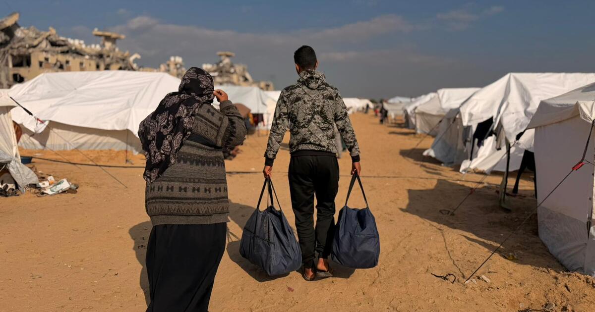
Humanitarians race to protect families from winter storms
OCHA reports that humanitarians in Gaza continue to help the most vulnerable families as harsh winter conditions leave hundreds of thousands of…
Humanitarians race to protect families from winter storms
OCHA reports that humanitarians in Gaza continue to help the most vulnerable families as harsh winter conditions leave hundreds of thousands of…

Brianna Dix: Jalen McMillan
This may be recency bias but I think Jalen McMillan is going to have another breakout game, continuing the trend he set against the Dolphins in Week 17. Last week, McMillan posted his first 100-yard game…

Orono, ME – The University of Maine men’s basketball team opens up America East play on Saturday, Jan. 3 as they…


Will Smith is being sued by a violinist from his 2025 tour, who claims the rapper and actor exhibited “predatory behaviour” and was “deliberately grooming and priming” him for “further sexual exploitation”. Brian King Joseph is also…

As cesarean delivery (C-section) rates continue to rise worldwide, experts at NYU Langone Health are highlighting a surgical technique that may help lower the risk of long-term complications. The endometrium-free closure technique…

Sunspots are active regions where the Sun’s magnetic field contorts itself into loops and arcs. Solar imaging captures these phenomena, providing both dramatic imagery and scientific insight. Credit: Mark…
Corporate
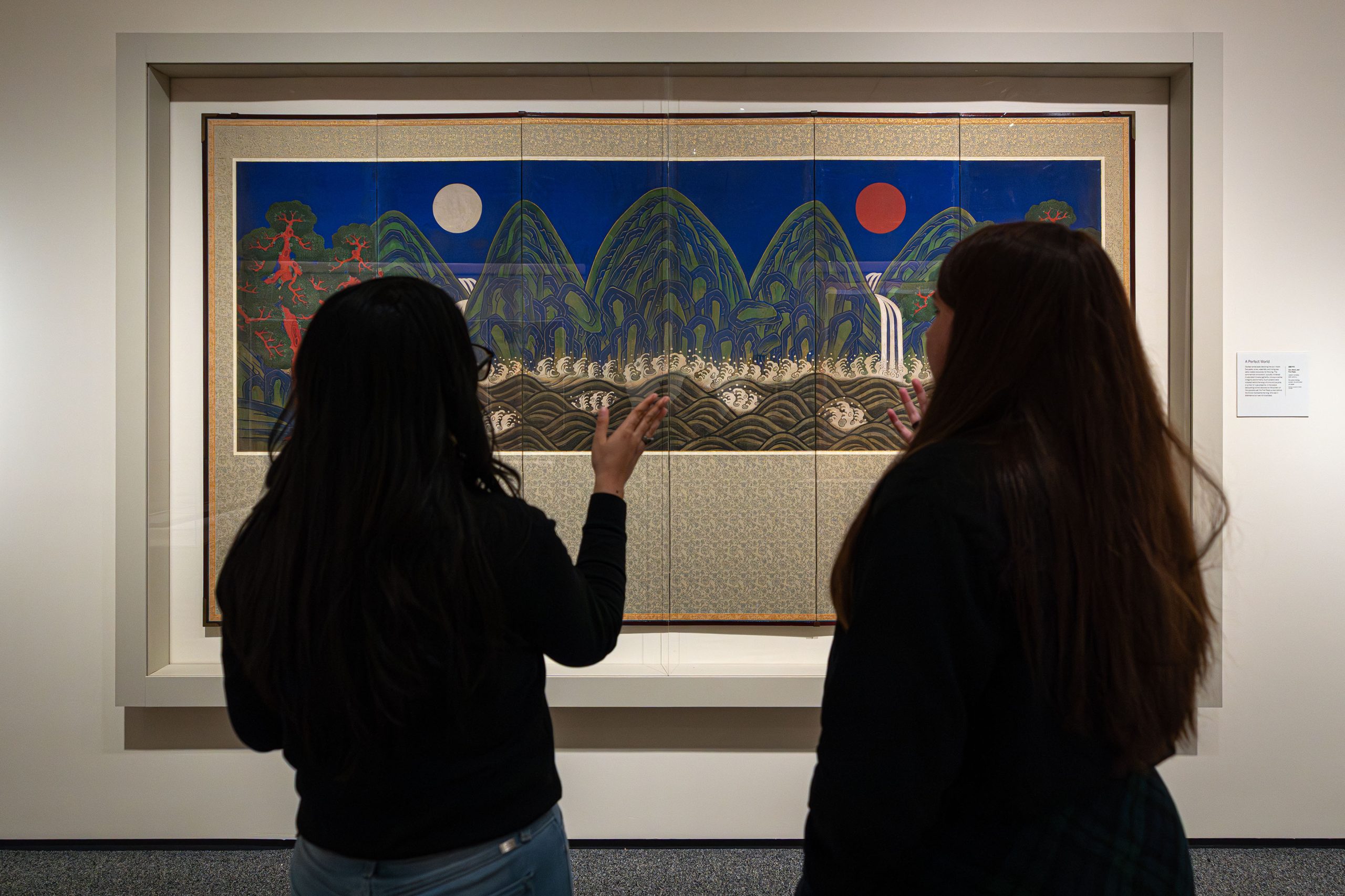
Creators visited the Korean Treasures exhibition with their Samsung Galaxy smartphones in hand to experience historical artworks
1/2/2026
© Colleen J. Dugan/National Museum of…