AI is moving rapidly, becoming a critical component in everything from Google searches to content creation. It’s also eliminating jobs and flooding the internet with slop. Thanks to the massive popularity of ChatGPT, now every major tech company…
Author: admin
-
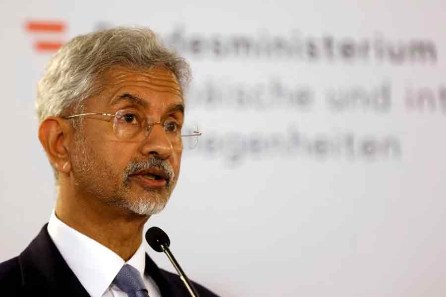
‘Good and Bad Neighbours’: Jaishankar Explains India’s Neighbourhood Policy Amid Bangladesh Unrest
External Affairs Minister S Jaishankar on Friday outlined India’s approach towards its neighbours, drawing a distinction between “good neighbours” and those that pose security challenges. Speaking at an event at IIT Madras,…
Continue Reading
-

Will Smith sued by tour violinist alleging sexual harassment and wrongful termination
Musician Brian King Joseph accuses Smith of “predatory behavior” during the ‘Based on a True Story: 2025’ tour
Will Smith has been sued by a violinist…
Continue Reading
-
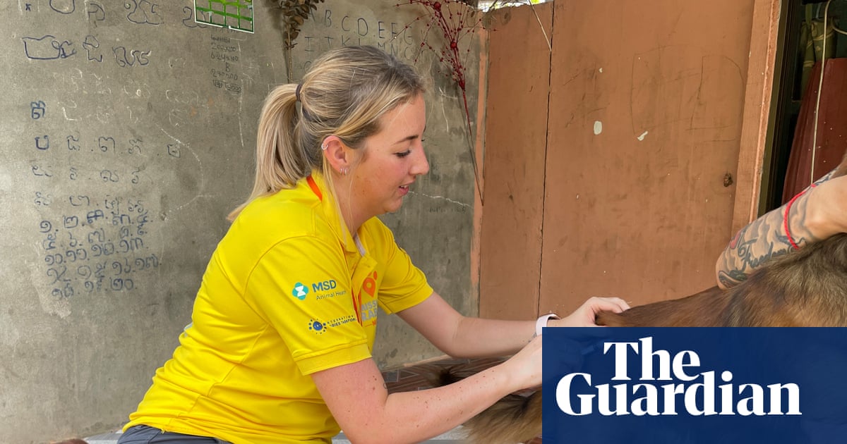
‘I need to help’: Barnsley woman’s rabies death inspires dog-vaccinating mission | Infectious diseases
It was just a scratch. Among all the feelings and thoughts that she has had to wrestle with since the summer, disbelief is the emotion that Robyn Thomson still struggles with the most. “You never think it would happen to you,” said Robyn….
Continue Reading
-

France to investigate deepfakes of women stripped naked by Grok – POLITICO
PARIS — French authorities will investigate the proliferation of sexually explicit deepfakes generated by artificial intelligence platform Grok on X, the Paris prosecutor’s office told POLITICO.
French…
Continue Reading
-
INCERGO S.A. ADVANCES MERGER WITH VISUAL SEMICONDUCTOR BY ISSUING SHARES AND SHOWCASES GLASSES-FREE 3D DISPLAYS AT THE CONSUMER ELECTRONICS SHOW IN LAS VEGAS
LUXEMBOURG and FREMONT, Calif., Jan. 2, 2026 /PRNewswire/ — INCERGO S.A. (ICG) today announced continued progress in its merger with Visual Semiconductor Inc. (VSI), including the…
Continue Reading
-

Launch of the new exclusive BMW Group brand.
Munich. The transfer of the Alpina trademark rights
on 1 January 2026 marks the launch of BMW ALPINA as an exclusive
standalone brand under the BMW Group umbrella. The initial focus will
be on brand activation. Key elements of the BMW ALPINA brand are its
unique balance of maximum performance and superior ride comfort,
combined with hallmark driving characteristics. This is complemented
by an exclusive portfolio of bespoke options and custom materials
along with unmistakeable details.Tradition meets future.
Given the storied history of the brand, the BMW Group is aware
of its tremendous responsibility and committed to making the new BMW
ALPINA chapter one that meets the highest expectations. The new
wordmark provides a first indication. It radiates clarity, calm and
confidence. Positioned at the centre of the rear, it underlines the
standalone character of the new exclusive brand and its independent
personality within the BMW Group. It is consciously inspired by the
brand’s asymmetrical wordmark from the 1970s. The elegant,
contemporary design provides a link between history and future.New direction for modern exclusivity.
BMW ALPINA vehicles are manufactured according to rigorous
standards for materials selection and craftsmanship. They fulfil the
most demanding expectations in terms of visuals, acoustics and feel.
Moreover, buyers can choose from a remarkable portfolio of bespoke
options. These ensure that every BMW ALPINA has its own, unmistakeable
character. Each vehicle will be an exclusive object for connoisseurs
in pursuit of the extraordinary, without compromises on performance,
comfort and individuality.www.bmwalpina.com
Continue Reading
-
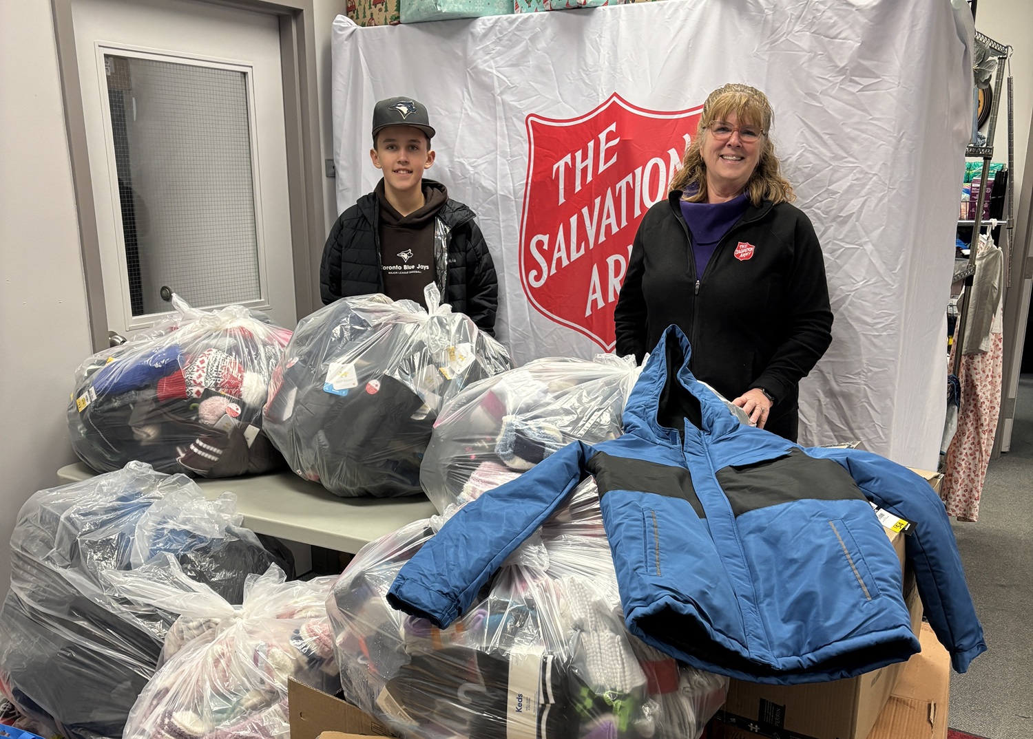
Young Donor Teams Up with The Salvation Army to Keep Children Warm
What started with a single $5 donation has now become an annual tradition for Owen Hewett, a 12-year-old from Napanee, Ont., who is passionate about ensuring less fortunate children have appropriate winter wear each Christmas.
When Owen was three, his dad, Darren, mentioned that there were families who couldn’t afford to provide their children with proper cold-weather attire. Moved after hearing that, Owen decided to donate $5 for mittens.
“I felt bad for kids who had cold hands and ears all the time, so I wanted to help,” Owen shares.
A Yearly Tradition
His father thought it would be a one-time donation, but as they enter the 10th year of their annual fundraiser, that has clearly not been the case.
“A friend of mine who is a local business owner donated $50 towards our efforts that first year. At that moment, we thought it would be a good idea to raise funds every year and here we are 10 years later,” Darren says.
Each year, donations increase, and the initiative continues to gain momentum. In 2025, Owen collected just over $4,400 to purchase mittens, toques and other cold-weather essentials for children in need.
“Usually around mid-November, we record a video reminding people about the fundraiser. My dad posts it on Facebook and the donations just start to come in,” Owen adds.
Partnership with The Salvation Army
To ensure these items reach children from families experiencing financial insecurity, Owen has partnered with The Salvation Army in Napanee in the past couple of years to assist with distribution.
“We reached out to Abigail Mills at The Salvation Army and asked her if our donation would fit any initiative they were doing. She said they were collecting snowsuits and boots, but they didn’t have mittens, so we decided that is exactly how we wanted to help,” Darren says.
Owen’s donation of hats and mitts helped The Salvation Army, in part, to provide winter gear for 250 local children this year.
Large fundraisers such as Owen’s help The Salvation Army redirect additional funds toward other programs and services within the community.
Community Impact
Although Owen has not had the opportunity to meet any of the kids he has helped, he has heard from school staff who described his initiative as heartwarming.
Despite the high cost of living in Napanee in recent years, the generosity of its people has remained strong.
“No matter how much people may struggle, they still always help this cause. It’s amazing,” Darren shares.
“We tend to get an additional $500 each year,” Owen says. “The custodian at my school donated this year.”
Looking ahead, for Owen, the goal remains the same.
“I just want to see people with enough winter clothes and be warm,” he says.
By Juan Romero
Continue Reading
-
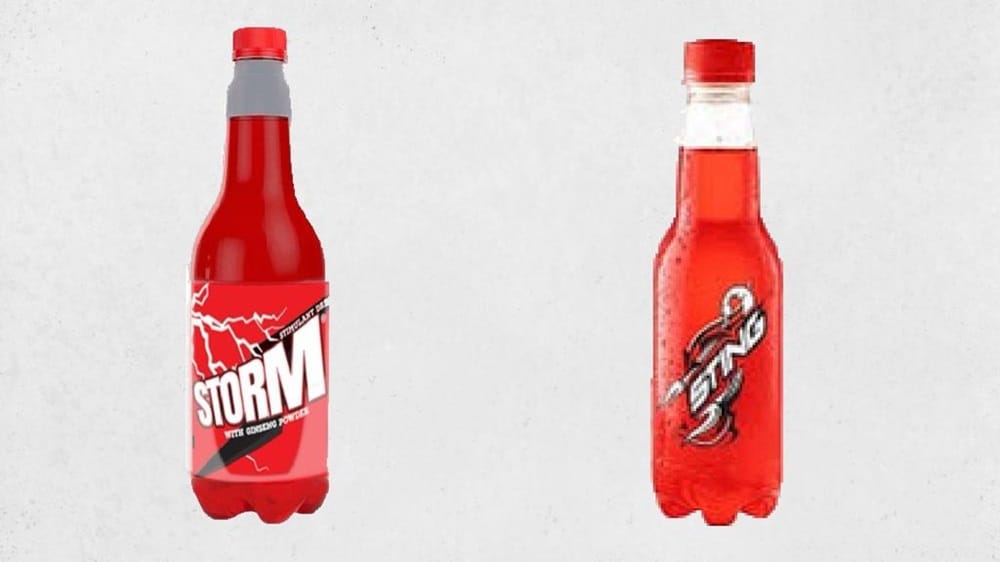
CCP Slaps Rs. 150 Million Fine on Mezan Beverages for Deceptive Marketing of Energy Drink
The Competition Commission of Pakistan (CCP) has imposed a penalty of Rs. 150 million on Mezan Beverages (Private) Limited for deceptive marketing practices, after finding that the company’s “Storm” energy drink closely imitated…
Continue Reading