- Australia bans Iranian tourists with valid visas for six months BBC
- ‘Massive betrayal’: Iranian nationals with valid tourist visas blocked from entering Australia for six months The Guardian
- Australia bans visitors from Iran amid war in the…
Author: admin
-

Australia bans Iranian tourists with valid visas for six months – BBC
-

AOUSD Has Announced OpenUSD v26.03 with Support for 3D Gaussian Splats
Another key addition is that OpenUSD can now be compiled to WebAssembly, and a new wasmFetchResolver example demonstrates loading and interacting with USD scenes directly in a web browser via HTTP protocol, which lowers the barrier for web-based…
Continue Reading
-

‘Significant progress’ made towards agreement with IMF, say sources
ISLAMABAD (Mudassar Ali Rana) – A significant progress has been made toward a staff level agreement between Pakistan and the International Monetary Fund (IMF) under the Extended Fund Facility and the Resilience and…
Continue Reading
-

Jimmy Fallon Show MC Apologizes for BTS North Korea Joke – 조선일보
- Jimmy Fallon Show MC Apologizes for BTS North Korea Joke 조선일보
- BTS Return to ‘The Tonight Show’ with 2 Performances, Surprises …and Slippers? People.com
- Racist remarks against BTS? ARMY slams Jimmy Fallon show over North Korea remark
Continue Reading
-
PM Shehbaz reaffirms commitment to advance CPEC 2.0 – RADIO PAKISTAN
- PM Shehbaz reaffirms commitment to advance CPEC 2.0 RADIO PAKISTAN
- PM Shehbaz, Chinese envoy discuss CPEC, economic ties The Express Tribune
- Pakistan, China pledge strong ties ahead of 75th anniversary of diplomatic relations Dunya News
- CM Punjab…
Continue Reading
-

Long-Term Outcomes of GLP-1 Therapies in Type 1 Diabetes
Use of GLP-1 receptor agonists—with or without glucose-dependent insulinotropic polypeptide (GIP)—may be associated with improved clinical outcomes in patients with Type 1 diabetes, according to a large retrospective analysis published in
Continue Reading
-
China’s experimental satellite constellation lays foundation for future lunar exploration -Xinhua
BEIJING, March 26 (Xinhua) — The Technology and Engineering Center for Space Utilization (CSU) of the Chinese Academy of Sciences (CAS) announced on Wednesday that the three-satellite constellation based on the Distant Retrograde Orbit (DRO)…
Continue Reading
-
Transcript: Investors try to stay ahead of Trump’s Iran moves – Financial Times
- Transcript: Investors try to stay ahead of Trump’s Iran moves Financial Times
- Oil traders bet millions ahead of Trump’s Iran talks post BBC
- Futures Markets Saw Trading Spike Before Trump’s Iran Post Bloomberg.com
- Oil, stock trading spiked before Trump’s Iran remarks: media Dawn
- Traders placed $580mn in oil bets ahead of Donald Trump’s social media post on Iran talks Financial Times
Continue Reading
-
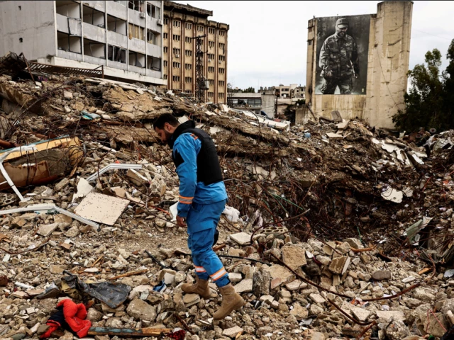
Trump threatens to unleash hell if Iran rejects latest US proposal
Araqchi says no formal negotiations have taken place, only messages exchanged through intermediaries
A paramedic walks among the rubble at a site damaged in an Israeli strike, amid escalating hostilities between Israel and Hezbollah, as the…
Continue Reading
-
Swing for Joy: Starbucks Korea Teams Up with the KBO to Celebrate Spring Baseball Season : Starbucks Stories Asia – Starbucks
- Swing for Joy: Starbucks Korea Teams Up with the KBO to Celebrate Spring Baseball Season : Starbucks Stories Asia Starbucks
- KBO collaborates with Starbucks → New product collection and baseball stadium concept launching “sequentially released…
Continue Reading