Psychedelic drugs such as psilocybin and MDMA may help repair the brain’s insulating myelin layer, according to new research.

New research by Elsevier suggests that psychedelic drugs could help repair damage to the brain’s…
Psychedelic drugs such as psilocybin and MDMA may help repair the brain’s insulating myelin layer, according to new research.
New research by Elsevier suggests that psychedelic drugs could help repair damage to the brain’s…

This surge, however, exposed cracks in the foundations. On 30 January 2026, the SFC issued a strongly worded circular1 putting sponsors firmly on notice. The message is clear: quality over volume. SFC warned that sponsors’ gatekeeping role “may have been eroded in their eager pursuit of deal volume” and urged all to “avoid overcommitment”. Sponsors should take heed since enforcement action may follow.
The SFC identified serious deficiencies in some sponsor work: poor listing document preparation, over-reliance on external experts and third parties without proper competency checks, insufficient capacity of sponsor principals to supervise deal teams and participate in the listing engagements, attempts to appoint unqualified sponsor principals, and staff lacking requisite knowledge and experience. The SFC’s concern is that some sponsors may be adopting a “process-driven approach” over substantive due diligence. Specific case examples and details of their potential non-compliance are set out in the Appendix to the circular.2
All sponsors were required to report to the SFC on the number of their principals and active engagements, plus staff who have not passed relevant licensing examinations within the prescribed timeframe.The SFC also identified a critical category: “Sponsors with Strained Principals” i.e. any sponsor that has designated any principals to simultaneously supervise or participate in six or more active IPO engagements. The SFC generally regards Sponsors with Strained Principals as lacking adequate resources, unless under very exceptional circumstances with valid justifications to the SFC’s satisfaction. Sponsors must monitor this threshold carefully.In addition 13 sponsors that received a December 2025 joint letter from the SFC and SEHK (Concerned Sponsors), plus Sponsors with Strained Principals, should expect on-site inspections “in the near future”.
The following sponsors must complete comprehensive internal reviews within three months:
These internal reviews should be signed off by the Managers-In-Charge of the Overall Management Oversight (OMOs) of the sponsors.
All individuals engaging in IPO sponsor work must now pass HKSI LE Papers 1 and 16 before starting IPO sponsor work. An individual who is currently engaged in sponsor work, but who has not passed the requisite examination even after the six-month period, should be removed from transaction teams immediately.
The SFC has said that it will not hesitate to act. It has warned that overly lengthy listing documents (expected not to exceed 300 pages for the main body), poor drafting, or incomplete responses to regulators’ comments may result in suspension of the vetting process (as at 31 December 2025, vetting of 16 listing applications remain suspended). In addition, listing applications may be returned where they are not substantially complete.
For persistent underperformers, the SFC may also restrict business scope and the number of permitted active listing engagements. Serious cases will attract SFC investigation and/or disciplinary action against sponsors, principals, as well as management (including directors and key group personnel) who are accountable for the sponsor’s failures.
Footnotes:
[1] https://apps.sfc.hk/edistributionWeb/gateway/EN/circular/doc?refNo=26EC4
[2] https://apps.sfc.hk/edistributionWeb/api/circular/openAppendix?lang=EN&refNo=26EC4&appendix=0

Northerly states in the USA are likely to have increased displays of the Northern Lights tonight, Friday March 6, 2026 and into the weekend.
Several northern US states and Canada are in line for aurora displays this weekend, potentially making…

Prime Minister Shehbaz Sharif on Friday presided over a high-level meeting on petroleum products, directing provincial governments to take strict legal action against those hoarding the commodities.
According to a handout by the Prime…

This recording is a celebration of what a fruitful relationship between a composer and an orchestra can be, as well as a souvenir of uneasy times. The Hallé co-commissioned Huw Watkins’ second symphony, having premiered his first; it was…

Italian design studio Pininfarina has channelled its expertise in sports cars into the design of its first phone, created with Chinese technology brand Infinix.
Debuted…
BOOK NOW!
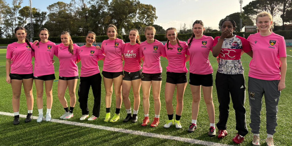
A group of young rugby players who met at Northampton Saints’ world‑leading residential camp have gone on to form their own international Sevens team – and are already making waves on the European stage.
The Legion of Sisters 7s…

A computer file that stores information on how scientists synthesise and characterise metal–organic frameworks (MOFs) could standardise the reporting of such materials. These files offer a potential way to reduce the discrepancy between methods…

The MacBook Air has been Apple’s entry-level offering since 2011, but as of this week, it’s now a mid-tier upgrade. On March 4, the company announced that it’s launching an all-new budget laptop called the MacBook Neo, which…