- When should kids get a smartphone? Study links owning one before age 12 to health risks TheDailyNewsOnline.com
- Giving a kid a phone before this age can be especially harmful, research suggests The Washington Post
- Experts Warn Against Giving…
Author: admin
-

When should kids get a smartphone? Study links owning one before age 12 to health risks – TheDailyNewsOnline.com
-

Quiz: How well do you remember 2025’s clean energy…
In this free online forum, we move beyond national talking points to dive deep into regional action. We’ll bring together the experts who are succeeding on the front lines in diverse regions like Southwest, Southeast, and Midwest regions. Don’t…Continue Reading
-
No shortage of wheat in country: Tanveer – RADIO PAKISTAN
- No shortage of wheat in country: Tanveer RADIO PAKISTAN
- Federal Minister for National Food Security and Research Rana Tanveer Hussain chairs a meeting of the National Wheat Oversight Committee. Associated Press of Pakistan
- Govt to exit wheat…
Continue Reading
-
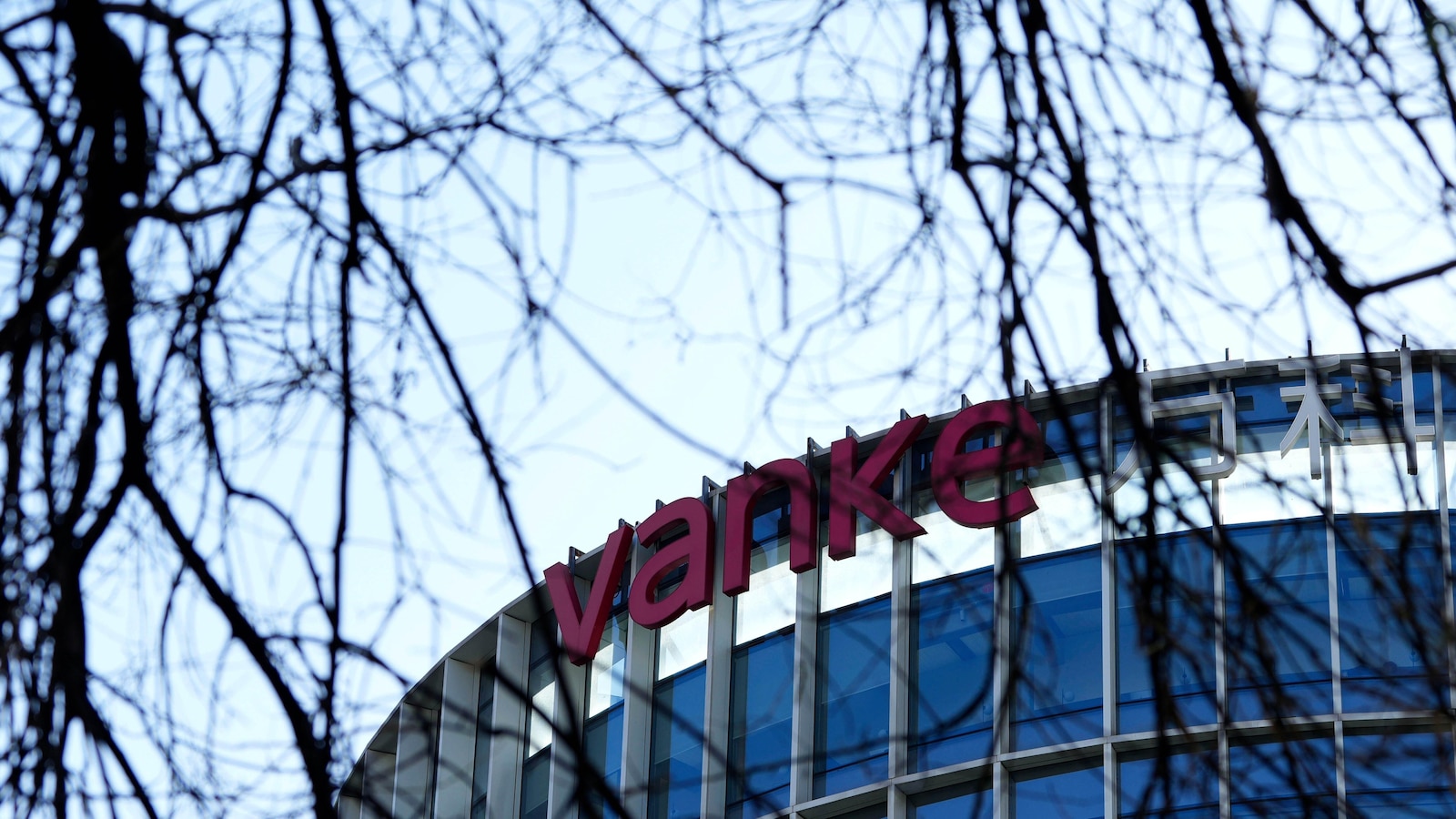
China Vanke’s near-default exposes fragility of the faltering recovery in the property industry
HONG KONG — State-backed property developer China Vanke, once the country’s largest homebuilder by sales, narrowly avoided defaulting on a 2 billion yuan ($284 million) bond last week as the painfully slow recovery in China’s property market drags on.
The Chinese developer also was seeking to delay repayment of another 3.7 billion yuan ($530 million) of onshore debt due on Dec. 28, with bondholders agreeing to extend the deadline to February.
Years after the downturn in the housing market began, Chinese developers are still struggling to regain their footing, despite a slew of government policies meant to revive the industry. Weak investment and housing prices have shaken investor confidence, spilling into the broader economy since millions of homeowners are stuck with apartments worth far less than what they paid for them.
Instead of the huge driver of prosperity that it once was, the property market is weighing on the economy.
Although Vanke’s bondholders have approved extensions for repayments of its debt, the risk of a default remains.
About a third-owned by Shenzhen Metro, a state-owned railway, publicly listed Vanke’s finances are a mess. Its revenue fell 27% from a year earlier in the latest July-September quarter, and several of its onshore bonds were suspended from trading after prices plunged.
The developer owes more than $50 billion, less than the more than $300 billion in debt racked up by China Evergrande, one of the first property dominos to fall when it defaulted in 2021 after the government cracked down on excessive borrowing in the industry.
Analysts say Vanke, founded in the 1980s in the southern boomtown of Shenzhen, may be testing the limits of state support for property developers in reviving the industry, which once accounted for more than a quarter of total economic activities in China.
More than four years after the downturn began, China’s property sector has yet to recover. The situation varies from city to city, but overall home prices have fallen by 20% or more from their peak in 2021.
The decline has continued, with new home sales falling 11.2% by value year-on-year in the first 11 months of 2025, according to official statistics. Property investments fell nearly 16% from a year earlier.
The slump has caused massive layoffs, hurting overall consumer confidence and spending.
“The continued slide in the property market remains one of the most significant risks to China’s efforts to shift to a domestically demand-driven growth model,” wrote Lynn Song, chief economist for Greater China at ING Bank, in a recent commentary.
China Evergrande, once deemed “too big to fail” as one of the country’s largest developers, ran into trouble in 2021 and eventually was forced into liquidation. Many other Chinese developers also defaulted and in some cases were restructured. Tough measures to fight Covid-19 during the pandemic took a toll as construction projects were suspended.
Restoring confidence in the property sector may take years, economists at Morgan Stanley say, and Vanke’s woes will only further weigh on its real estate market outlook. Economists at Morningstar say home prices are unlikely to rebound until 2027 due to excess supply, despite repeated pledges by regulators to stabilize the real estate market.
While Vanke’s debt is way smaller than Evergrande’s was, a default would sting: It had been considered one of the financially sounder real estate developers in China.
Shenzhen Metro Group, which is controlled by the Shenzhen government, has provided more than 29 billion yuan ($4 billion) in shareholder loans to Vanke so far this year to help with its debt repayments, according to S&P Global.
That’s not enough to repay its full obligations. Vanke reported 60 billion yuan ($8 billion) of cash by the end of September 2025, against short-term debts of about 151 billion yuan ($21 billion), Fitch Ratings said.
“This is one of the most significant, quasi state-backed developers that may be defaulting (on) their repayment,” said Foreky Wong, a founding partner at Fortune Ark Restructuring.
S&P Global, one of the world’s main rating agencies, recently downgraded Vanke to “selective default,” saying it viewed the extension of its bond repayment period as a distressed debt restructuring “tantamount to a default.” Fitch Ratings also downgraded Vanke’s rating to “restricted default”.
Vanke — which employed more than 120,000 people as of last year — still faces hundreds of millions of dollars more of debt repayments in 2026. S&P said it faces more than 9.4 billion yuan of bonds maturing over the next six months.
A default by Vanke could spill over into the wider real estate sector, making it more difficult for non-state owned developers to get help, said Jeff Zhang, an analyst at Morningstar.
“Without a strong commitment by the Shenzhen government on the bailout, we think Vanke’s liquidity profile should remain fragile,” Zhang said.
Continue Reading
-

Wall Street ticks lower in light trading ahead of the final opening bell of 2025
Wall Street drifting lower before the opening bell, heading for the fourth day of losses, on the final day of trading for 2025, a banner year for markets that was driven by both optimism and uncertainty.
Futures for the S&P 500 and the Dow Jones Industrial Average each ticked down 0.1% early Wednesday. Nasdaq futures were off 0.2%.
Institutional investors are largely closed out of their positions for the year, so trading is expected to be extremely light. U.S. markets will be closed on Thursday for New Year’s Day.
The S&P 500 is up more than 17% this year as investors embraced artificial intelligence technology, both in the sector and its potential across almost all other sectors.
The AI frenzy that drove markets in 2025 did not come without concerns. Chief among them is the worry that artificial intelligence technology may not produce enough profits and productivity to make all the investment worth it. That could keep the pressure on AI stocks like Nvidia and Broadcom, which were responsible for much of the market’s gains this year.
And it’s not just AI stocks that critics say are too pricey. Stocks across the market still look expensive after their prices climbed faster than profits.
On top of concerns that stocks are overvalued, the ongoing impact of a wide-ranging U.S.-led trade war threatens to add more fuel to inflation in the U.S. While the Federal Reserve has cut its benchmark lending rate three times to close out the year, inflation remains solidly above the central bank’s 2% target.
Fed officials have cited concerns over a weakening labor market as their motivation for cutting rates. Arriving later Wednesday is the Labor Department’s most recent data on weekly jobless claim applications, which are viewed as a proxy for layoffs.
The Fed has signaled more caution moving forward. Minutes from its December meeting reflect the divisions within the central bank as it deals with uncertainty about the threats facing the economy.
Wall Street is betting that the Fed will hold interest rates steady at its next meeting in January.
Sung Won Sohn, professor of finance and economics at Loyola Marymount University, believes uncertainty is brewing for global markets because of inflation, labor shortages and questions about where interest rates might be headed.
“Central banks must tread carefully, and financial markets will likely experience continued volatility as expectations shift,” he said.
“For businesses, investors, and policymakers alike, flexibility, risk management, and close attention to economic signals will be essential in navigating the challenges ahead.”
Trading in precious metals continued to be volatile as the year winds down. Silver swung back to a big loss, giving back more than 8% early Wednesday after Tuesday’s gain of more than 10%. Following Friday’s 7.7% jump, silver lost nearly 9% on Monday. It’s still up more than 140% this year.
Gold fell 1.5% Wednesday morning but is still up 66% in 2025.
Elsewhere, global stock markets including Germany, Japan and South Korea were closed Wednesday for the New Year’s holidays, while trading was mixed in those that remained open.
France’s CAC 40 lost 0.5% by midday, while Britain’s FTSE 100 shed 0.2%.
Earlier in Asia, the Hang Seng index dipped 0.9% to 25,630.54, while the Shanghai Composite rose 0.1% to 3,968.84. The Taiex in Taiwan jumped 0.9% to 28,963.60. In Australia, Sydney’s S&P/ASX 200 dipped less than 0.1% to 8,714.30.
Tokyo trading was set to be closed for the New Year’s holidays on Thursday and Friday and scheduled to reopen on Monday. In South Korea, trading was scheduled to be closed on Thursday.
U.S. crude picked up 31 cents to $58.26 per barrel. Brent crude, the international standard, added 28 cents to $61.61 per barrel. Yet crude, unlike some other commodities this year, has been falling in prices. A barrel of crude costs 19% less today than it did at the start of the year, and gas prices are down 6% to 7% nationally, or about 20 cents per gallon.
Continue Reading
-

Scientists are publishing more than ever with AI. But not all papers measure up, study finds
As scientists increasingly rely on artificial intelligence for writing, coding and even generating ideas, a new study examines how AI is reshaping academic research.
What once sounded like academic gossip now reflects a real and measurable…
Continue Reading
-
Terminal Handling Charges Update – Muara, BN
In order, to keep providing you with our global services, Maersk is revising Terminal Handling Service – Origin & Terminal Handling Service – Destination for the scope World to / from Muara, BN for specific commodities with effective price calculation date 1st Feb 2026 until further notice.
The tariff amount is detailed as follows:
Surcharge code
Origin
Destination
Container basis
Currency
ALL_20_DRY / ALL_40_DRY
Surcharge code
OHC
Origin
Muara, BN ( BNMUP)
Destination
World
Container basis
Per container
Currency
BND
ALL_20_DRY / ALL_40_DRY
335 / 495
Surcharge code
DHC
Origin
World
Destination
Muara, BN ( BNMUP)
Container basis
Per container
Currency
BND
ALL_20_DRY / ALL_40_DRY
335 / 495
The above tariff will be applicable for the below HS Codes.
HS Code
Commodity Description
HS Code
004610
Commodity Description
New Electric Vehicle, solely lithium Bettery Powered
HS Code
004613
Commodity Description
Used Electric Vehicle, solely lithium Bettery Powered
HS Code
004614
Commodity Description
Damaged Electric Vehicle, solely lithium Bettery Powered
HS Code
004615
Commodity Description
Combined New/Damaged or Total Loss Non-Electric Vehicles and Electric Vehicles.
HS Code
004616
Commodity Description
New Electric Vehicle, solely powered by non-lithium Betteries
HS Code
004617
Commodity Description
Used Electric Vehicle, solely powered by non-lithium Betteries
HS Code
004618
Commodity Description
Damaged or Total Loss Electric Vehicle, solely powered by non-lithium Betteries
* Non-SPOT booking – The above rate is retrieved based on PCD. PCD = Price Calculation Date. For non-FMC, PCD refers to the scheduled departure date of the first water leg at the time of booking confirmation for non-spot bookings. For FMC, PCD is last container gate-in date for non-spot bookings.
* SPOT booking – The above rate is retrieved based on 1st vessel ETD at booking confirmation for Spot bookings.
For your reference, we have also included the levels and rate structure for some sample corridors from Los Angeles, US to Muara, BN from 01-Feb-26 until further notice. These may be subject to future Change; however, we will make sure to notify you accordingly.
Los Angeles, US to Muara, BN
Dry Container
Surcharge Code
20 DRY
40 DRY
40 HDRY
45 HDRY
Surcharge Code
BAS – Basic Ocean Freight
20 DRY
642 USD
40 DRY
735 USD
40 HDRY
735 USD
45 HDRY
1035 USD
Surcharge Code
DDF – Documentation fee – Destination
Per Bill of Lading
20 DRY
50 BND
40 DRY
50 BND
40 HDRY
50 BND
45 HDRY
50 BND
Surcharge Code
DHC – Terminal Handling Service – Destination
20 DRY
335 BND
40 DRY
495 BND
40 HDRY
495 BND
45 HDRY
N/A
Surcharge Code
OHC – Terminal Handling Service – Origin
20 DRY
335 BND
40 DRY
495 BND
40 HDRY
495 BND
45 HDRY
N/A
Reefer and Special Container
Surcharge Code
20 REEF
40 HREEF
20 Special
40 Special
Surcharge Code
BAS – Basic Ocean Freight
20 REEF
N/A
40 HREEF
3506 USD
20 Special
642 USD
40 Special
735 USD
Surcharge Code
DDF – Documentation fee – Destination
Per Bill of Lading
20 REEF
50 BND
40 HREEF
50 BND
20 Special
50 BND
40 Special
50 BND
Surcharge Code
DHC – Terminal Handling Service – Destination
20 REEF
N/A
40 HREEF
N/A
20 Special
335 BND
40 Special
495 BND
Surcharge Code
OHC – Terminal Handling Service – Origin
20 REEF
N/A
40 HREEF
N/A
20 Special
335 BND
40 Special
495 BND
- The above rates are also subject to other applicable surcharges, including local charges and contingency charges.
- These rates are unaffected by, and do not affect, any tariff notified, published or filed in accordance with local regulatory requirements.
- For trades subject to the US Shipping Act or the China Maritime Regulations, quotations or surcharges that vary from the Maersk Line tariff shall not be binding on Maersk Line unless included in a service contract or service contract amendment that has been filed with the Federal Maritime Commission (FMC) or the Shanghai Shipping Exchange, as applicable.
If you have any questions, please feel free to reach out to our local representatives on Maersk.com
Continue Reading
-

Can bad oral bacteria travel to the brain and trigger Parkinson’s disease? | Health News
According to new research, Parkinson’s disease may be influenced by bacteria that originate in the mouth.
A study published recently in Nature Communications shows that an oral bacterium can settle in the gut and produce a chemical…Continue Reading
-
Pakistani FM to visit China, hold 7th round of China-Pakistan Foreign Ministers’ Strategic Dialogue-Xinhua
BEIJING, Dec. 31 (Xinhua) — Pakistani Deputy Prime Minister and Foreign Minister Mohammad Ishaq Dar will visit China and hold the Seventh Round of China-Pakistan Foreign Ministers’ Strategic Dialogue from Jan. 3 to 5, 2026, a Chinese foreign…
Continue Reading