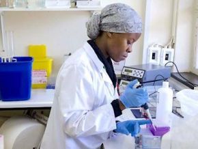
An experimental breath test successfully sorted out type 2 diabetes patients from healthy people, based on their exhalations, Penn State researchers report. File Photo by Arshad Arbab/EPA
Detecting diabetes might soon be as easy as breathing into a device, a new study says.
An experimental breath test sorted out type 2 diabetes patients from healthy people, based on their exhalations, researchers report in the September issue of the Chemical Engineering Journal.
“This sensor only requires that you exhale into a bag, dip the sensor in and wait a few minutes for results,” senior researcher Huanyu “Larry” Cheng, an associate professor of engineering science and mechanics at Penn State, said in a news release.
If validated, this test could prove simpler than the complicated blood testing and lab work now required to diagnose diabetes, researchers said.
Of the 37 million adults with diabetes in the U.S., about 1 in 5 isn’t aware that they have the condition, researchers said in background notes.
The sensor detects acetone, a chemical byproduct of energy production in the human body.
Everyone’s breath contains acetone, but elevated acetone levels indicate that a person has diabetes, researchers said.
Researchers exposed the sensor to the breath of 51 people with type 2 diabetes and 20 healthy volunteers, all of whom exhaled into aluminum foil bags.
The sensor responded differently to people with type 2 diabetes, results showed.
Further, the sensor response tracked with the participants’ blood sugar levels. This shows that with work, the sensor might be powerful enough to help track blood sugar levels on a regular basis, rather than just as a simple test for diabetes, Cheng said.
“If we could better understand how acetone levels in the breath change with diet and exercise, in the same way we see fluctuations in glucose levels depending on when and what a person eats, it would be a very exciting opportunity to use this for health applications beyond diagnosing diabetes,” Cheng said.
Researchers next plan to improve the sensor so it can be used directly under a person’s nose or attached to the inside of a mask, to make it easier to use.
More information
The American Diabetes Association has more on diabetes diagnosis.
Summary: A new study shows that overactivation of dopamine-producing neurons in the brain can trigger their degeneration, mirroring what happens in Parkinson’s disease. Using a mouse model, researchers found that chronic activation disrupted daily activity patterns, damaged neuronal axons, and eventually killed cells in the substantia nigra.
These findings align with molecular changes observed in human Parkinson’s brain tissue, linking excess neuron activity to decreased dopamine production and cell death. The results suggest that therapies targeting neuron activity could help protect vulnerable cells and slow the disease’s progression.
Key Facts
Neuron Vulnerability: Dopamine neurons in the substantia nigra are most affected by overactivation.
Cellular Changes: Overactive neurons reduce dopamine production, leading to degeneration.
Therapeutic Potential: Drugs or deep brain stimulation could adjust activity and protect cells.
Source: Galdstone Institutes
Certain brain cells are responsible for coordinating smooth, controlled movements of the body. But when those cells are constantly overactivated for weeks on end, they degenerate and ultimately die.
This new observation made by scientists at Gladstone Institutes may help explain what goes awry in the brains of people with Parkinson’s disease.
Within a few days of overactivating dopamine neurons, the animals’ typical cycle of daytime and nighttime activities became disrupted. Credit: Neuroscience News
Researchers have long known that a particular subset of neurons die as Parkinson’s disease progresses, but they aren’t sure why.
The new work, published in the scientific journal eLife, shows that in mice, chronic activation of these neurons can directly cause their demise.
The scientists hypothesize that in Parkinson’s, neuron overactivation could be triggered by a combination of genetic factors, environmental toxins, and the need to compensate for other neurons that are lost.
“An overarching question in the Parkinson’s research field has been why the cells that are most vulnerable to the disease die,” says Gladstone Investigator Ken Nakamura, MD, PhD, who led the study. “Answering that question could help us understand why the disease occurs and point toward new ways to treat it.”
Too Much Buzz
More than 8 million people worldwide are living with Parkinson’s disease, a degenerative brain disease that causes tremors, slowed movement, stiff muscles, and problems walking and balancing.
Scientists know that a set of neurons that produce dopamine and support voluntary movements die in people with Parkinson’s. Many lines of evidence also suggest that the activity of these cells actually increases with disease, both before and after degeneration begins. But whether this change in activity can directly cause cell death is poorly understood.
In the new study, Nakamura and his colleagues tackled this question by introducing a receptor specifically into dopamine neurons in mice that allowed them to increase the cells’ activity by treating the animals with a drug, clozapin-N-oxide (CNO). Uniquely, the scientists added CNO to the animals’ drinking water, driving chronic activation of the neurons.
“In previous work, we and others have transiently activated these cells with injections of CNO or by other means, but that only led to short bursts of activation,” says Katerina Rademacher, a graduate student in Nakamura’s lab and first author of the study.
“By delivering CNO through drinking water, we get a relatively continuous activation of the cells, and we think that’s important in modeling what happens in people with Parkinson’s disease.”
Within a few days of overactivating dopamine neurons, the animals’ typical cycle of daytime and nighttime activities became disrupted. After one week, the researchers could detect degeneration of the long projections (called axons) extending from some dopamine neurons. By one month, the neurons were beginning to die.
Importantly, the changes mostly affected one subset of dopamine neurons—those found in the region of the brain known as the substantia nigra, which is responsible for movement control—while sparing dopamine neurons in brain regions responsible for motivation and emotions. This is the same pattern of cellular degeneration seen in people with Parkinson’s disease.
A Link to Human Disease
To gain insight into why overactivation leads to neuronal degeneration, the researchers studied the molecular changes that occurred in the dopamine neurons before and after the overactivation. They showed that overactivation of the neurons led to changes in calcium levels and in the expression of genes related to dopamine metabolism.
“In response to chronic activation, we think the neurons may try to avoid excessive dopamine—which can be toxic—by decreasing the amount of dopamine they produce,” Rademacher explains.
“Over time, the neurons die, eventually leading to insufficient dopamine levels in the brain areas that support movement.”
When the researchers measured the levels of genes in brain samples from patients with early-stage Parkinson’s, they found similar changes; genes related to dopamine metabolism, calcium regulation, and healthy stress responses were turned down.
The research did not reveal why activity of the dopamine neurons might increase with Parkinson’s disease, but Nakamura hypothesizes that there could be multiple causes, including genetic and environmental factors. The overactivity could also be part of a vicious cycle initiated early in disease.
As dopamine neurons become overactive, they gradually shut down dopamine production, which worsens movement problems. Remaining neurons work even harder to compensate, ultimately leading to cell exhaustion and death.
“If that’s the case, it raises the exciting possibility that adjusting the activity patterns of vulnerable neurons with drugs or deep brain stimulation could help protect them and slow disease progression,” Nakamura says.
Funding: The work was supported by Aligning Science Across Parkinson’s (ASAP-020529) through the Michael J. Fox Foundation for Parkinson’s Research (MJFF), the National Institutes of Health (RO1NS091902, F31NS137765), the Joan and David Traitel Family Trust and Betty Brown’s Family, a Burroughs-Wellcome Fund Award, the Hillblom Foundation, and a Berkelhammer Award for Excellence in Neuroscience.
About this Parkinson’s disease research news
Author: Julie Langelier Source: Gladstone Institutes Contact: Julie Langelier – Gladstone Institutes Image: The image is credited to Neuroscience News
Original Research: Open access. “Chronic hyperactivation of midbrain dopamine neurons causes preferential dopamine neuron degeneration” by Ken Nakamura et al. eLife
Parkinson’s disease (PD) is characterized by the death of substantia nigra pars compacta (SNc) dopamine (DA) neurons, but the pathophysiological mechanisms that precede and drive their death remain unknown.
The activity of DA neurons is likely altered in PD, but we understand little about if or how chronic changes in activity may contribute to degeneration.
To address this question, we developed a chemogenetic (DREADD) mouse model to chronically increase DA neuron activity and confirmed this increase using ex vivo electrophysiology.
Chronic hyperactivation of DA neurons resulted in prolonged increases in locomotor activity during the light cycle and decreases during the dark cycle, consistent with chronic changes in DA release and circadian disturbances.
We also observed early, preferential degeneration of SNc projections, recapitulating the PD hallmarks of selective vulnerability of SNc axons and the comparative resilience of ventral tegmental area axons.
This was followed by the eventual loss of midbrain DA neurons. Continuous DREADD activation resulted in a sustained increase in baseline calcium levels, supporting a role for increased calcium in the neurodegeneration process.
Finally, spatial transcriptomics from DREADD mice examining midbrain DA neurons and striatal targets, and cross-validation with human patient samples, provided insights into potential mechanisms of hyperactivity-induced toxicity and PD.
Our results thus reveal the preferential vulnerability of SNc DA neurons to increased neural activity and support a potential role for increased neural activity in driving degeneration in PD.
The doubles team of time and biology is, of course, undefeated. But they don’t win at a consistent pace. Venus Williams can look aged as a 40-year-old and then spry as a 45-year-old—as she did in her three-set loss to Karolína Muchová in the first round at the U.S. Open on Monday night. Now it’s Novak Djokovic’s turn to roll back time.
In search of his 25th career major singles title—pretty much his sole motivator right now, as he concedes—Djokovic did not play a match between Wimbledon and his first rounder at the U.S. Open on Sunday night. When he took the court against California’s Lerner Tien, a player precisely half his age, Djokovic looked more like a man nearing 40 who hadn’t played in six weeks than a legitimate contender. Compounded by a gnarly foot blister, he looked a half-step slow and a few kilos heavier than usual, and twigged together a straight-set win.
But the operative word is “win.” This is a survive-and-advance business. In his second-round match, Djokovic faced another young American in 22-year-old Zachary Svajda. The 38-year-old once again looked sluggish in the opening set, before shaking off his slow start and claiming the following three sets, and the match, gutting out the win.
Water bottle half full: Djokovic got the opportunity to shake off the tennis equivalent of ring rust and play himself into this tournament—a tournament he has won four times. He now finds himself with an extra day of rest and into the Round of 32, having pushed aside time and biology once again.
Microneedle-based (MN) vaccine systems are emerging as viable options for future vaccination efforts, according to a study published in Vaccine.1 While study researchers noted significant limitations to implementing them, MN-based systems showed the ability to facilitate precise vaccine administration, heightened immune responses, and enhanced stability in vaccine formulation.
“While conventional vaccine administration approaches face numerous challenges, the development and application of MN technology could provide effective alternatives that enhance vaccine stability, safety, and accessibility,” wrote authors of the study. “Vaccine delivery systems based on MNs, resulting from the fusion of MN and vaccine technologies, facilitate the direct administration of vaccines to the skin surface using micrometer-sized needles, serving as an innovative alternative to conventional injection techniques.”
When it comes to vaccination, researchers have been grappling with the best approaches for developing safe, effective, and non-costly immunization schedules to protect community health. Many of these recent developments are notable surrounding discussions on the COVID-19 pandemic and the mass vaccination efforts used throughout that time period.
The application of MN-based vaccine technology holds advantages, disadvantages, hurdles, but most importantly, promise for the development of more accessible and effective vaccine administration. | image credit: Péter Mács / stock.adobe.com
READ MORE: How Retail Pharmacy is Transforming Immunization Services, Access
According to McKinsey & Company, vaccine development was seemingly stalled throughout 2019.2 Since then, however, public health in the US and elsewhere experienced unprecedented events during the pandemic, which resulted in hospitalizations, deaths, and most notably—in the context of this study—significant vaccine hesitancy. With the pandemic activating health care professionals and researchers to act on global vaccine uptake outside of COVID-19, vaccine hesitancy continued to increase.3
While a variety of reasons have emerged regarding patients’ reasons for expressing this hesitancy, pain and fear are 2 specific factors that have led to the development of MN-based vaccines.
“As an emerging vaccine delivery tool, microneedles overcome the problems associated with routine needle vaccination, which can effectively deliver vaccines rich in antigen-presenting cells to the epidermis and dermis painlessly, inducing a strong immune response,” wrote authors of a study published in Pharmaceutics.4 “In addition, microneedles have the advantages of avoiding cold chain storage and have the flexibility of self-operation, which can solve the logistics and delivery obstacles of vaccines, covering the vaccination of the special population more easily and conveniently.”
Despite the significant promise in MN-based vaccine development, adult and childhood immunization rates have continued to decline or show insignificant levels since the pandemic has waned.5,6 Amid these issues and aforementioned vaccine development, researchers of the current study aimed to provide an end-to-end perspective on MN-based vaccine strategies.
“This review considers the rational design of MN systems and the development of MN strategies, including material selection and formulation with vaccines, as well as preclinical and clinical validation,” continued authors of the study.1 “Moreover, this review aims to provide an integrated, end-to-end perspective on the development of MN strategies through a consideration of cost, scalability, safety, and regulatory considerations.”
In their extensive breakdown of MN vaccine technology, researchers started by exploring the 5 main groups of MN-based vaccine delivery: coated, dissolving, hollow, solid, and hydrogel-forming MNs. While significant research is needed on MN technology in general, let alone the respective MN groups, these 5 options provide ample alternatives for vaccine administration as MN technology is further developed.
They then delved into how health care leaders will be expected to apply MN vaccine technology that is safe, effective, and accessible.
“The primary objective of MNs is to enhance patient comfort and ensure safety by offering a less painful option than conventional needle and syringe injections,” they wrote.1 “MNs penetrate the skin by creating micrometer-sized openings in the outer skin layer to enable drug delivery with minimal pain and irritation, avoiding interaction with nerve endings.”
According to the researchers’ review, the application of MN-based vaccine technology provides a myriad of potential advantages. If implemented into health care properly, MN vaccines can assist in self-administration capabilities, increasing immunization access, reducing pain from vaccines, enhancing vaccine acceptance, and decreasing needle-related injuries or infections.
However, with these new opportunities and advantages, there are certainly challenges to MN-based implementation in vaccine administration.
“The fusion of vaccination technology with MN technology is associated with several technological hurdles,” continued the authors.1 “These include issues with structural integrity, material selection, the challenge of ensuring precise dosage, design complexity for a range of uses, obstacles in terms of small-scale production, obstacles in terms of cost management, concerns about sterilization, and concerns about safety.”
While there are certainly concerns regarding MN vaccines’ costs, scalability, safety, and regulations, the use of this technology for boosting immunization rates and decreasing vaccine hesitancy is still in its beginning stages. With so much promise in bolstering public health behind these new approaches, researchers are dedicated to overcoming existing challenges and implementing the technology where it may be useful.
“The integration of MN technology with vaccine development offers a promising avenue for refining vaccine delivery and broadening immunization efforts,” they concluded.1 “By overcoming existing hurdles and leveraging the capabilities of MN-based systems, a more effective and accessible vaccination strategy can be developed, which contributes to global health initiatives.”
READ MORE: National Immunization Awareness Month
Don’t get left behind: Sign up today for our free Drug Topics newsletter and get the latest drug information, industry trends, and patient care tips delivered straight to your inbox.
References
1. Koçer AT, Durasi E, Kuscu E, et al. Piercing the future of vaccination: the revolutionary role of microneedle-based systems in healthcare advancements. Vaccine. 2025;63:127612. https://doi.org/10.1016/j.vaccine.2025.127612
2. Sabow A, Heller J, Conway M, et al. Beyond the pandemic: the next chapter of innovation in vaccines. McKinsey & Company. May 16, 2024. Accessed August 27, 2025. https://www.mckinsey.com/industries/life-sciences/our-insights/beyond-the-pandemic-the-next-chapter-of-innovation-in-vaccines
3. New data indicates declining confidence in childhood vaccines of up to 44 percentage points in some countries during the COVID-19 pandemic. UNICEF. April 20, 2023. Accessed August 27, 2025. https://www.unicef.org/rosa/press-releases/new-data-indicates-declining-confidence-childhood-vaccines-44-percentage-points-some
4. Feng YX, Hu H, Wong YY, Yao X, He ML. Microneedles: an emerging vaccine delivery tool and a prospective solution to the challenges of SARS-CoV-2 mass vaccination. Pharmaceutics. 2023 Apr 27;15(5):1349. doi: 10.3390/pharmaceutics15051349.
5. Immunization coverage. WHO. July 15, 2024. Accessed August 27, 2025. https://www.who.int/news-room/fact-sheets/detail/immunization-coverage
6. Trends in global adult vaccination. IQVIA. July 18, 2023. Accessed August 27, 2025. https://www.iqvia.com/insights/the-iqvia-institute/reports-and-publications/reports/trends-in-global-adult-vaccination
Punjab Education Secretary Khalid Nazir Watoo has denied rumours circulating on social media about extended school holidays until September 5. In a statement issued on Wednesday, he clarified that the notification about school closures, which has been circulating online, is fake.
“There has been no such notification issued by the Education Department regarding additional holidays,” he said. He further urged the public not to pay attention to these false claims, stating that no decision has been made regarding school closures.
He also recommended that the public check for authentic information on the official Facebook page of the School Education Department, where updates and announcements are made. The situation across Punjab has worsened as floodwaters swell following India’s release of water into eastern rivers.
The Pakistan Army has been deployed to assist in large-scale rescue and relief operations, with the National Disaster Management Authority (NDMA) confirming that more than 210,000 people have been safely evacuated from vulnerable areas, with no casualties reported so far.
Read More: Punjab floods worsen after Indian water release
Water levels in major rivers and reservoirs remain critically high, with inflows surpassing 1.2 million cusecs and floodwaters continue to move downstream into Punjab.
Authorities reported both large-scale evacuations and extensive rescue efforts as concerns mounted over the safety of communities along the Chenab, Ravi and Sutlej rivers.
According to the Water and Power Development Authority, the Indus River at Tarbela recorded an inflow of 240,000 cusecs and outflow of 245,400 cusecs.
At Mangla on the Jhelum, inflow was 34,000 cusecs and outflow 8,000 cusecs. Chashma saw inflows of 326,600 cusecs and outflows of 329,000 cusecs, while at Head Marala on the Chenab, inflows reached 107,500 cusecs against 89,500 cusecs outflow.
Your peripheral nervous system (PNS) is crucial to navigating daily life. It lets you walk, controls your eye movements, and rings your brain’s alarms when you step on a Lego brick. Yet researchers have never built a complete map of this essential network in any mammalian body.
Now a study published in Cell shows a complete, three-dimensional map of every single nerve fiber threading through a mouse. It completes the first-ever mammalian “connectome,” a flowchart of an entire nervous system, beyond just the well-researched brain and spinal cord.
“High-Speed Mapping of Whole-Mouse Peripheral Nerves at Subcellular Resolution,” by Mei-Yu Shi et al., in Cell, Vol. 188, No. 14; July 10, 2025 (CC BY 4.0)
“Mapping of the PNS has been a neglected component of mapping the connectome in animal and human brain studies,” says John Darrell Van Horn, a brain and data science researcher at the University of Virginia, who was not involved in the study.
The research team began by making the bodies of 16 mice as visually transparent as possible, removing fat, calcium, and other materials that block light. They then used a custom combined slicing tool and microscope to take images of each of the bodies 400 microns at a time, which took about 40 hours per mouse—providing data the researchers say would otherwise have taken months or years to collect.
Nerves (blue) wind through a mouse’s head.
“High-Speed Mapping of Whole-Mouse Peripheral Nerves at Subcellular Resolution,” by Mei-Yu Shi et al., in Cell, Vol. 188, No. 14; July 10, 2025 (CC BY 4.0)
The scientists genetically modified seven of the mice to have fluorescent neurons; as expected, this caused mostly the head to light up. In four of the mice, the team applied a technique called immunostaining, which uses antibodies to target and color specific proteins—in this case, those in the body’s sympathetic nervous system, which controls “fight or flight” responses. In the remaining five mice, the researchers tested a method using viruses to measure the full length of nerve projections known as axons. They specifically focused on tracing the vagus nerve, which contains projections threading in from thousands of individual neurons. The team found that each vagus nerve fiber connected to only one organ in the gut, rather than branching to many different organs as some had predicted. (Its path through the stomach and part of the small intestine is visualized in the topmost image.)
“By revealing the precise projection patterns and organ-specific targeting of different peripheral nerves, these maps will provide a structural framework for understanding how the PNS mediates body physiology,” says co-author Guo-Qiang Bi, a biophysicist at the University of Science and Technology of China.
Sympathetic nerves (green) within a mouse’s kidney.
“High-Speed Mapping of Whole-Mouse Peripheral Nerves at Subcellular Resolution,” by Mei-Yu Shi et al., in Cell, Vol. 188, No. 14; July 10, 2025 (CC BY 4.0)
The researchers hope to apply this method to human tissue next to help plan precision surgeries. Van Horn says the work could also inspire therapies for nerve-related disorders such as chronic pain. “It moves us closer to the precision mapping of the entire mammalian connectome and the diseases that affect it, not just the part between the ears.”
Research has long pointed to a link between poor gum health and a higher risk of cardiovascular disease — and now, a new trial suggests that treating severe gum disease may reduce the narrowing of a major artery over time in otherwise healthy people.
Likely by reducing inflammation, this routine oral hygiene procedure may be an unsung way of minimizing declines in blood vessel functioning.
“I was very overwhelmed when I looked at the data the first time,” study co-author Dr. Marco Orlandi, a clinical research periodontist at University College London, told Live Science.
Around 40% of U.S. adults ages 30 and older have some level of gum disease, known as periodontitis, a chronic inflammatory condition whose progression leads to wobbly teeth, tooth loss and persistent bad breath. As the disease worsens, small pockets around the teeth that cannot be reached by a toothbrush or floss expand and fill with plaque and bacteria.
There is now an abundance of research linking periodontitis to a higher risk of various health outcomes, including Alzheimer’s disease, colon cancer and rheumatoid arthritis. A key area with mounting evidence is the association between severe gum disease and cardiovascular disease, with previous studies finding that the management of gum disease is linked to improved blood vessel function.
Now, a clinical trial published Aug. 19 in the European Heart Journal has found that treating periodontitis slows the thickening of the inner two layers of the carotid arteries, found on each side of the neck, in otherwise healthy adults. The thickness of those artery walls is a key marker for cardiovascular disease risk.
Related:Keratin extracted from sheep’s wool repairs teeth in breakthrough
Get the world’s most fascinating discoveries delivered straight to your inbox.
By targeting inflammation rather than other artery health factors, like cholesterol, “the impact of what we are doing comes without going through the classic risk pathway” for cardiovascular disease, Orlandi said.
To test whether treating periodontitis actually causes the carotid arteries to thicken less over time, Orlandi and his team of periodontists and cardiologists conducted a gold-standard trial at a dental hospital in central London. The trial was randomized, meaning participants were randomly placed in a treatment group or a comparison group that didn’t receive the intensive gum treatment.
An ultrasound screen showing the 2D image of the common carotid segment. The central black area is the channel within the carotid blood vessel, and the more defined layers on each side of this central channel are the artery walls. The periphery of the image shows the soft tissues surrounding the artery. (Image credit: Marco Orlandi)
First, the researchers took ultrasounds of the carotid arteries of 135 people with severe periodontitis, to establish a baseline level of thickness. They also measured how much the arteries dilated when blood flow increased — a measure of blood vessel functioning — and took blood samples to pinpoint markers of inflammatory and oxidative stress. All of the individuals were healthy besides having gum disease.
Next, the participants were randomly divided into either the treatment group or the control group. Care was taken to ensure there was a roughly even split between the two groups in terms of the participants’ periodontitis severity, smoking status and family history of cardiovascular disease.
In the treatment group, patients received intensive periodontitis treatment: a thorough clean of the whole mouth and a deep clean below the gumline to remove plaque and tartar. The control group received a simple scale and polish, more akin to a regular dental cleaning that doesn’t include a deep clean of the gums.
The participants were then followed for two years, and they each received further dental treatments at regular intervals throughout that time. The researchers also reassessed the carotid artery at the one-year and two-year marks, and took blood samples and measured blood-vessel function at five time points.
They found that the thickness of the carotid arteries’ innermost linings was lower for individuals who received intensive treatment than for those in the control group. This difference was “comparable to what has been seen with lifestyle interventions and some pharmacological agents in similar populations,” study co-author Dr. Francesco D’Aiuto, a clinical research periodontist at University College London, told Live Science in an email.
The treated patients also had better blood vessel function and lower levels of inflammatory and oxidative stress markers in their blood, which are known to contribute to the narrowing of artery walls — a condition called atherosclerosis.
Although many factors drive atherosclerosis, “our results reinforce the view that untreated periodontitis is a modifiable risk factor for vascular ageing and possibly cardiovascular events,” D’Aiuto said.
However, a key limitation of the research is that it was conducted in only one location, so there is a chance that the findings partly come down to quirks of the location or the people in the sample, Orlandi said.
Another limitation, said Dr. Maurizio Tonetti, a clinical and research periodontist at the University of Hong Kong who was not involved in the research, is that all of the participants in the trial were healthy other than having periodontitis. As such, these results should not be interpreted as evidence that if someone with atherosclerosis has their gum disease treated, the health of their arteries will improve, he told Live Science.
Even so, the findings “are bringing the concept of these [intensive gum disease] interventions into the preservation of wellness,” Tonetti said.
“For many, many years, dentists have been focusing on the teeth, forgetting the rest of the body, and physicians have been focusing on the body, forgetting that there are teeth,” he said. “They are really two worlds that have been separated and need to go back together for the benefit of patients.”
This article is for informational purposes only and is not meant to offer medical advice.
TAMPERE (Finland) – Hosts Finland were made to work hard, but eventually got over the line to beat Sweden 93-90 in front of 11,865 fans in Tampere.
They had the star power of Lauri Markkanen with a team-high 28 points, but also a pivotal contribution from 18-year-old Miikka Muurinen down the stretch as the game went down to the wire.
Turning Point
Both teams matched each other, blow for blow and bucket for bucket, in a fiercely intense battle. Sweden edged ahead at 75-72 in the fourth, but Finland responded, thanks to consecutive scores from teenager Muurinen.
Pelle Larsson would narrow the gap to 83-81 with 3:44 remaining, but a triple from the wing by Mikael Jantunen in the next possession handed the hosts an 86-81 advantage. However, Sweden, led by the evergreen Ludwig Hakanson, retook the lead before Finland made sure they sank their all-important free throws at the most critical of times.
TCL Player of the Game
When Finland needed their star man, he delivered. Lauri Markkanen scored the majority of his first-half points at the free-throw line, as Sweden made life as difficult as they could.
Then an emphatic dunk to start the third gave his nation the needed boost, and despite more pressure from their neighbors in yellow, Markkanen helped Finland through the storm to lead all scorers with 28 points.
Edon Maxhuni added 15 points for the victors in what was a physical first test with Sasu Salin adding 11 points to 7 assists. Ludvig Hakanson led Sweden with 28 points including six triples.
Stats Don’t Lie
In a pulsating encounter that featured 20 lead changes, the most significant lead of the game was eight points, which Finland established in the first quarter, leading 21-13 with two minutes to play.
Despite Sweden shooting the ball better than their counterparts at an impressive 63 percent, they were let down by their free throw shooting with eight misses from 17 attempts.
Bottom Line
The hosts passed their first test on home court. They will face off against Great Britain on Friday, looking to improve to 2-0 against a spirited GB side that is looking to bounce back. Sweden’s next test will come against the world champions, Germany, who dominated their opener against Montenegro.
They Said
For more quotes, tune in to the official post-game press conference!
Sadhguru explains that a grain in its natural state is structurally composed of three components, which includes endosperm, germ, and bran. He states in the blog that the primary component of the endosperm is starch, which serves as the major energy supply for the germinating seed. The endosperm however, is relatively scarce when it comes to vitamins, minerals, fibre, or phytochemicals. The bran and germ in contrast, are rich in a majority of these nutrients including B vitamins, amino acids, phytochemicals, and minerals such as calcium, magnesium, potassium, sodium and iron. He also informs that before grains are commercially sold, they are often refined to improve texture and shelf life. Known as refined grains, these grains go through a process where the bran and the germ are separated and discarded, leaving only the starchy endosperm. The result is a grain from which most of the nutrients, minerals and dietary fibre have been lost.